![]()
![]()
![]()
Use LEFT and RIGHT arrow keys to navigate between flashcards;
Use UP and DOWN arrow keys to flip the card;
H to show hint;
A reads text to speech;
124 Cards in this Set
- Front
- Back
|
Master controlling and communication system of the body |
Nervous System |
|
|
Functions of the Nervous System |
1. Sensory Input 2. Integration 3. Motor Output |
|
|
Two Divisions of Nervous System |
1. Central Nervous System 2. Peripheral Nervous System |
|
|
Central Nervous System consists of: |
1. Brain
2. Spinal Cord |
|
|
Peripheral Nervous System consists of: |
Everything outside the CNS. Nerves that extend from the brain and spinal cord |
|
|
What are the two function sub divisions of the PNS? |
1. Sensory Division (Afferent)
2. Motor Division (Efferent) |
|
|
What sub division of the PNS consists of nerve fibers that convey impulses TOWARD the CNS from sensory receptors |
Sensory Division (AFFERENT=TOWARD) |
|
|
What sub division of the PNS consists of nerve fibers that transmits impulses from CNS to effector organs
|
Motor Division (EFFERENT=AWAY) |
|
|
What are the two subdivisions of the PNS Motor Division? |
1. Somatic Nervous System 2. Autonomic Nervous System |
|
|
What system is referred to as the voluntary nervous system and is composed of nerve fibers that conduct impulses from CNS to skeletal muscles? |
Somatic Nervous System |
|
|
What system is referred to as the involuntary nervous system and consists of visceral motor nerve fibers that regulate the activity of smooth muscle, cardiac muscles, and glands |
Autonomic Nervous System |
|
|
What are the two subdivisions of the Autonomic Nervous System? |
1. Sympathetic Division 2. Parasympathetic Division |
|
|
Which subdivision of the Autonomic Nervous System mobilizes body systems during activity, and controls the fight or flight response? |
Sympathetic Division |
|
|
Which subdivision of the Autonomic Nervous System conserves energy and promotes house-keeping functions during rest? |
Parasympathetic Division |
|
|
Excitable nerve cells that transmit electrical signals |
Neurons |
|
|
Supporting cells that surround and wrap around the more delicate neurons? |
Neuroglia |
|
|
What are the four types of Neuroglia in the CNS? |
1. Astrocytes 2. Microglia 3. Ependymal 4. Oligodendrocytes |
|
|
"Star Cells" Abundant and Versatile Support and brace neurons to nutrient supply line Recycle released neurotransmitters |
Astrocytes |
|
|
Thorny, veiny, processes Monitor health of neurons Can perform macrophage duties and phagocytize dead neurons or neuronal debri |
Microglial Cells |
|
|
"Wrapping garment" Line central cavities of brain and spinal cord Ciliated |
Ependymal Cells |
|
|
Line up along the thicker nerve fibers in the CNS and wrap their processes tightly around the fibers Myelin Sheath |
Oligodendrocytes |
|
|
What are the two kinds of neuroglia in the PNS?
|
1. Satellite Cells 2. Schwann Cells |
|
|
Cells that surround neuron cell bodies located in the PNS The "astrocytes" of the PNS |
|
|
|
Cells that surround all the nerve fibers in the PNS. Form myelin sheath around the thicker nerve fibers |
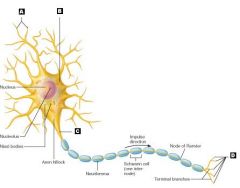
Schwann Cells |
|
|
What is the structural unit of the nervous system? |
Neuron |
|
|
What are the three special characteristics of neurons? |
1. Extreme Longevity 2. Amitotic (can't divide) 3. High Metabolic Rate |
|
|
What are clusters of cell bodies in the CNS |
Nuclei
|
|
|
What are clusters of cell bodies in the PNS |
Ganglia |
|
|
Bundles of neuron processes in the PNS are called |
Nerves |
|
|
Bundles of neuron processes in the CNS are called |
Tracts |
|
|
Movement along the axon away from the cell body
|
Anterograde |
|
|
Movement along the axon towards the cell body |
Retrograde |
|
|
Increases transmission speed of nerve impulses Only covering axons |
Myelin Sheath |
|
|
What are gaps between myelin sheath |
Nodes of Ranvier |
|
|
What is white matter in the brain?
|
Myelinated Fibers |
|
|
What is the gray matter in the brain?
|
Nerve cell bodies |
|
|
What are the three structural classifications of Neurons? |
2. Bipolar 3. Unipolar |
|
|
|
Multipolar Neurons |
|
|
These neurons have two process; an axon and a dendrite |
Bipolar Neurons |
|
|
These neurons have a single short process that emerges from the cell body and divides T-Like into proximal and distal brances |
Unipolar Neurons |
|
|
What are the 3 functional classifications of neurons? |
2. Motor Neurons (efferent) 3. Interneurons |
|
|
Which neurons transmit impulses from sensory receptors in the skin or internal organs toward or into the CNS |
Sensory Neurons |
|
|
Which neurons carry impulses away fromt eh CNS to the effector organs (muscles and glands) of the body |
Motor Neurons |
|
|
Which neurons lie between motor and sensory neurons in neural pathways and shuttle signals through CNS pathways where integration occurs |
Interneurons |
|
|
Interneurons make up over __% of the neurons in the body?
|
99% |
|
|
What is a decrease in membrane potential? |
Depolarization |
|
|
What is an increase in membrane potential? |
Hyperpolarization |
|
|

|
|
|
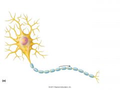
A. Dendrites B. Cell Body C. Axon D. Axon Terminals |
|
|
What is also known as the cell body of a neuron?
|
Soma |
|
|
The rough ER of a neuron is also known as the... |
Nissl Bodies (chromatophilic substance) |
|
|
A junction that mediates information transfer from one neuron to the next or from a neuron to an effector cell
|
Synapse
|
|
|
A potential difference in a resting neuron
|
resting membrane potential |
|
|
What two factors generate the resting membrane potential?
|
2. Differences in the permeability of the plasma membrane to those ions |
|
|
Changes in membrane potential can produce two types of signals:
|
2. Action Potentials |
|
|
What type of potentials are short lived, localized changes in membrane potential that can be either depolarizations or hyperpolarizations |
Graded Potentials (local potentials) |
|
|
A brief reversal of a membrane potential with a total amplitude of about 100mV
|
Action Potential |
|
|
What are the stages of an action potential? |
2. Depolarization 3. Repolarization 4. Hyperpolarization |
|
|
What stage in an action potential has all Na+ and K+ gates closed?
|
1. Resting State
|
|
|
What state in an action potential starts when Na+ channels open. During this stage the stimulation site reaches a critical level called threshold. |
2. Depolarization |
|
|
What stage in an action potential has the Na+ channels closing and the K+ channels opening. |
3. Repolarization |
|
|
What stage in an action potential has some of the K+ channels remaining open and all the Na+ channels reset |
4. Hyperpolarization |
|
|
Period from the opening of the Na+ channels until the Na+ channels begin to reset to their original resting state. No stimulus, no matter the size, can get the neuron to respond and start another action potention
|
Absolute refractory Period |
|
|
Period of time when a stimulus that would normally generate an action potential is no longer sufficient, but an exceptionally strong stimulus can start it
Interval following the absolute refractory period |
Relative refractory Period |
|
|
This type of conduction is 30 times faster than continuous conduction. |
Saltatory Conduction
|
|
|
Part of the axon that contains many tiny, membrane-bounded sacs called synaptic vesicles |
Axon terminal |
|
|
Tiny, membrane bounded sacs in the axon terminal containing thousands of neurotransmitter molecules |
synaptic vesicles |
|
|
Stages of chemical synapse transmitting signals to another neuron |
1. Action potential arrives at axon terminal 2. voltage-gated Ca2+ channels open and Ca2+ enters the axon terminal 3. Ca2+ entry causes synaptic vesicles to release neurotransmitter by exocytosis 4. Neurotransmitter diffuses across the synaptic cleft and binds to receptors on postsynaptic membrane 5. binding of neurotransmitter opens ion channels, creating grading potentials 6. neurotransmitter effects are terminated |
|
|
The means by which each neuron communicates with others to process and send messages to the rest of the body |
Neurotransmitter |
|
|
This embryonic structure is the beginning of the brain and spinal cord
|
Neural Tube |
|
|
What are the three primary brain vesicles? |
2. Mesencephalon (midbrain) 3. Rhombencephalon (hindbrain) |
|
|
What two divisions does the Forebrain divide into? |
2. Diencephalon |
|
|
The hindbrain constricst, forming which two divisions? |
2. Myelencephalon |
|
|
The telencephalon sprouts two lateral swellings that become what?
|
The two cerebral hemispheres, known as the cerebrum |
|
|
What forms the superior part of the brain, and accounts for about 83% of total brain mass? |
The cerebral hemispheres |
|
|
Elevated ridges of tissue that mark nearly the entire surface of the cerebral hemispheres
|
Gyri |
|
|
Shallow grooves that separate the gyri |
Sulci |
|
|
Deep grooves that separate large regions of the brain? |
fissures |
|
|
What separates the cerebral hemispheres? |
longitudinal fissure |
|
|
What separates the cerebral hemispheres from the cerebellum below? |
Transverse Cerebral Fissure |
|
|
What separates the frontal lobe from the parietal lobe? |
Central Sulcus
|
|
|
What separates the occipital lobe from the parietal lobe? |
parietal-occipital sulcus |
|
|
What sulcus outlines the flaplike temporal lobe, and separates it from the parietal and frontal lobes? |
Lateral Sulcus |
|
|
What helps form the floor of the lateral sulcus, and is the fifth lobe? |
Insula |
|
|
What are the three basic regions of the cerebral hemispheres?
|
2. Internal White Matter 3. Basla Nuclei |
|
|
Where conscious mind is found Enables us to be aware of ourselves, our sensations, to communicate, to remember, understand, and initiate voluntary movements |
Cerebral Cortex |
|
|
What are the motor areas found in the frontal lobe?
|
premotor cortex broca's area frontal eye field |
|
|
-located in precentral gyrus of frontal lobe -control precise or skilled voluntary movement -contain pyramidal cells, and pyramidal tracts |
Primary Motor Cortex
|
|
|
-Anterior to the precentral gyrus in the front lobe -Helps plan movement -Selects and sequences basic motor movements -coordinates the movement of several muscle groups simultaneously or sequentially |
Premotor Cortex |
|
|
-Anterior to the inferior region of the Premotor Area -Considered to be present in the left hemisphere only -Motor speech area that directs muscles involved in speech |
Broca's Area |
|
|
-Superior to Broca's Area -Controls voluntary movement of the eyes |
Frontal Eye Field |
|
|
-Resides in the postcentral gyrus of parietal lobe -Neurons receive information from general sensory receptors in the skin -Just posterior to the primary motor cortex |
Primary Somatosensory Cortex |
|
|
-Integrate sensory inputs (temp, pressure, etc) relayed by primary somatosensory cortex to produce an understanding of an object being felt |
Somatosensory Associate Cortex |
|
|
-Buried deep in the calcarine sulcus -Largest cortical sensory area -receives visual information that originals on the retina of the eye |
Primary Visual Cortex |
|
|
-Communicates with primary visual cortex to sue past visual experiences to interpret visual stimuli -Enables recognition |
Visual Association Area |
|
|
-Interprets the impulses from sound energy in the inner ear as pitch, loudness, and location |
Primary Auditory Cortex |
|
|
-Permits the perception of the sound stimulus -Memories of sounds are stored here |
Auditory Association Area |
|
|
-Piriform lobe area -Afferent fibers from smell receptors in nasal cavities send impulse along this area |
Olfactory Cortex |
|
|
-Region involved in perceiving taste stimuli |
Gustatory Cortex |
|
|
-Including: upset stomach, full bladder , etc |
|
|
|
What broad cortex allows ps to give meaning to the information we receive, store it in memory, tie it to previous experience and knowledge, and decide what action to take |
Multimodal Association Cortex |
|
|
-Located in frontal lobe -Most complicated cortical region of all -Involved in: intellect, cognition, recall, and personality -Develops slowly and relies on positive and negative feedback |
Anterior Associate Area (Prefrontal Cortex) |
|
|
The anterior associate area is also known as the... |
prefrontal cortex |
|
|
-Large region encompassing parts of the temporal, parietal, and occipital lobes -Plays a role in recognizing faces and patterns, and binding different sensory inputs into a whole -Understanding written and spoken language |
Posterior Associate Area |
|
|
-Cingulate gyrus, parahippocampal gyrus, hippocampus -Provides emotional impact that makes scene important to us |
Limbic Associate Area |
|
|
Division of labor for each hemisphere of the brain, where each has it's own abilities not completely shared by its partner |
Lateralization |
|
|
Designates the hemisphere that is dominant for language |
cerebral dominance |
|
|
Composed of neuron cell bodies, dendrites, associate glia and blood vessels, but no fiber tracts. Contains billions of neurons arranged in six layers, and accounts for 40% of total brain mass |
Cerebral cortex: Gray Matter
|
|
|
Responsible for communication between cerebral areas and between the cerebral cortex and lower CNS centers -Consists largely of myelinated fibers |
Cerebral White Matter
|
|
|
Fibers that connect different parts of the same hemisphere |
Association Fibers |
|
|
Fibers that connect corresponding gray areas of the two hemipsheres -Allows the two hemispheres to function as a coordinated whole |
Commissural Fibers |
|
|
Largest commissure in brain Lies superior to the lateral ventricles Deep within the longitudinal fissure |
Corpus Callosum |
|
|
Fibers that either enter the cerebral cortex from lower brain or cord centers -Motor output leaves the cerebral cortex through these fibers |
Projection Fibers
|
|
|
Third basic region of each hemisphere -Group of subcortical nuclei |
Basal Nuclei |
|
|
What does the basal nuclei of the cerebral cortex consist of? |
1. Caudate Nucleus
2. Putamen 3. Globus Pallidus |
|
|
Plays a role in cognitions and emotion -Have no direct access to motor pathways -Filters out incorrect or inappropriate responses |
Basal Nuclei |
|
|
What are the five lobes of the cerebrum? |
1. Frontal 2. Parietal 3. Temporal 4. Occipital 5. Insula |
|
|
Forms the central core of the forebrain and surrounded by cerebral hemispheres |
Diencephalon |
|
|
What does the diencephalon consist of? |
1. thalamus 2. hypothalamus 3. epithalamus |
|
|
-Makes up 80% of the diencephalon -THE relay station for information coming into the cerebral cortex -Afferent impulses from all senses and all parts of the body converge on this -Information is sorted out and edited -plays key role in mediating sensation, motor activities, cortical arousal, learning, and memory |
Thalamus |
|
|
-Main visceral control center of the body -Vitally important to overall body homeostasis -Chief Roles: control autonomic nervous system, initiate physical responses to emotion, regulate body temperature, regulate food intake, regulate water balance, regulate sleep-wake cycles, control endocrine system functions |
Hypothalamus |
|
|
Emotional part of the brain |
Limbic System |
|
|
-Most dorsal portion of the diencephalon -Pineal Gland extends from the posterior border -Helps regulate sleep-wake cycle |
Epithalamus
|
|
|
Regions of the brain stem |
1. Midbrain 2. pons 3. Medulla Oblongata |