Reading...
![]()
Play button
![]()
Play button
![]()
Use LEFT and RIGHT arrow keys to navigate between flashcards;
Use UP and DOWN arrow keys to flip the card;
H to show hint;
A reads text to speech;
60 Cards in this Set
- Front
- Back
|
BLOOD SUPPLY
|
- receives 20% of blood
- flow to brain via: INTERNAL CAROTID AND VERTEBRAL ARTERIES. - Blood can be shifted from one brain regulator to another - Glucose: E substrate neuron that metabolizes for ATP... but is not stored in brain so needs constant blood supply. |
|
|
BBB: Blood Brain Barrier
|
Regulates substance exchange between BLOOD & BRAIN
-Formed by : ASTROCYTES + TIGHT JUNCTION between capillary endothelial calls and cap. BM.. - permeable to lipid soluble and carrier transported subs. - BBB is the reason why some meds are hard to administer in brain - can be damaged by inflammation and trauma. |
|
|
CSF: Cerebrospinal fluid
liquid that surrounds CNS |
Functions:
1. Mechanical protections: -shock absorbing, -buoyancy: effective weight of brain way less than the actual weight. 2. Homeostatic function: - must maintain CSF pH bc it affects pulmonary ventilation, blood flow. - also deals with transporting hormones. 3. Circulation: - acts as a medium for exchange of nut. & waste products b/w blood and Nervous tissue. |
|
|
Blood- CSF barrier
|
by tight junction between the ependymal cells
This decreases harmful materials from blood into CSF. |
|
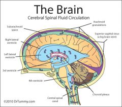
CSF circulation
|
Ventricles: 4 CSF filled chambers in brain (continous).
2 are lateral: large, one for each cerebral hemisphere 1 is 3rd ventricle: midsagittal plane; inferior to corpus collosum 1 is 4th ventricle: smal triangle shaped; between pons and cerebellum. Process: LV > intervent. foraminia > 3rd. > Cerebral aqueduct > 4th > central canal> apertures (lateral (to subarachnoid) or median) >subarachnoid space> arachnoid villi (allow CSF to enter bloodstream bc it is protrudes to...) > dural venous sinuses> blood> LV |
|
|
Clinical
|
Normal: CSF production rate = CSF reabsorption rate
BUT: If ^ CSF prod. or low reabs. (caused by obstruction (blockage)) ---> HYDROCEPHALUS (water in head) HYDROCEPHALUS: high pressure due to high CSF in ventricle In childhood: enlarged head In adult: venticular dilation and compressed brain tis. |
|
|
Clinical cont'd
|
Concussion: temp loss of consciousness bc of blow to head
CONTUSION: brain bruise SUBDURAL HEMATOMA: bleeding into subdural space, not as rapid - this is why they tell people not to sleep EPIDURAL HEMATOMA: FAST; bleed bw dura & skull; usually middle meningeal a. bleed. MENINGITIS: by bacterial or viral infection; lumbar puncture is the diagnosis. |
|
|
The Brain Stem
|
- medulla, pons, and midbrain
- bw s.c and diencephalon - produces programmed behaviors for survival - pathway for fiber tracts bw s.c and cerebrum |
|
|
MEDULLA
|
- superior part of s.c but wider
- starts from foramen magnum and extends to pons. - contains motor and sensory tract. -has last 5 cranial nerve nuclei (VIII, IX, X, XI, XII) -contains medullary "centers" = neurons controlling heart rate, respiration, bv diameter, coughing, swallowing, vomiting |
|
|
"Pyramids" of medulla
|
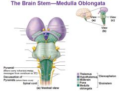
-2 large columns of motor neuron tracts on ventral anterior surface
- they are separated by anterior median fissure - has CORTICOSPINAL TRACTS: control voluntary limb movement |
|
|
Decussation of pyramids
|
Look at previous image
at junction of medulla with the s.c where 90% of corticospinal axon cross with message from cerebrum to s.c EACH SIDE OF BRAIN CONTROLS VOLUNTARY MOVEMENT ON OPPOSITE SIDE OF BODY... everything has crossed over at s.c |
|
|
"OLIVES"
|
- oval like structure lateral to the pyramids
- contains "inferior olivary nuclei" responsible for sending axon to cerebellum. |
|
|
Pons... the bridge
|
- pontine nuclei= synaptic RELAY STATION bw cerebrum and cerebellum
- coordination of voluntary motor output. - has Ascending/sensory ( stimulus to PNS TO CNS) and DESCENDING/motor tracts - helps control breathing using: pneumotaxic and apenustic (promotes inhalation) areas. (antagonize each other) Cranial nerve nuclei: V, VI VII, VIII |
|
|
Midbrain
(mesencephalon) |
b/w pons and diencephalon
contains cerebral aqueduct (plays a part in CSF circulation). -made up of cerebral peduncles, substantia nigra, tegmentum, and tectum |
|
|
Cerebral peduncles
|
- ventral surface of midbrain
- contain corticospinal, coticopontine, and corticobulbar motor neuron tracts CORTI-... |
|
|
Substantia nigra (XS) (seen only when cross section the midbrain)
|
- black nucleus bw peduncles and tegmentum
- dark bc if high level of melanin DOPAMINE - motor center containing dopamine neurons that extend to the basal nuclei > thus help with controling muscles activity. PARKINSON DISEASE: caused loss of neuron association/ loss of dopamine |
|
|
Tegmentum (XS)
|
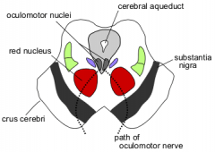
- has red nucleus ---> rubrospinal tract origination. (motor control pathway)
- receives axons from cerebrum and cerebellum--> some movement coordination. |
|
|
Tectum
|
- dorsal part of midbrain
- corpora quadrigemina 4 rounded bodies (colliculi) in the tectum: Superior Collicili: Upper pair; some optic tract input > VISUAL REFLEXES INFERIOR COLLICULI: lower pain; AUDITORY REFLEXES Cranial nerve of mid brain : III and IV |
|
|
Reticular formation
|
arrangement of white and gray matter from upper s.c to BS to inferior diencephalon
- neurons have both ascending (sensory) and descending (motor) functions: 1. Somatic motor control: muscle tone regulation 2. Autonomic input: parasympathetic and sympathetic 3. arousal: ascending fibers activate the cerebral cortex |
|
|
RAS: Reticular activating system
|
regulates arousal and sleep-wake transition
- prevents sensory overload Damage can cause coma. NO input from olfactory receptors. |
|
|
Cerebellum
|
posterior to medulla and pons (seperated by 4th ventricle)
- highly folded... thus higher surface area - Tentorium cerebelli causes lower pressure on cerebellum bc it separates the cerebellum from occipital. Vermis: connects the two hemisphere of cerebellum at a central area Cerebellar cortex: superficial gray mater, has folds called folia, separated by grooves (sulci) Arbor vitae: tracts of white matter, deep of gray matter. |
|
|
Cerebellar connection ( 3 paired tracts)
|
1. Superior cerebellar peduncles - axons connecting to midbrain and thalamus
2. Middle cerebellar peduncles - pons connection 3. Inferior cerebellar peduncles - medulla connection |
|
|
Cerebellar Function
|
checks and compares if the muscle is doing what it is told by the cerebral cortex, corrects it otherwise
Damage= uncoordinated movements (ATAXIA) and low muscle tone. How to test: finger to nose and heel to shin test. |
|
|
Diencephalon (2nd division of forebrain)
|
- superior to BS midbrain
surrounded by cerebral hemisphere surrounds the 3rd ventricle. Thalamus, hypothalamus, and epithalamus. |
|
|
Thalamus ( must cut the brain to see)
|
80% of diecephalon
-2 oval masses of cell bodies (each organized as 7 groups of nuclei) -intermediate mass: neuron bridge connecting the oval masses (passes through the 3rd ventricle) -each mass "embedded" in a cerebral hemisphere, underlies cortex, and lateral ventricle, and each mass is bound laterally by an "internal capsule" |
|
|
Internal capsule
|
White matter connecting thalamus with cerebral cortex
|
|
|
Thalamus function
|
GATEWAY to cerebral cortex
MAJOR RELAY startion for most SENSORY input nuclei integrate sensory, motor, emotions, and learning Information that reaches thalamus doesn't need to reach the cerebral cortex (thus won't reach conscious level) so thalamus decides what reaches cerebral cortex. For example: when you are studying, you see bunch of things around you, but you don't concentrate on that because thalamus won't let it reach the conscious level. |
|
|
Hypothalamus (HT)
|
- inferior to thalamus
- extends from the anterior "optic chiasm" to the posterior "mammillary bodies" (visible in ventral view) -infundibulum = attaches the pituitary to the HT -merges into midbrain of brain stem -composed of many nuclei organized into 4 main regions (preoptic, supraoptic, tuberal, and mammillary) |
|
|
HT function
|
ANS control
hormone production regulation of emotion and behavior (heart of limbic system) regulation of eating and drinking control of body temp sleep wake cycle regulation... regulation of circadian rhythms and consciousness. |
|
|
Epithalamus
|
dorsal part of dienecephalon
thin roof over 3rd ventricle has pineal gland also has habenula : relay system for limbic system (emotional responses to odor). |
|
|
Pineal gland
|
in epithalamus
a midline marker, makes melatonin , which induces sleep. HIGH in teens regulate circadian rhythms (sleep) |
|
|
Circumventricular organs
|
- at the walls of 3rd ventricle
BBB is absent allow brains to monitor and respond to blood composition BUT may be the entry sites for pathogens into brain (HIV) |
|
|
CEREBRUM
|
-seat of intelligence
-right and left hemispheres -cerebral cortex, cerebral white matter, and basal nuclei |
|
|
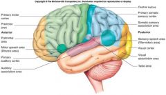
Cerebral Cortex
|
-outer surface of gray matter
-contains billions of neuron -folding of cortex - increased surface area (a large part of cortex is not visible because hidden in the fissures and sulci) -pattern variation from one brain to another BUT some are constant enough to serve as landmarks --> lobes (frontal, parietal, occipital, temporal, and insula) |
|
|
Anatomy of cerebral cortex
|
Gyrus: fold on surface of cortex, separated by grooves
Fissures... Deep grooves Sulci... Shallow grooves |
|
|
Separation
|
Longitudinal fissure: rt and lt hemisphere division
Central sulcus: 90deg to L.F. Frontal and parietal division Parieto occipital sulcus: seperates parietal and occipital lobes Transverse cerebral fissure: separates cerebral hemisphere from cerebellum |
|
|
Function of each lobes
|
Frontal: motor! Motivation, judgement, aggression, decision making (most developed in human).
Parietal: sensory info processing, like feeling pain Occipital: visual info Temporal: hearing, learning and memory, emotion, visual recog Insula: deep inside in lateral fissure |
|
|
Cerebral white matter
|
Myelinated axon tracts
Association tracts: connect area of cerebral cortex in the same Hemisphere Commissural tracts: Connect area of cerebral cortex in the opposite hemisphere (Transfer learned information from one hemisphere to another) Projection tracts: Descending and ascending tract. Internal capsule Has both ascending and descending axon |
|
|
Basal nuclei (not ganglia bc It's located in the central nervous system not PNS)
|
3 nuclei deep within each hemisphere
1. Globus pallidus: lateral to thalamus 2. Putamen: lateral to GP P + GP = lentiform nucleus 3. Caudate: C shaped ( head body and tail); + putamen= corpus striatum Substantia nigra and subthalamic nuclei Are functionally linked to basal nuclei |
|
|
Function of BN
|
Control subconscious skeletal muscle contraction
Regulate muscle tone Facilitate some muscle contraction while inhibiting others thus Fine motor control Damage can cause muscular rigidity And termors... Parkinson's disease Other roles: Involved in attention and memory planning and act with limbic system... Dysfunction of both can cause psychiatric disorder |
|
|
Limbic system (Oldest component of cerebrum)
|
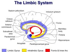
Surrounds thalamus and corpus callosum
Cingulate gyrus, Hippocampus, Dentate gyrus, septal nuclei, olfactory bulb, Mammillary bodies, Fornix, And amygdala Involved in emotional responses memory and smell |
|
|
Clinical notes
|
Lesion or rumors can can emotional disturbance: Such as uncontrollable laughter or cry
Temporal lobe tumor can cause aggression or memory loss Psychosomatic illness : LS connects to HT > Emotion induced illness.... Thus stress causes sickness Amnesia is lesion in hippocampus Korsakoff's syndrome: Amnesia caused by vitamin B12 (thiamine) Deficiency...due to alcoholism or starvation... This affects the limbic system structures |
|
|
Generalization about cortex
|
1. Three functional areas
2. Each hemisphere is concerned with opposite side of the body 3. Some lateralization (Control by one hemisphere or the other) |
|
|
Functional areas: 1deg Motor area
|
Precentral gyrus ( of frontal lobe)
-Control skeletal muscle Somatotopy: Corresponding between groups of neurons in the gyrus and muscle on the opposite side of the body (contralateral) - broca's speech area : Located in frontal lobe near lateral sulcus - motor homunculus is Disproportional. High area means finer control.... Mouth and hand |
|
|
2. Sensory areas
|
1deg Somatosensory area
Postcentral gyrus (Parietal lobe) Receives general sensory information such as temperature touch pressure pain from opposite side of the body Also: somatotopy: Sensory homunculus |
|
|
Special sensory areas
|
information sent somewhere other than somatosensory area
1. 1° visual area - posterior occipital lobe 2. 1° auditory area - superior temporal lobe 3. 1° gustatory area - taste, base of postcentral gyrus in parietal lobe 4. 1°olfactory area (smell) medial temporal lobe |
|
|
3. Association areas
|
All areas of the cortex had except the primary (1deg)
Surrounds the sensory and the motor areas Processes and Interpret info |
|
|
Premotor area = motor Association area
|
Anterior to primary motor area
It's where the voluntary muscle contraction is initiated.... Causes muscles to contract in sequence which is based on learned events |
|
|
Somatosensory association area
|
Posterior to primary sensory area in parietal lobe
Receives input from primary sensory area Remembers object which means allows determination of objects space and texture Determination of body position |
|
|
Visual association area
|
At occipital lobe
Posterior to somatosensory areas Allows identification of objects that you see Facial recognition is part of inferior temporal lobe |
|
|
Auditory Association area
|
Part of the temporal lobe
Allows us to realize whether sound is this speech music or just noise |
|
|
Frontal association area
|
Aka Prefrontal Area
Intellectual activities such as judgment |
|
|
Hemispheric lateralization
|
Functionally asymmetrical
Both Hemisphere share performance of many function but they're also specialize Left hemisphere: speech grammar problem-solving reasoning analytical Right hemisphere processing information in more integrated way such as imagination, artistic, recognize complex visual and music patterns |
|
|
Characterization of left and right Hemisphere
|
Cerebral dominance : the left hemisphere dominance for language which is 97%
Unilateral inattention: Lesion of inferior parietal lobe Ignores the stimuli coming from contralateral side of the body or the opposite side of the body Lateralization develops with age and it's more distinct in male so they have a harder time speaking after getting a stroke |
|
|
Language
|
Broca's: Generates motor program causing muscles to produce speech
Wernicke's: Recognizes spoken and written language. Damage can cause fluent aphasia (word salad) (Language defect hard time understanding) |
|
|
Affective language area
|
Gives emotion to speech
Damage can cause aprosodia : Flat emotionless speech |
|
|
Brain waves
|
Action potential of brain neurons
Recorded by electrode on scalp... Recording is called EEG.... Helps the diagnosis of brain disorder and studies normal brain function. Example: Epileptic seizure |
|
|
Cranial nerves
|
Nerve fiber: Axon of Single neuron
Nerve: Structure made up of a collection of Axon in PNS Connective tissue covering the nerves: Endoneurium, perineurium, and Epineurium. Nerve fiber classifications: afferent: receptor to CNS Efferent: CNS to effector |
|
|
Classification of nerves
|
mixed nerve : contain both sensory (afferent) fibers and motor (efferent) fibers
Sensory (1,2,8) Motor (3,4, 6, 11, 12) |
|
|
Mnemonic of 12 pairs of cranial nerves
|
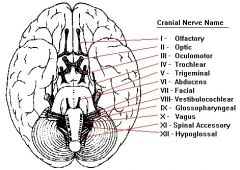
oh oh oh to touch and feel a(V) girl's V sure helps
some say money matters but my brother says big brains matter more |