![]()
![]()
![]()
Use LEFT and RIGHT arrow keys to navigate between flashcards;
Use UP and DOWN arrow keys to flip the card;
H to show hint;
A reads text to speech;
47 Cards in this Set
- Front
- Back
|
Blood
|
Fluid CT tissue. Carries oxygen, nutrients, waste.
|
|
|
Blood : cell components
|
cellular component:
leukocytes, erythrocytes, and thrombocytes |
|
|
Blood: Matrix component
|
plasma
|
|
|
Blood : fibrous component
|
fibrin
|
|
|
LTT vs RTT (SST) vs GTT
|
lavender top: whole blood
RTT: serum GTT: Plasma |
|
|
Plasma
|
liquid minus blood cells
55% top layer |
|
|
Buffy coat
|
leukocytes (middle layer)
|
|
|
Erythrocytes
|
Red blood cells (bottom layer) 45%
|
|
|
Serum
|
liquid minus blood cells and clotting elements
|
|
|
Clot
|
blood cells enmeshed in fibrin
|
|
|
Hematopoiesis 3 types & where |
Hematopoiesis (mostly red marrow, continuous. Can be in yellow and spleen and liver in crisis but alway in fetus), Erythropoiesis , thrompoiesis, leukopoiesis |
|
|
Erythropoiesis
|
(multiple maturation steps, rate controlled by hormones.
EPO released from kidney in response to hypoxia) |
|
|
Thrombopoiesis
|
production of platelets.
Stem cell to megakaryocyte, pieces of megakaryocyte released as platelets, multi-nucleated megakaryocyte stays in marrow |
|
|
Leukopoiesis
|
formation of WBC's.
Granulopoiesis, lymphopoiesis, monopoiesis |
|
|
Erythrocytes
|
highly specialized.
No nucleus, no mitochondria, no ribosomes. RBC's continue to use plasma glucose until depleted. Transport oxygen and carbone dioxide, maintain shape. 20-30 days in mice, 150 in large animals |
|
|
senescence
|
process of aging, enzyme activity decreases , membrane loses deform-ability,
1% of cells removed daily, extracelluar vs intracelluar |
|
|
extravascular hemolysis
|
90%.
Splenic macrophages remove RBCs from circulation Cell membranes rupture and release hemoglobin (Hb) Hb degraded to amino acids, iron, and heme Amino acids return to liver to be recycled in new proteinsIron returns to bone marrow to be recycled in new RBCs Heme degraded to bilirubin Bilirubin attaches to albumin and goes to the liver Bilirubin is conjugated to glucuronic acid and excreted into the intestines as bile pigment Bacteria converts bilirubin to urobilinogen Some is reabsorbed Excreted as urobilin in urine or stercobilin in feces |
|
|
Intravascular hemolysis
|
Destruction takes place within blood vessels
RBC ruptures within a vessel Hb released into bloodstream Hb binds to protein in plasma Travels to macrophages in liver – proceeds as with extravascular hemolysis Excess unconjugated hemoglobin (hemoglobinemia) is carried to kidneys and eliminated in urine (hemoglobinuria) |
|
|
CBC
|
Hct – volume of RBCs expressed as % (vs. PCV)
Hemoglobin RBC count Mean corpuscular volume (MCV) – average volume of individual RBCs Red cell distribution width (RDW) – numerical expression of variations in RBC sizes – anisocytosis Reticulocyte count – count of immature RBCs Leukocyte count (WBC) & differential Platelet count – thrombocytosis, thrombocytopenia |
|
|
Hemoglobin: 2 types |
Oxyhemoglobin
Deoxyhemoglobin |
|
|
Blood smears |
wright's stain, diff-quick stain. New methylene blue (reticulocyte count - immature erythrocytes) |
|
|
Platelets |
thrombocytes. pieces of cytoplasm from megakaryocytes (bone marrow). non-nucleated round/oval, size varies by species 5-7 day lifespan liver produces thrombopoietin macrophages remove old platelets from circulation blue to purple granules (contain calcium & clotting factors) |
|
|
hemostasis
|
platelet adhesion and aggregation. thrombin.
coagulation cascade (13 enzymes including fibrin) |
|
|
Clotting disorders: 3 |
Immune-mediated Thrombocytopenia (ITP) -
petechiae, ecchymosis(eccymosis is small petechiae) von Willebrand Disease (vWD) (dobey's, excessive or extensive bleeding) Anticoagulant rodenticides |
|
|
White blood cells |
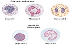
Granulocytes -
Agranulocytes -
|
|
|
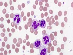
Granulocytes: |
prominent granules when stained Eosinophils – acidic stain – appear red Basophils – basic stain – appear blue Neutrophils – don’t pick up either stain well – appear colorless or faintly violet |
|
|
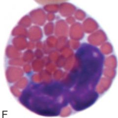
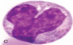
Neutrophils |
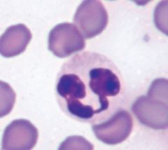
40-75% of circulating leukocytes most abundant type in dogs, cats, horses immature neutrophils are band cells mature cells are polymorphonuclear (PMN) replaced 2 1/2 times a day |
|
|
Hypersegmented Neutrophils – |
Anemia |
|
|
Neutrophil function (3) |
granulocyte early inflammatory response Diapedesis – process of neutrophils going from circulation to tissue spaces |
|
|
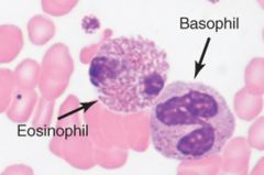
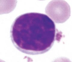
Eosinophils |
granulocyte segmented nucleus - usually 2 lobes larger than neutrophil allergy and parasites shown: horse |
|
|
Basophils |
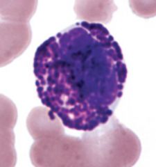
granulocyte multi-lobed nuceli least phagocytic of the granulocytes histamine and heparin, similiar to mast cells (IgE) heartworm |
|
|
lymphocytes |
Agranulocytes Round or oval nucleus live in lymphoid tissue and circulate between tissues and blood 3 types: T lymphocytes (T cells) B lymphocytes (B cells) Natural Killer cells (NK cells) |
|
|
T Cells
|
lymphocyte
most abundant lymphocyte in blood processed in thymus *cell mediated immunity* activate B cells |
|
|
Agranulocyte types |
lymphocyte monocyte |
|
|
B Cells |
lymphocyte inactive b cells are in lymphoid tissue antibody production each cell produces ONE antibody for ONE antigen Epitope - surface receptors fit only one antigen shape |
|
|
Humoral immunity |
B cell recognizes an antigen by forming an antigen antibody complex b cells transform into plasma cell and produces, stores and releases antibodies |
|
|
Natural Killer Cells
|
granular lymphocytes ID and kill virus-infected cells bind to cell and stimulates apoptosis (doesnt injest) 2 types of receptors : KAR and KIR (killer activating and killer inhibiting receptor) stimulated by cytokines (interleukins and interferons) |
|
|
memory cells |
T and B cells can become memory cells clones of original lymphocyte wait in lymphoid tissue for antigen faster and stronger response |
|
|
monocytes |
nuclei can vary in shape inflammatory response known as macrophages after entering tissue clean up cellular debris |
|
|
lymphatic system |
system of ducts, fluid LYMPH, and lymphoid organs/tissues System picks up fluid leaked from capillaries (due to hydrostatic and osmotic pressures), filters it through lymph nodes, and deposits it back in the circulatory system near the heart |
|
|
lymphatic functions (4) |
removes excess fluid (prevents edema) transports waste material filtration of lymph protein transport |
|
|
Lymph |
transparent liquid mostly lymphocytes chyle is lymph from digestive system |
|
|
lymphoid organs |
thymus bursa of fabricius peyer's patches also; spleen, lymph nodes, tonsils |
|
|
Thymus
|
prominent in young animals produces mature T cells cells leave thymus and travel to lymph tissue bursa of fabricius - above cloaca |
|
|
peyer's patches
|
Wall of small intestine (GALT, gut-associated lymphoid tissue) Structure and function varies among species Activate B cells to produce antibodies |
|
|
Lymph nodes |
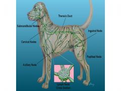
filters traps antigens and other foreign bodies |
|
|
tonsils |
No capsule (unlike LNN) |