Reading...
![]()
Play button
![]()
Play button
![]()
Use LEFT and RIGHT arrow keys to navigate between flashcards;
Use UP and DOWN arrow keys to flip the card;
H to show hint;
A reads text to speech;
67 Cards in this Set
- Front
- Back
|
Purpose of having folded pattern of cortex?
|
allows for an increase in the SA of the cortical gray matter without a corresponding increase in size of the cranial vault
|
|
|
Compare
Gyrus Sulcus Fissure |
Gyrus - elevation on surface of hemisphere
Sulcus - depression or groove between gyri Fissure - a large and constant sulcus may be called a fissure, but the 2 terms are sometimes used interchangeably |
|
|
In which fissure can you visualize the insula?
|
opening lateral fissure
|
|
|
What are the boundaries of the frontal lobe?
|
On lateral surface:
posterior boundary: central sulcus Inferior boundary: lateral sulcus On medial surface: posterior: arbitrary line from central sulcus to CC inferior: CC/cingulate gyrus (limbic lobe) |
|
|
What are the boundaries of the parietal lobe?
|
on lateral surface:
anterior: central sulcus interior: lat sulcus and an arbitrary line directed posteriorly from the lateral sulcus Posterior: upper half of an arbitrary line connecting parieto-occiptal sulcus with the pre-occipital notch Medial surface: Anterior: frontal lobe Posteror/inferior: parieto-occipitosulcus and cingulate gyrus |
|
|
What are the boundaries of the temporal lobe?
|
on lateral surface:
superior: lateral sulcus and it posterior projection posterior: lower portion of arbitrary line connecting the parieto-occital sulcus and the pre-occipital notch on medial surface: imaginary line joining the parieto-occipital sulcus and the pre-occipital notch |
|
|
What are the boundaries of the occiptal lobe?
|
lateral surface: line joining the parieto-occipital sulcus to the pre-occiptal notch
medial: posterior borders of the parietal and temporal lobes |
|
|
4 functional groups of the cortex?
|
Motor areas
Sensory areas Language areas Frontal association areas |
|
|
General role of association areas of the cortex
|
produce a meaningful perceptual experience of the world, enable us to interact effectively, and support abstract thinking and language.
|
|
|
Components of the primary motor cortex
|
Precentral gyrus
Bordered by: Precentral sulcus + Central sulcus |
|
|
Consequence of lesion to primary motor area
|
UMN signs
|
|
|
Location of the premotor or motor association areas
|
includes:
anterior part of precentral gyrus parts of sup, middle and inf frontal gyri |
|
|
Consequences of a lesion to the premotor or motor association area
|
Apraxia = deficits in learned, skilled motor activity, in absence of paralysis.
|
|
|
Location of primary somatosensory area
|
postcentral gyrus
|
|
|
Consequence of lesion to primary somatosensory area
|
- decreased awareness of sensory info
- reduced proprioception, touch and pain sensation |
|
|
Location of the somatosensory association area
|
superior parietal lobules, extending onto medial surface
|
|
|
Consequence of lesion to somatosensory association area
|
Tactile agnosia = Impaired ability to recognize or identify objects by touch alone.
|
|
|
Location of the primary auditory area
|
superior surface of superior temporal gyrus - Heschl's gyri (aka transverse temporal gyri)
|
|
|
Consequences of a lesions to primary auditory area
|
- subtle impairment in hearing
- cannot localize sounds - usually okay because bilateral input |
|
|
Location of auditory association area
|
superficial temporal gyrus and area posterior to primary auditory area in lateral sulcus
|
|
|
Consequences of a lesions to auditory association area
|
Deficit in sound interpretation (even though can hear normally)
|
|
|
Location of primary visual area
|
in walls of posterior part of calcarine sulcus, extending onto lateral surface
|
|
|
Consequences of lesion to primary visual area
|
blindness in opposite visual field
|
|
|
Location of visual association area
|
surround primary visual are on medial and lateral surfaces
|
|
|
Consequences of lesion to visual association area
|
- Visual agnosia = unable to recognize object in opposite visual field, despite intact vision
- also, deficit in pursuit or tracking in IPSILATERAL eye |
|
|
Location of the primary taste area
|
- on insula and adjacent medial surface of parietal-frontal operculum at base of centra sulcus (operculum = lid or cover, overlying cortical area)
- can be visualized deep in lateral fissure btw temporal and frontal lobes |
|
|
Location of secondary taste area
|
orbital cortex of frontal lobe and amygdala
|
|
|
Function of secondary taste area
|
taste information is integrated with olfactory information
|
|
|
Functions of frontal association areas
|
aka prefrontal cortex
- concerned with complex aspects of behaviour (ie affect, personality, attention) - extensive connection with dorsomedial (DM) nucleus of thalamus |
|
|
Consequences of lesion to frontal association cortex
|
- change in emotion, motivation, personality, initiative, judgement, conentration, social behaviour, carelessness of appearance/dress
|
|
|
Location of motor speech area of broca
|
part of inferior frontal gyrus of dominant hemisphere (usually left)
* upside down U shape near intersection of premotor area and lateral sulcus |
|
|
Function of broca's area
|
expression of speech
|
|
|
Consequences of lesion to broca's area
|
Nonfluent aphasia:
- cannot get words out properly even though normal comprehension - aware of problem |
|
|
Location of sensory speech are of Wernicke
|
- posterior part of the superior temporal gyrus, with extensions around the posterior end of the lateral sulcus into the parietal region (DOMINANT hemisphere - usually in left)
* armpit of lateral fissure |
|
|
Function of wernicke's area
|
reception of speech (comprehension)
|
|
|
Consequences of lesion to wernicke's area
|
Fluent aphasia
speech fluent but nonsensical - usually unaware of the problem |
|
|
Consequence of lesion to language are of NON dominant hemisphere
|
Aprosodia
= deficit in intonative, rhythm |
|
|
Types of association fibers
|
Short: connect cortical areas in adjacent gyri
Long: pass bw cortical areas that are further removed from each other |
|
|
Superior longitudinal fasciculus
|
aka arcuate fasciculus (because arches over lateral fissure)
- located above the insula - connects: frontal <-> parietal <-> temporal lobes - connects wernickes and broca's areas |
|
|
Consequence of lesion to arcuate fasciculus to speech
|
Conduction aphasia
Speech fluent but paraphasia word substitutions errors (ex treen instead of train) * due to sensory from broca not being properly passed to broca |
|
|
Inferior occipitofrontal fasciculus
|
- runs below insula
- connects: frontal <-> temporal <-> occipital lobes |
|
|
Unicate fasciculus
|
- part of inferior occipitofrontal fasciculus
- connect frontal and temporal lobe |
|
|
Superior occipitofrontal fasciculus
|
- fibers fun adjacent and perpendicular to corpus collosum along most of its course
- connect: occipital and frontal lobes |
|
|
Cingulum
|
- runs beneath cingulate gyrus and parahippocampal gyrus
- connects areas of limbic cortex with each other` |
|
|
Commisural fibers
|
- connect R and L hemispheres
- largest one: corpus collosum |
|
|
Connections made with body of CC
|
parietal lobes
posterior part of 2 frontal lobes |
|
|
Connections made with splenium of CC
|
connects occipital lobes
posterior temporal lobes |
|
|
Connections made with genu of CC
|
fibers originating in or proceeding to frontal areas
|
|
|
Connections made with rostrum of CC
|
thin shelf of fibers projecting backwards in genu
|
|
|
Connection made within radiations of CC
|
fibers fan out as they project to all parts of cortex
forceps minor : at anterior end foreceps major : at posterior end |
|
|
Anterior commissure
|
connects:
two anterior temporal lobes and olfactory bulbs |
|
|
Posteior commissure
|
connects the two pretectal nuclei
|
|
|
Projection fibers
|
thalamus <-> cortex
descending fibers to: striatum, brainstem and spinal cord |
|
|
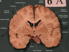
Internal capsule
|
compact bundle formed from gathering of projection fibers
|
|
|
Limbs of internal capule
|
anterior limb: cuts bw head of caudate and lenticular nucleus
posterior limb: cuts bw thalamus and lenticular nucleus Genu = where two limbs meet |
|
|
What is the lenticulate nucleus?
|
= putamen + globus pallacius
|
|
|
Location of basal nuclei and thalamus in relation to internal capsule
|
- caudate nucl. and thalamus always medial
- palladium and putamen always lateral |
|
What type of information is processed by the thalamus?
|
- all sensory information (except olfaction)
- basal ganglia - cerebellum |
|
|
Major sensory relay nuclei of the thalamus + functions
|
- Ventral posterior (VPL, VPM) = somatosensory
- Medial geniculate nucleus (MGN) = hearing - Lateral geniculate nucleus ( LGN) - vision |
|
|
Major motor nuclei of the thalamus and their functions
|
- ventral lateral (VL)
- Ventral anterior (VA) * connect with basal ganglia and cerebellum |
|
|
Thalamic Limbic nuclei ?
|
Limbic nuclei in anterior thalamus connects with cingulate gyrus
|
|
|
MCA supplies..
|
most of lateral surface of the brain
|
|
|
ACA supplies..
|
medial surface of frontal and parietal lobes
* overlap onto lateral surface and supply thin border of the cortex |
|
|
PCA supplies...
|
medial surface of temporal and occipital lobes
* overlap onto lateral surface and supply thin border of the cortex |
|
|
Which vessels in brain are particularly susceptible to high BP?
|
- inferior choroidal aa.
- deep penetrating arteries of the MCA |
|
|
What provides most blood supply to uncus?
|
Anterior choroidal arteries ( typically off int. carotid but sometimes arise from MCA)
|
|
|
What supplies internal capsule and basal ganglia?
|
Lenticulostriate arteries ( medial off ACA or lateral off MCA)
|