![]()
![]()
![]()
Use LEFT and RIGHT arrow keys to navigate between flashcards;
Use UP and DOWN arrow keys to flip the card;
H to show hint;
A reads text to speech;
295 Cards in this Set
- Front
- Back
|
Be able to describe the structural organization of the human body (from atom toorganism) |
Chemical Level (atoms), Cellular Level, Tissue Level, Organ Level, System Level, Organismal Level |
|
|
Anatomical Position |
Standing erect, Facing observer, Eyes facing forward, Feet flat and facing forward, Upper limbs at side with palms facing forward. |
|
|
Prone |
lying face DOWN |
|
|
Supine |
lying face UP |
|
|
Superior/cephaled |
toward the head or upper part of a structure |
|
|
Inferior/caudal |
away from the head or lower part of a structure |
|
|
Anterior/ventral |
toward the front of the body |
|
|
Posterior/dorsal |
toward the back of the body |
|
|
Medial |
closer to midline
|
|
|
lateral |
farther from midline |
|
|
intermediate |
in between 2 structures |
|
|
ipsilateral |
on same side of the body |
|
|
contralateral |
on opposite side of the body |
|
|
Proximal |
closer to attachment of limb to trunk or closer to origin of a structure |
|
|
Distal |
farther from attachment of limb totrunk, farther from the origin of a structure |
|
|
Superficial/external |
toward the body surface |
|
|
Deep/internal |
away from the body surface |
|
|
major body cavities
|
cranial, thoracic, abdominopelvic |
|
|
membranes of the thoracic cavity |
membranes of the lungs membranes of the heart |
|
|
membranes of the lungs |
Parietal Pleura (right and left) Visceral Pleura (right and left) Pleural Cavity |
|
|
membranes of the heart |
Parietal Pericardium Visceral Perdicardium Pericardial Cavity |
|
|
membranes of the abdominopelvic cavity |
peritoneum |
|
|
membranes of the peritoneum
|
Parietal Peritoneum Visceral Peritoneum Peritoneal Cavity |
|
|
serous membrane |
double layered membrane |
|
|
parietal layer |
thin layer that lines the walls of the cavities |
|
|
visceral layer |
thin layer that covers the organs (viscera) |
|
|
abdominpelvic quadrants |
right upper right lower left upper left lower |
|
|
which abdominopelvic quadrant would you find a majority of the liver and the stomach? |
left upper |
|
|
3 main parts of the cell |
plasma/cell membrane (plasmalemma) cytoplasm nucleus |
|
|
active transport |
Substances move AGAINSTthe concentration/electrical gradient and require energy in the form of ATP. |
|
|
types of active transport |
primary secondary vesicular |
|
|
passive transport |
Substances move DOWNthe concentration/electrical gradient. (Meaning it moves to area of LOWERconc.) |
|
|
types of passive transport |
Simple diffusion Channel-mediated facilitated diffusion Carrier-mediated facilitated diffusion Osmosis |
|
|
isotonic solution |
soln. that contains the same [solutes] as thecytosol. |
|
|
hypotonic solution |
soln. containing a lower [solute]than the cytosol. This type of soln causes the cellto SWELL. |
|
|
hypertonic solution |
soln. containing a higher [solute]than the cytosol. This type of soln causes the cellto SHRINK |
|
|
lipid bilayer |
FluidMosaic Model Made upof: phospholipids,cholesterol, and glycolipids Bilayer: result of amphipathicproperties Hydrophobic tails Hydrophilic heads Separates cytosol from ECF |
|
|
centrosomes |
Contain centrioles and pericentriolar material. Centrioles composed of 9 microtubule triplets organized into acircular pattern. Function: organizing center for mitotic spindle (cell division)and for microtubule formation in nondividing cells. |
|
|
cilia |
Short, hairlike projections. Composed mostly of microtubules. Function: move fluid along the cells surface |
|
|
flagella |
Similar to cilia, but longer. Composed mostly of microtubules. Function: move entire cell; ex: sperm |
|
|
ribosomes |
Found on outer surface of nucleus, within endoplasmic reticulum andfloating freely. Composed of rRNA and proteins. Function: protein synthesis. |
|
|
endoplasmic reticulum |
Network of membranes organized into flattened sacs |
|
|
rough ER |
continuous with nuclear membrane; CONTAINS RIBOSOMES Function: synthesizes secretory proteins, membrane proteins aswell as other proteins. |
|
|
smooth ER |
extends from rough ER; LACKS RIBOSOMES. Function: synthesizes fattyacids (FA’s) and steroids; participates in detoxification |
|
|
golgi complex |
Composed of cisternae, flattened membranous sacs with bulbous endpoints. Cis face is side that faces RER; Transface is side that faces the plasma membrane. Function: contains enzymes that act to modify, sort &package proteins into vesicles. |
|
|
lysosomes |
Membrane-enclosed vesicles that form from the membrane of the golgicomplex. Composed of many enzymes- digestive & hydrolytic. Function: digest substances that enter cell. |
|
|
peroxisomes |
Similar to lysosomes, but smaller. Function: Contains oxidases, enzymes which removehydrogen from a variety of organic substances and aid in detoxification ofcells |
|
|
proteosomes |
Constructed fromproteins stacked in ring formation. Containproteases, enzymes that breakdown proteins. Function: destroysunnecessary, damaged, or faulty proteins. |
|
|
mitochondria |
Known as the POWERHOUSE of the cell due to its massive generation ofATP. Consists of an outer double-membrane, internal folds known as cristae,and mitochondrial matrix. Different from other organelles in that mitochondria contain their ownDNA. *Mitochondrial genes are inherited ONLY from your mother. Function: generation of ATP (energy) & has an importantrole in apoptosis |
|
|
define cancer |
group of diseases characterized by uncontrolledor abnormal cell division As a result atumor (neoplasm) develops. tumors may be benign or malignant, cancerous tumors are malignant |
|
|
malignant tumors |
have the ability mestastasize |
|
|
benign tumors |
do not have the ability mestastasize (e.g. warts) |
|
|
location of: simple cuboidal epithelium |
Surface of ovary Lines anterior surface of lens of the eye and formspigmented epithelium at posterior surface of retina of the eye Lines kidney tubules and smaller ducts of many glands Makes up secreting portion of some glands,such as thyroid gland and ducts of some glands suchas pancreas. |
|
|
location of: ciliated psuedostratified columnar epithelium |
Lines respiratory tract from nasal cavity throughbronchi. Eustachian tubes. |
|
|
location of: keratinized stratified squamous epithelium |
Keratinized variety forms superficial layer of skin(epidermis) *Keratin= waterproof protein |
|
|
location of: transitional epithelium |
Lines ureters, bladder, urethra. Has elastic properties allowing surface cells to take on varying shapes. |
|
|
location of: Adipose CT |
Wherever areolar connective tissue is located |
|
|
location of: Dense regular CT |
Tendons- attach muscle to bone Ligaments- attach bone to bone Aponeuroses- broad tendons that attach muscle to muscle or muscle to bone |
|
|
location of: Dense irregular CT |
Fascia Reticular region of dermis (skin) Fibrous pericardium of heart Periosteum of bone Perichondrium of cartilage Joint capsules Heart valves Membrane capsules around various organs |
|
|
location of: Fibrocartilage |
IVDs Menisci Pubic symphysis |
|
|
four cells of the epidermis |
Keratinocytes Melanocytes Langerhans cells Merkel cells |
|
|
Keratinocytes |
make up 90% of the cells. They produce keratin - a tough fibrous protein that provides protection. |
|
|
Melanocytes |
produce the pigment melanin that protects against damage by ultraviolet radiation. |
|
|
Langerhans cells |
are macrophages that originated in the red bone marrow. They are involved in the immune responses. |
|
|
Merkel cells |
function in the sensation of touch along with the other adjacent tactile discs (receptors). |
|
|
layers of the epidermis |
The epidermis is composed of fourlayers in thin skin, and five layers in thick skin. Stratum corneum Stratum lucidum Stratum granulosum Stratum spinosum Stratum basale |
|
|
melanin |
impart colors to the skin Melanin is produced by melanocytes in the stratumbasale. The # of melanocytes is the same in everyone, it is the amount ofmelanin that differs |
|
|
2 types of melanin |
Eumelanin (brown to black) Pheomelanin (yellow to red) |
|
|
what are freckles? |
Freckles are clusters of concentrated melanintriggered by exposure to sunlight |
|
|
what are nevi? |
Localized overgrowth ofmelanocytes= nevus (mole) |
|
|
two regions of dermis |
papillary region reticular region |
|
|
papillary region |
consists of areolar CT containing thincollagen and elastic fibers, dermal papillae, corpuscles of touch and freenerve endings. |
|
|
reticular region |
consists of dense irregular CT containingcollagen and elastic fibers, adipose cells, hair follicles, nerves, sebaceous(oil) glands, and sudoriferous (sweat) glands. |
|
|
first-degree burn |
involves only the epidermis. It ischaracterized by mild pain and erythema (redness) but no blisters and skinfunctions remain intact. |
|
|
second-degree burn |
destroys the epidermis and part of the dermis -some skin functions are lost. Redness, blister formation, edema, and painresult |
|
|
third-degree burn |
is a full-thickness burn (destroys theepidermis, dermis, and subcutaneous layer). Most skin functions are lost, andthe region is numb because sensory nerve endings have been destroyed |
|
|
compact bone |
has few spaces, is stronger than spongy, and containsosteons. Intricate system provides routes for nutrients and oxygen as well asthe removal of waste |
|
|
spongy bone |
lighterthan compact bone, trabecular, no osteons, and spaces filled with red bonemarrow. |
|
|
bone remodeling |
The continuous replacement of oldbone tissue by new bone tissue via resorption and deposition ofminerals and collagen fibers. Resorption= OSTEOCLASTS Depositon= OSTEOBLASTS 5% of total bone is beingremodeled at any given time. Remodeling occurs at differentrates in different areas of the body. Remodeling occurs in response toinjury, exercise, diet, etc. |
|
|
types of bone fractures |
open (compound), comminuted, greenstick, pott, colles |
|
|
open (compound) fracture |
broken ends of bone protrude through skin |
|
|
comminuted fracture |
bone is splintered, crushed or broken into pieces |
|
|
Greenstick fracture |
partial fracture, only one side broken; only occurs in children |
|
|
Pott fracture |
fracture at distal end of fibula |
|
|
Colles’ fracture |
fracture at distal end of radius |
|
|
describe the steps of bone repair |
Formation of fracture hematoma w/in 6-8 hours ofinjury. Fibrocartilaginous callus formation- fibroblasts enter fracture site toproduce collagen fibers and chondroblasts produce fibrocartilage. Takes ~3 weeks. Bony callus formation- osteoblasts produce trabeculae of spongybones. Takes 3-4 mo. Final Phase= bone remodeling |
|
|
bone disorders |
fractures rickets osteomalacia osteogenesis imperfecta (brittle bone disease) osteoporosis osteomyelitis osteosarcoma |
|
|
rickets |
Defective mineralization of bones due to deficiency orimpaired metabolism of vitamin D, phosphorus or calcium.Bones more likely to bend. |
|
|
osteomalacia |
Softening of bones, often caused by a vitamin Ddeficiency. Bones more likely to break. |
|
|
osteogenesis imperfecta (brittle bone disease) |
A geneticbone disorder characterized by fragile bones that break easily. A person isborn with this disorder and is affected throughout his or her life time. |
|
|
osteoporosis |
Most common bone disease Bones lose mass - both organic matrix and mineral Highest incidence in elderly white and asian women;african americans and hispanics also susceptible Increased frequency of fractures ( about 40% of 50 yearold women will fracture a bone during their remaining lifetime) Greatest risk in post-menopausal women Estrogen stimulates osteoblast Also occurs in amenorrheal younger women Actonel, Fosamax - new treatment |
|
|
osteomyelitis |
infection in a bone |
|
|
osteosarcoma |
bone cancer that commonly occurs in children |
|
|
TMJ joint |
Combined hinge and planar joint Formed by condylar process of mandible and mandibularfossa of temporal bone Meniscus present (fibrocartilaginous) *TMJ disorder |
|
|
TMJ disorder |
pain and inflammation of the tempormandibular joint. potential causes – injury, arthritis, bruxism (grinding teeth) |
|
|
shoulder joint |
Ball and socket joint Formed by head of humerus and glenoid cavity of scapula Glenoid labrum, - fibrocartilage around edge of glenoidcavity4 bursae present *Rotator cuff injury *Disolcation *Torn Glenoid labrum |
|
|
Rotator cuff injury |
strain or tear in rotator cuff muscles |
|
|
Disolcation |
most common is inferior displacement of humeral head |
|
|
Torn glenoid labrum |
can lead to dislocation, common in pitchers and weight lifters |
|
|
elbow joint |
Hinge joint Formed by trochlea & capitulum of humerus,trochlear notch of ulna, and head of radius. *Tennis elbow (lateral epicondylitis) *Golfers elbow (medial epicondylitis |
|
|
tennis elbow |
lateral epicondylitis |
|
|
golfers elbow |
medial epicodylitis |
|
|
hip joint |
ball and socket joint formed by head of femur and acetabulum of coxal (hip) bone |
|
|
knee joint |
Hinge joint. Consists of 3 joints: Tibiofemoral (laterally) Tibiofemoral (medially) Patellofemoral 2 menisci (medial & lateral) 3 bursae |
|
|
organization of CNA |
Brain + Spinal Cord Source of thoughts, emotions andmemories |
|
|
organization of PNS |
Nervous tissue outside of CNS Divided into: -- Somatic NS- voluntary -- Autonomic NS- involuntary - Sympathetic - Parasympathetic -- Enteric NS- “brain gut”-involuntary |
|
|
multipolar neurons |
several dendrites + one axon found in brain, spinal cord, motor neurons |
|
|
bipolar neuron |
one main dendrite + one axon found in retina, inner ear, and olfactory area of brain |
|
|
unipolar neuron |
dendrites + axon fused together found in ganglia of spinal & cranial nerves |
|
|
which neuroglia are found in the CNS? |
Astrocytes Oligodendrocytes Microglia Ependymal |
|
|
which neuroglia are found in the PNS? |
Schwanncells Satellitecells |
|
|
is myelin found in grey or white matter? |
white |
|
|
what function does myelin serve? |
lipid & protein covering thatallows nerve impulse conduction to occur at a quicker rate. Unmyelinated axons transmitimpulses more slowly |
|
|
electrical synapses |
AP conducted between adjacent neurons via gap junctions(tunnels connecting cytosol of 2 cells). Faster communication than is seen at chemical synapses. Have ability to coordinate (synchronize) the activityof groups of neurons or mm fibers - Ex: heartbeat, digestion of food through GI tract |
|
|
chemical synapses |
Unlike electrical synapses, chemical synapses areseparated by a synaptic cleft. Conducts impulse across synaptic cleft using aneurotransmitter (Ach in mm contraction). Post-synaptic neuron receives chemical signal fromneurotransmitter and produces a potential. This need to relay information across the synapse makeschemical signal transduction occur at a slower rate. |
|
|
the speed of the action potential propagation depends on what three things? |
Amount of myelination: more myelin = faster Axon diameter: larger = faster Temperature: warmer = faster |
|
|
what is summation? |
process by which graded potentials are added together. Greater summation= greaterchance of reaching threshold, therefore greater chance of creating an action potential |
|
|
2 types of summation |
spatial temporal |
|
|
spatial summation |
Summation of postsynaptic potentials triggered by simultaneous stimuli arising fromDIFFERENT LOCATIONS |
|
|
temporal summation |
Summation of postsynaptic potentials triggered bystimuli from the same location,but at DIFFERENT TIMES. |
|
|
how do continuous and saltatory conduction differ? |
CONTINUOUS- occurs along UNMYELINATEDaxons SALTATORY- occurs along MYELINATED axons - AP “jumps” from node to node,allowing the AP to travel much faster - Saltare (latin): to hop or leap |
|
|
how is a neurotransmitter action stopped? |
As previously discussed with muscle contraction,neurotransmitters must be removed in order for the synapse to return to anormal state. This may be accomplished in several ways: - DIFFUSION of NT away from synaptic cleft - ENZYMATIC DEGRADATION - CELLULAR REUPTAKE of NT |
|
|
acetylcholine |
Released mostly from PNS neurons. Can be eitherEXCITATORY or INHIBITORY. Its action depends on the type of receptor receivingthe acetylcholine. Inactivated by acetylcholinesterase |
|
|
norepinephrine |
*catecholamine role in arousal, dreaming and mood regulation |
|
|
epinephrine |
*catecholamine released by adrenal medulla;actions similar to those seen with sympathetic stimulation |
|
|
dopamine |
*catecholamine role in emotions, addiction, pleasure.Parkinson’s disease: degeneration of neurons responsible for releasingdopamine. |
|
|
substance p |
* neuropeptide neurotransmitter in pain pathways (mediates ourperception of pain) |
|
|
enkephalins and endorphins |
* neuropeptide endogenous morphine-likesubstances. Both are structurally similar to morphine and bind to morphinereceptors. Modulate pain by inhibiting release of substance P. Runner’shigh. Natural child birth |
|
|
nitric oxide |
Excitatory neurotransmitter with widespreadeffects on body. Causes relaxation of mm cells, resulting in vasodilation:Increase in blood vessel diameter. Act to lower BP and cause erection in males.Basis of the drug Sildenafil (Viagra). Toxic in high quantities! |
|
|
sympathetic NS |
Fight-or-flight response ↑ alertness, ↑ heart rate, ↑respiratory rate, pupildilation, dry mouth, etc. Origin of neurons: Thoracolumbar outflow: lateral hornof gray matter of spinal cord segments T1-L2 Location of ganglia: Ganglia within a few cm of CNS:alongside vertebral column (paravertebral ganglia) and anterior tovertebral column (prevertebral ganglia) |
|
|
parasympathetic NS |
Rest-and-digest response ↑ salivation, lacrimation,urination, digestion & defecation ↓ heart rate, respiratory rate anddiameter of pupils (constriction) Origin of neurons: Craniosacraloutflow: brainstem nuclei of cranialnerves III, VII, IX and X; lateral horn of spinal cord segments S2-S4 Location of ganglia: Ganglia in orclose to visceral organ served (intramural = terminal) |
|
|
which transmitters are released by sympathetic preganglionic neurons? |
Ach |
|
|
which transmitters are released by sympathetic postganglionic neurons? |
norepinephrine (NE) |
|
|
which transmitters are released by parasympathetic preganglionic neurons? |
Ach |
|
|
which transmitters are released by parasympathetic postganglionic neurons? |
Ach |
|
|
how many total pairs of spinal nerves are there? |
31 |
|
|
how many total pairs of cervical nerves are there? |
8. nerves c1-c7 exit above vertebrae. |
|
|
how many total pairs of thoracic nerves are there? |
12 |
|
|
how many total pairs of lumbar nerves are there? |
5 |
|
|
how many total pairs of sacral nerves are there? |
5 |
|
|
how many total pairs of coccygeal nerves are there? |
1 |
|
|
what type of signal does the anterior root (spinal nerves) carry? |
motor |
|
|
what type of signal does the posterior root (spinal nerves) carry? |
sensory |
|
|
components of the reflex arc |
Sensory Receptor Sensory Neuron Integrating Center Motor Neuron Effector |
|
|
3 dura mater extensions |
Falx Cerebri Falx Cerebelli Tentorium Cerebelli |
|
|
Falx Cerebri |
separates hemispheres of CEREBRUM |
|
|
Falx Cerebelli |
separates hemispheres of CEREBELLUM |
|
|
Tentorium Cerebelli |
separates cerebrum from cerebellum |
|
|
three meningeal layers and the spaces found in/around meninges |
*made up of connective tissue (inside -> outside) pia mater subarachnoid space - potential space arachnoid mater subdural space dura mater |
|
|
pia mater |
contains CSF |
|
|
arachnoid mater |
contains interstitial fluid |
|
|
dura mater |
contains fat, nerves, vessels |
|
|
What are dermatomes? |
Sensory neurons supply all the skin of the body. Each spinal nerve serves a specific area- these areasare known as DERMATOMES Certain areas have considerable overlap which minimizesloss of sensation |
|
|
Be able to describe the location of each majordermatomal area |
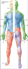
green - C blue - Thoracic orange - Lumbar purple - Sacral |
|
|
which structures make up the brain stem? |
medulla oblongata, pons, midbrain |
|
|
which structures make up the diencephalon? |
thalamus, hypothalamus, epithalamus |
|
|
where are substantia nigra found? |
midbrain |
|
|
what is the connection between substantia nigra and parkinson's disease? |
substantia nigra release dopamine and are the nuclei lost in parkinson's disease |
|
|
functions of the cerebellum |
Fine motor control Balance and equilibrium. Posture & balance Coordination Eye movement Cognitive functions- attention, language/musicprocessing. |
|
|
how does alcohol affect the cerebellum? |
Alcohol significantly effects the cerebellum. Chronic consumption leads to nutritional deficiencies,in particular vitamin B1 (thiamine), to which the cerebellum is very sensitive. |
|
|
what is basal nuclei (basal ganglia)? |
Collection of gray matter locatedwithin each cerebral hemisphere next to the thalamus. |
|
|
what is the major role of basal nuclei (basal ganglia)? |
Play important role in the control of postureand voluntary movement |
|
|
what gland is found in the epithalamus? |
pineal gland |
|
|
what hormone does the pineal gland secrete and what role does it play? |
secretes melatonin which is associate with sleep. |
|
|
stretch reflex |
Contraction of skeletal mm in response to stretching ofthe muscle. Monosynaptic feedback mechanism to control musclelength Can be elicited by tapping on tendons attached tomuscles. --> Elbow, wrist, knee, and ankles. At same time as monosynaptic reflex, a polysynapticreflex arc to the ANTAGONISTIC muscle is activated. 3 neurons (sensory, interneuron, motor 2 synapses Interneuron is inhibitory, causing the antagonisticmuscle to relax when the primary muscle contracts. |
|
|
tendon reflex |
Feedback mechanism that controls muscle tension (asopposed to length).Causes relaxation before tendon is torn. (i.e. droppinga weight that is too heavy) Less sensitive than stretch reflex, but can overridestretch reflex if tension is significant. The sensory neuron arising from the tendon organ alsosynapses with an excitatory interneuron. The interneuron synapses with a motor neuron, causingsimultaneous contraction of the ANTAGONISTIC muscle. |
|
|
vessels that supply blood to the brain |
internal coratoid and vertebral arteries |
|
|
vessels that drain blood from the brain |
internal jugular veins |
|
|
what happens if there is a disruption in blood flow to/from the brain? |
For ~1-2 min = neuron function impaired For ~4 min = permanent injury |
|
|
what can cross the Blood-Brain Barrier? |
Oxygen,CO2, alcohol and other lipophilic substances cross freely. Glucosevia active transport. Creatinine,urea, ion cross very slowly. |
|
|
what cannot cross the Blood-Brain Barrier? |
Proteins and antibiotics |
|
|
What is the limbic system? |
Consists of many different parts,including: - Cingulate gyrus - Amygdala of basal nuclei - Mammillary bodies of hypothalamus - Certain nuclei of the thalamus - Olfactory bulbs - Fornix Often referred to as “emotionalbrain”. Also plays role in memory. |
|
|
left hemisphere |
reasoning, numerical skills,scientific skills, speech, writing, sign language Damage = aphasia (disturbance in language formation) |
|
|
right hemisphere |
musical and artistic abilities,pattern perception, facial recognition, emotion, smells, images, taste Damage= monotonous voice |
|
|
hemispheric lateralization |
left hemisphere and right hemisphere Much variation from one person to the next. Less pronounced in females, possibly related to broaderposterior corpus callosum. |
|
|
Meniere's disease |
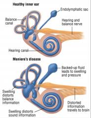
Results from ↑ amount of endolymph that enlarges the membranouslabyrinth. Symptoms: hearing loss + tinnitus + vertigo Almost total destruction of hearing may occur over aperiod of years. |
|
|
describe the physiology of hearing |
Sound waves strike the TM causing it to vibrate The vibrations are transmitted through the middle earbones to the oval window Fluid pressure waves are transmitted down the scalavestibuli, through the helicotrema, down the scala tympani, and eventually tothe round window Fluid pressure waves are transmitted to cochlear ductcausing the basilar membrane to vibrate, which moves the inner ear hair cellsagainst the tectorial membrane. Bending of the hair cells ultimately leads togeneration of nerve impulses. Each segment of the basilar membrane is “tuned” for aparticular pitch – high-pitched near the oval window; low-pitched near thehelicotrema |
|
|
NREM Stage 1 |
lasts 1-7 min: stage between wakefulness and sleep |
|
|
NREM Stage 2 |
light sleep; first stage of true sleep;fragments of dreams |
|
|
NREM Stage 3 |
occurs 20 minutes after falling asleep;moderately deep sleep; drop in body temp & BP |
|
|
NREM Stage 4 |
deepest level of sleep If sleepwalking occurs it isduring this stage |
|
|
NREM |
For 7-8hr night of rest, you willhave 3-5 episodes of REM sleep equaling 90-120 minutes. Most dreaming occurs during REMsleep. Brain blood flow & oxygen usehigher during REM sleep than during intense activity while awake. REM & NREM alternatethroughout the night with REM occurring about every 90 minutes. |
|
|
In what part of sleep does most of the dreaming occur? |
REM sleep. Brain blood flow & oxygen use higher duringREM sleep than during intense activity while awake. |
|
|
How many stages of NREM are there? And lasting for how long? |
4. < 1 hour. |
|
|
Where are cold receptors found? |
stratum basale |
|
|
where are warm receptors found? |
dermis |
|
|
what is referred pain? |
pain that the brain interprets ascoming from an area other than its origin |
|
|
First-Order Neurons |
Conduct impulses from receptors to the brain stem orspinal cord. Cranial nerves go to the brain stem. - Sensory impulses from face, mouth, teeth, eyes. Spinal nerves go to the spinal cord. Sensory impulses from neck, trunk, limbs and posterioraspect of head |
|
|
Second-Order Neurons |
Conduct impulses from brain stemand spinal cord to the THALAMUS. The axons of second-order neuronswill DECUSSATE (cross-over) in the brain stem or spinal cord beforeascending to the thalamus. - In other words, the sensory inputfrom one side of the body will go to the thalamus on the opposite side of thebrain. |
|
|
Third-Order Neurons |
Conduct impulses from thalamus to the primarysomatosensory area of the cortex on the SAME side |
|
|
What is the blood brain barrier? |
Tight junctions between endothelial cells ofblood vessels in NS. |
|
|
What is the function of the blood brain barrier? |
Astrocyte processes wrap around capillaries toprotect CNS neurons from harmful substances in blood by secreting chemicalsthat maintain the permeability of the endothelial cells of the capillaries. |
|
|
Where is CSF? |
Liquid that bathes the CNS. Reabsorbed by arachnoid villi. CSF isproduced & absorbed at approximately the same rate |
|
|
What is CSF primarily composed of? |
Primarily water but also carries O2,glucose, cations, anions,WBCs, etc. |
|
|
How does CSF flow through the brain? |
through cavities known as ventricles. - lateral ventricle x2 - third ventricle - fourth ventricle |
|
|
Where is CSF produced? |
by choroid plexuses |
|
|
where is CSF absorbed? |
arachnoid villi. |
|
|
What are parathyroid hormones (PTH) and how are they involved in how calcium in the blood is regulated? |
they are secreted by parathyroid glands; INCREASES bloodcalcium levels. operate via negative feedback system |
|
|
What is calcitriol and how is it involved in how calcium in the blood is regulated? |
activeform of vitamin D whose formation is stimulated by PTH; INCREASES blood calciumlevels |
|
|
What is calcitonin and how is it involved in how calcium in the blood is regulated? |
secretedby parafollicular cells of thyroid gland; inhibits osteoclastic activitythereby DECREASING blood calcium levels |
|
|
what happens when blood levels of calcium decrease? |
osteoclasts help to release calcium |
|
|
what happens when blood levels of calcium increase? |
osteoblasts help to absorb calcium |
|
|
Mechanoreceptors |
detect mechanical stimuli (egtouch, pressure, etc. |
|
|
Thermoreceptors |
detect changes in temperature |
|
|
Nociceptors |
detect painful stimuli |
|
|
Photoreceptors |
detect light striking the retina |
|
|
Chemoreceptors |
detect chemicals in mouth, nose and bodily fluids |
|
|
Osmoreceptors |
detect changes in osmotic pressure of bodily fluids |
|
|
list the sensory receptors that are grouped by the type of stimulus detected by receptor |
Mechanoreceptors Thermoreceptors Nociceptors Photoreceptors Chemoreceptors Osmoreceptors |
|
|
Sensory receptors grouped by microscopic structure |
Free nerve endings Encapsulated Separate Cells |
|
|
Free Nerve Endings |
bare dendrites that lack structural specialization. Ex: pain, temperature, tickle, itch, some touch. |
|
|
Encapsulated |
dendrites enclosed in CT capsule with distinct microscopic structure Ex: pressure (Pacinian corpuscle), vibration, some touch |
|
|
Separate cells |
receptors for special sense; cells which synapse with sensory neurons Ex: hair cells for hearing, gustatory receptor cells in taste buds, photoreceptors for vision. |
|
|
Sensory receptors grouped by location of the receptors |
exteroreceptors interoreceptors proprioreceptors |
|
|
Exteroreceptors |
located at/near external body surface; monitor EXTERNAL environment Ex: hearing, vision, smell, taste, touch, pressure, vibration, temperature, pain |
|
|
Interoreceptors |
aka visceroreceptors; located in bv, organs, mm, nervous system; monitor INTERNAL environment NOT CONSCIOUSLY PERCEIVED |
|
|
Proprioreceptors |
located in mm, tendons, joints, and inner ear. Provide info about body position, mm length & tension |
|
|
3 main layers of skin |
epidermis (avascular) dermis (vascular) sub Q (hypodermis) |
|
|
stratum corneum |
20layers of dead keratinocytes |
|
|
Stratum lucidum |
present only in thick skin (fingertips, palms, soles) |
|
|
Stratum granulosum |
are filled with granules of keratin. |
|
|
Stratum spinosum |
layer of 8–10 keratinocytes |
|
|
Stratum basale |
the deepest layer, undergoes continual cell division |
|
|
basic spinal cord anatomy |
Spinal cord extends from medullaoblongata to the superior border of L2. The meninges extend down to S2 |
|
|
major landmarks of the spinal cord |
Cervical enlargement Lumbar enlargement Conus medullaris Filum terminale |
|
|
Cervical enlargement |
nerves for upper limbs |
|
|
Lumbar enlargement |
nerves for lower limbs |
|
|
Conus medullaris |
where spinal cord ends |
|
|
Filum terminale |
extension of pia mater that fuses with arachnoid & dura mater to anchor spinal cord to coccyx |
|
|
ABSOLUTErefractory period |
absolutely no action potentials can be generated. |
|
|
RELATIVE refractory period |
a larger-than-normal action stimulus can generate action potential |
|
|
what are graded potentials |
usedin short distance neuron electrical signaling communication |
|
|
what are action potentials |
used in long distance neuron electrical signaling communication |
|
|
Whatis the “All or Nothing” principle? |
The all-or-none law is the principle that the strength by which a nerve or muscle fiber responds to a stimulus is independent of the strength of the stimulus. If that stimulus exceeds the threshold potential, the nerve or muscle fiber will give a complete response; otherwise, there is no response. |
|
|
what is neurolemma? |
the thin sheath around a nerve axon (including myelin where this ispresent). |
|
|
what is the function of neurolemma? |
found ONLY in PNS, allows for regeneration of axon Neurolemma (alsoknown as neurilemma or sheath of Schwann (Schwann's Sheath))is the outermost nucleated cytoplasmic layer of Schwann cells that surroundsthe axon of the neuron. It forms the outermost layer of the nerve fiber in theperipheral nervous system. |
|
|
how is white matter organized in the CNS? |
tracts |
|
|
how is white matter organized in the PNS? |
nerves |
|
|
what is the cauda equine? |
Spinalnerves gather inferiorly near filum terminale à cauda equina (“horse’s tail”) |
|
|
4 properties of muscular tissue |
excitability contractility extensibility elastcitity |
|
|
excitability |
Abilityof muscular tissue to respond to stimuli. Respondsto stimuli by generating action potentials. Propertyof both muscle and nerve cells. |
|
|
contractility |
Abilityof muscular tissue to contract in response to an action potential. Duringcontraction, the muscles may or may not shorten. |
|
|
Extensibility |
Ability of muscular tissue to stretch. Smooth muscle is subjected to the greatest amount of stretching |
|
|
Elasticity |
Ability of muscular tissue to return to original size post-contraction. |
|
|
Beable to describe all the layers that must be penetrated in order to reach themuscle. |
Stratum Corneum Stratum Lucidum Stratum Granulosum Stratum Spinosum Stratum Basale Basement Membrane Papillary Dermis Reticular Dermis Hypodermis/Subcutaneous Layer Fascia MUSCLE |
|
|
three layers of CT |
epimysium, perimysium, endomysium |
|
|
epimysium |
denseirregular CT , surrounds groups of fascicles. |
|
|
Perimysium |
dense irregular CT, surrounds groups of muscle fibers separating them into FASCICLES. |
|
|
Endomysium |
mostly reticular fibers, surrounds muscle fibers |
|
|
contractile proteins |
myosin (thick)- contains ATP binding siteon head actin (thin)- contains binding sites formyosin |
|
|
regulatory proteins |
tropomyosin troponin |
|
|
what is glaucoma? |
Aqueoushumor drained into canal of schlemm. Blockage of drainage can lead toglaucoma Acondition of increased pressure within the eyeball causing gradual loss ofsight |
|
|
four major plexuses |
cervical brachial lumbar sacral |
|
|
cervical plexus |
formed primarily by anterior rami of nerves C1-C4 |
|
|
Brachial |
Formed from anterior rami of nerves C5-C8 and T1 |
|
|
Lumbar |
Formed from anterior rami of nerves L1-L4 |
|
|
Sacral |
Formed from ant. Roots of nerves L4-L5 & S1-S4 |
|
|
connective tissue coverings |
endoneurium perineurium epineurium |
|
|
endoneurium |
convers individual acons |
|
|
perineurium |
covers groups of axons known as fascicles |
|
|
epinerium |
covers entire nerve Fuses with duramater as nerve passes through IVF |
|
|
Primary Somatosensory |
receives impulses for touch, pressure, vibration, itch, tickle, temperature, pain, proprioception |
|
|
Primary Motor Area |
controls voluntary skeletal muscle contractions |
|
|
major landmarks of the brain |
Brain Stem (continuousw/spinal cord) Medulla oblongata, Pons, Midbrain Cerebellum Diencephalon Thalamus, Hypothalamus,Epithalamus Cerebrum |
|
|
brain stem |
(continuous w/spinal cord) Medulla oblongata, Pons, Midbrain |
|
|
Diencephalon |
Thalamus, Hypothalamus, Epithalamus |
|
|
12 cranial nerves |
CN I: olfactory(S) - smelling CN II: optic(S) - vision CN III: oculomotor(M) – eye movement CN IV: trochlear(M) – eye movement CN V: trigeminal(B) CN VI: abducens(M) – eye movement CN VII: facial(B) – facial expression, taste CN VIII: vestibulocochlear(S) – hearing, equilibrium CN IX: glossopharygneal(B) – taste, swallowing CN X:vagus (B) – taste, swallowing, GI motility CN XI: accessory(M) – movement of head and pec girdle CN XII: hypoglossal(M) – speech and swallowing |
|
|
CN I |
olfactory (S) |
|
|
CN II |
optic (S) |
|
|
CN III |
oculomotor (M) |
|
|
CN IV |
trochlear (M) |
|
|
CN V |
trigeminal (b) |
|
|
CN VI |
abducens (M) |
|
|
CN VII |
facial (B) |
|
|
CN VIII |
vestibulocochlear (S) |
|
|
CN IX |
glossopharygneal (B) |
|
|
CN X |
vagus (B) |
|
|
CN XI |
accessory (M) |
|
|
CN XII |
hypoglossal (M) |
|
|
olfactory nerve |
smelling |
|
|
optic nerve |
vision |
|
|
oculomotor nerve |
eye movement |
|
|
abducens nerve |
eye movement |
|
|
facial nerve |
facial expression, taste |
|
|
vestibulocochlear nerve |
hearing, equilibrium |
|
|
glossopharyngeal nerve |
taste, swallowing |
|
|
vagus nerve |
taste, swallowing, GI motility |
|
|
accessory nerve |
movement of head and pec girdle |
|
|
hypoglossal nerve |
speech and swallowing |
|
|
What is RAS? |
The ascending portion is known as the reticular activating system (RAS). - Receives stimuli from many sensoryorgans (vision, auditory, etc) which project to cerebral cortex --> no receptors for sense of smell - Most important function: CONSCIOUSNESS Recall that the Reticular Activating System (RAS) has a huge role in consciousness,arousal, & wakefulness. - Stimulating the RAS results in increased activity of the cerebral cortex. (Stimuli: pain, touch & pressure on skin, movement of limbs, bright lights, loud noises) - No input from olfactory receptors à hence the need for smoke alarms!!! |
|
|
3 groups of integrative functions |
Sleep & Wakefulness Learning & Memory Emotional Responses (recall limbicsystem) |
|
|
lacrimal apparatus |
produces tears from lacrimal gland and drainsthem from the eye via lacrimal canaliculi and nasolacrimal duct inside nasalcavity |
|
|
flow of tears |
Lacrimal gland Lacrimal ducts Superior or inferiorlacrimal canal Lacrimal sac Nasolacrimal duct Nasal cavity |
|
|
What muscles are found in the ear? Whatis their function? |
Tensortympani and stapedius muscles – protect ear by limiting movement and vibrationof the TM and stapes, respectively |
|
|
Know where olfactory receptors are foundand what part of the receptors are triggered by odorants. What type of neuron(unipolar, bipolar, Multipolar) are olfactory receptors? Where do the axons ofthe olfactory receptors project? |
Each olfactory receptor is a bipolarneuron with its dendrite & axon projecting through the cribiform plate. Receptorsare only found in the superior portion of the nasal cavity. - 10-100MILLION receptors in nose - Olfactoryreceptors are considered first-order neurons. Olfactory hairsof the receptors are triggered by inhaled chemicals (odorants). - Transduction:conversion of a stimulus (odorant) into a graded potential in a sensoryreceptor. The graded potential triggered by theodorant results in nerve impulse(s). - Odorantbinds to olfactory receptor in plasma membrane of olfactory hair. - Olfactoryreceptor coupled to G protein which activates adenylate cyclase and subsequentproduction of the second messenger cAMP. - cAMPopens sodium channels resulting in depolarization and nerve impulse generation. - Axonsof olfactory receptors extend through olfactory foramina in cribiform plate. - Theseaxons form the olfactory nerves, which extend posteriorly to form the olfactorytract. - The axons of theolfactory tract then project to the primary olfactory area of the cerebralcortex, to the limbic system, or to the hypothalamus. |