![]()
![]()
![]()
Use LEFT and RIGHT arrow keys to navigate between flashcards;
Use UP and DOWN arrow keys to flip the card;
H to show hint;
A reads text to speech;
224 Cards in this Set
- Front
- Back
|
What's the difference between an exon and an intron? |
Exon = protein coding sequence |
|
|
What is an allele? |
Different versions of the same gene |
|
|
Explain the cell biology approach for studying cells |
Direct observation (microscopy), label and observe cell structures |
|
|
Explain the biochemical approach to studying cells |
Isolating and describing proteins |
|
|
Explain the genetics approach to studying cells |
Looking at mutant genes and proteins and studying their effects |
|
|
Explain the genomics approach to studying cells |
Looking at all the genes at the same time |
|
|
Explain the developmental biology approach to studying cells |
Looking at the differential gene expression and signals that lead to the mature organism |
|
|
What happens in G1? |
Recovery from mitosis, growth |
|
|
What happens in S phase? |
DNA is replicated |
|
|
What happens in G2 |
Pre-mitosis checkpoints |
|
|
What happens in M phase? |
Mitosis - chromosome segregation and division |
|
|
What happens in G0 phase? |
Permanent or temporary exit from the cell cycle (most cells are in this phase, fully differentiated, quiescent) |
|
|
What is a chromatid? |
One copy of a duplicated chromosome |
|
|
What are sister chromatids? |
identical copies of a chromosome joined by a centromere |
|
|
What are homologous chromosomes? |
Chromosome pairs, distinct, one from each parent |
|
|
What is the product of mitotic division? |
Two genetically identical daughter cells |
|
|
What type of chromosome alignment happens in mitosis? |
Independent alignment (not paired with its homologue)
Each sister chromatid separates and goes to a different cell making both of the daughters genetically identical |
|
|
What is chromatin? |
Proteins that bind DNA to help it condense |
|
|
What are cohesins? |
Proteins (like little rubber bands) that hold the chromosome together |
|
|
What is the centromere? |
repetitive DNA sequence that serves as a target for mitotic machinery (the bit in the middle of the chromosome 'X' shape) |
|
|
What is a kinetochore? |
Protein complex (one per chromatid) that links the centromere to the microtubules. Kinetochore proteins target the centromere to place itself |
|
|
What are the stages of mitosis? |
Interphase |
|
|
What happens during interphase? |
chromosome duplication and cohesion |
|
|
What happens during prophase? |
interphase microtubule breakdown replaced by mitotic asters Chromosome condensation |
|
|
What happens during prometaphase? |
Nuclear envelope breakdown Microtubules contact the kinetochore |
|
|
What happens during metaphase? |
Chromosomes align at metaphase plate |
|
|
What happens during anaphase? |
Cohesins degrade |
|
|
What happens during telophase? |
Assembly of contractile ring |
|
|
What happens during cytokinesis? |
Reformation of interphase microtubule array |
|
|
What is the goal of meiosis? |
To produce four genetically different daughter cells with half the number of chromosomes as the cell that undergoes the division (e.g. 2n cell --> 4 x 1n cells) |
|
|
What is the form of chromosome alignment in meiosis? |
Homologous chromosomes pair, recombination occurs and each duplicated chromosome separates. |
|
|
What is the basic process of meiosis |
duplication of chromosomes, pairing of homologous chromosomes, recombination, separation of homologous pairs (meiosis 1) (each daughter cell contains recombined version of either maternal or paternal chromosome), sister chromatids separate (meiosis 2), four haploid gametes form |
|
|
What is a kinase? |
An enzyme that adds a phosphate (activating or inhibiting) to a target |
|
|
What is a phosphatase? |
An enzyme that removes a phosphate group |
|
|
What is a cyclin? |
A family of proteins that control a cell's progression through the cell cycle by binding and activating cyclin dependent kinases |
|
|
At what stages of the cell cycle are cyclin and CDKs expressed? |
CDKs- present throughout cell cycle |
|
|
Does the cyclin or the kinase determine the specificity of the cyclin heterodimer? |
The cyclin |
|
|
What is ubiquitination? |
Ubiquitin protein ligases attach Ub to target proteins. Repeating this process multiple times (polyubiquitination) marks a protein for degradation by the proteasome (only K63 ubiquitination) |
|
|
What are the key complexes involved in the G1/S and the G2/M transition? |
G1/S = SCF (removes skp1 inhibitor from S cyclin complex) |
|
|
What is the difference between phosphorylation and ubiquitination in terms of permanence of effect? |
Phosphorylation is temporary and reversible Ubiquitination is permanent and irreversible |
|
|
What are some functions of G1 CDKs? |
Phosphorylate transcription factors help prepare G1/S CDKs |
|
|
Explain the basic transition at the G1/S boundary (in terms of the CDKs) |
An inhibitor of S CDKs determines the G1/S boundary. |
|
|
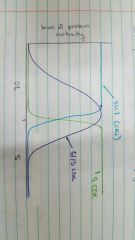
Graph the level of protein activity of sic1, S CDKs and G1/S CDKs |
|
|
|
What are some of the functions of S CDKs? |
Phosphorylation of numerous proteins that go on to replicate DNA Also help prepare cell for mitosis |
|
|
What are some ways to differentiate cells based on their positions in the cell cycle? |
- size (later in cycle = larger) - growth factors - drugs (can arrest cells at particular stages) |
|
|
What is cdc2? |
A cyclin dependent kinase that is key in the mitosis pathway |
|
|
What is the phenotype of Cdc2+, Cdc2- and Cdc2 dominant cells?
|
Cdc2+ = wild type (normal cells) Cdc2- = long cells (no mitosis) Cdc2 dominant = wee cells (premature mitosis) |
|
|
What is cdc13 |
Mitotic cyclin (binds to cdc2) |
|
|
What is cdc25? Does it drive or inhibit mitosis? |
CDC25 is a phosphatase --> DRIVES MITOSIS |
|
|
What is wee1? Does it drive or inhibit mitosis? |
WEE1 is a kinase --> INHIBITS MITOSIS |
|
|
What mutations could cause a long phenotype in S. pombe? |
Defecit of CDC25 (drives mitosis) |
|
|
What mutations could cause a wee phenotype in S.pombe? |
Overexpression of CDC25 (drives mitosis) |
|
|
Explain the basic process of wee1/cdc25 activation of the maturation promoting factor (MPF) (MPF = mitotic CDK complex) |
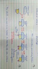
Mitotic cyclin and CDK come together to form an inactive MPF. WEE1 phosphorylates Y15 (tyrosine) (inhibitory phosphorylation). CAK then phosphorylates T161 (threonine) with an activating phosphorylation. CDC25 then removes the inhibitory phosphorylation and the MPF becomes active |
|
|
What are some of the functions of mitotic CDK complexes? |
Activaiton of - chromatin associated proteins - microtubule associated proteins - kinetochore proteins |
|
|
What is a cell cycle checkpoint? |
Cellular mechanisms that make sure a task has been completed before cell cycle progression |
|
|
Give an example mechanism of a cell cycle checkpoint |
ATR1 |
|
|
How do mitotic CDKs breakdown the nuclear envelope? |
The nuclear lamina supports the nuclear envelope. The lamina is made of lamins. Normally, lamins form a tetramer. Mitotic CDKs phosphorylate lamins, making them only able to form dimers. This results in the breakdown of the nuclear envelope. |
|
|
What are two major roles of APC/C? |
1. Metaphase-anaphase transition (securin-separase to remove cohesins) 2. Degrade mitotic cyclins in late anaphase (after chromatids have separated) |
|
|
What is PP2A? |
A phosphatase associated with the centromere that stops the cohesins close to it from being removed by phosphorylation --> meaning they have to be removed via the APC/C pathway |
|
|
Explain the basic process of the removal of cohesins near the centromere by APC/C |
Cohesins are composed of smc proteins and scc1. Separase is a protease that cleaves scc1. Securin binds to and inhibits separase. When all kinetochores bind to microtubules, CDC20 binds APC/C and polyubiquitinates securin leaving separase to be free to cleave the scc1 of the cohesin. |
|
|
Explain how APC/C degrades mitotic CDKs |
APC/C is an E3 ubiquitin ligase. It can either be bound to cdc20 or cdh1 to activate it. When bound to cdc20 it degrades securin. APC/C binds to cdh1 (changes its specificity) in late anaphase. This E3 complex then polyubiquitinates mitotic cyclins (as well as remaining cdc20 bound APC/C) to ensure that the cell doesn't undergo mitosis again and can have a period of growth |
|
|
What is necrosis? |
Unplanned cell death involving membrane rupture and the spilling of the cellular contents into the surrounding tissue |
|
|
What are some causes of necrosis? |
Toxins Burns Lack of circulation Inflammation |
|
|
What is apoptosis? |
Programmed cell death involving the cell splitting up into 'apoptotic blebs' to be consumed by macrophages |
|
|
What are some causes of apoptosis? |
DNA damage (e.g. UV exposure)
Structural removal of cells from tissue (eg. removing tadpole tail) Lack of growth factor (e.g. keeping useful, well connected neural junctions because they secrete growth factor) |
|
|
explain the basic apoptosis signalling pathway (C. elegans) |
Apoptosis signal --> EGL 1 binds to and inhibits CED-9 (which normally represses apoptosis) --> stimulates CED4 dimer release --> CED4 becomes octamer and joins with CED 3 to form the CED4/CED3 caspase holoenzyme which then leads to cell death |
|
|
What are the results of mutations in the apoptotic pathway? |
EGL 1 mutant: no apoptosis CED 3 mutant: no apoptosis |
|
|
What is the result of a CED9/CED3 double mutant? |
No apoptosis - becuase CED 3 is downstream of CED 9 (epistatic to it) and whatever is downstream is more important |
|
|
How do tumor cells develop sustained proliferative signalling? |
Mutations allowing growth factor receptors to fire independently of the GF ligand |
|
|
How do tumor cells evade growth supressors? |
Mutations e.g. the removal of the sic1 inhibitor on S CDKs which allow them to progress into S phase more quickly |
|
|
How can cancer cells enable replicative immortality? |
Telomeres have repeating terminal sequences. Replication of chromosomes shortens these sequences each time, limiting the amount of time a cell can divide. Telomerase extends the end of telomeres after replication. Telomerase is turned off in adult cells and cancer can turn it back on.
|
|
|
What is angiogenesis? |
The stimulation of blood vessel formation within a tumor to avoid necrosis |
|
|
What are the three different types of mutation in cancer cells? |
Gain of function: e.g. in pathways that promote growth Loss of function: e.g. in pathways that inhibit growth Dominant negative: e.g in proteins that form complexes (p53), one mutant part of the complex can turn off the whole thing |
|
|
What are proto-oncogenes? |
Genes that normally promote cell growth. Once mutated (generally gain of function), they become oncogenes. e.g. ras, myc, src |
|
|
What are tumor supressor genes? |
Genes that normally inhibit cell cycle progression. Generally loss of function mutations allow the cell to grow out of control e.g. p53, Rb, ATR |
|
|
How can cancer mutations occur? |
They can be hereditary (e.g. retinoblastoma), spontaneous or evironmental (e.g. carcinogens, UV) |
|
|
What is the biggest cancer risk factor? |
AGE - multiple mutations are required for cancers to form - DNA repair is less efficient with age |
|
|
What is metastasis? |
The spread of cancer cells from their site of origin and the start of secondary growth |
|
|
What is retinoblastoma? |
A heritable or spontaneous cancer in young children caused by a defect in the Rb gene |
|
|
What is the role of Rb in the cell cycle? |
Rb fits in the G1 phase of the cell cycle and is a key restriction point - i.e. if it is passed then the cell is committed to another phase of replication.
Rb is a transcriptional repressor of E2F. While it is in place, G1 CDKs cannot phosphorylate and activate the E2F transcription factor (which drives the expression of S phase genes). Thus, removal of Rb commits the cell to another phase of replication. |
|
|
What is hyperphosphorylation? |
Some proteins (sic1, Rb) must be phosphorylated multiple times for an action to occur. This means that the action can only happen when the CDK concentration is very high |
|
|
What is P16 and how does it relate to Rb? |
P16 is a CDK inhibitor which prevents phosphorylation and removal of Rb. This inhibitor thus inhibits activation of the E2F transcription factor and cell cycle progression. |
|
|
What is the expression of P16 driven by? |
Cell stress More stress = more P16 = no progression of cell cycle whilst conditions are bad |
|
|
How do cancer cells affect P16? |
Turn it off |
|
|
How do cancer cells affect the Rb pathway? |
Overexpress/amplify G1 CDKs Delete or inactivate Rb (retinoblastoma) |
|
|
What is p53? |
p53 is a transcription factor that can cause cell cycle arrest, senescence (permanent arrest) and apoptosis |
|
|
Explain the basic process of p53 activation in cells |
ATR can inhibit Mdm2 (which normally ubiquitinates p53 to keep it unstable). This makes p53 stable allowing it to detect conflict within the cell and activate cell cycle arrest/senescence/apoptosis |
|
|
Do p53 mutations cause cancer? |
No, but they do predispose a cell to cancer through a loss of tumor suppressor function |
|
|
What type of mutation is a p53 mutation? |
A dominant negative - p53 forms a tetramer so if the mutation produces just one defective protein then the entire complex is defecctive |
|
|
How do cancer cells target the p53 pathway? |
Deletion or inactivation of ATR Upregulation of Mdm2 Deletion or inactivation of p53 |
|
|
How does p53 inhibit cell growth? |
Upregulates CKI --> inhibiton of G1/S and G2/M progression via CDKs |
|
|
What is meant by the term amphipathic? |
Hydrophilic and hydrophobic elements to the same molecule |
|
|
What are some of the functions of cell membranes? |
Structural - protein modification Other - cell signalling - enzyme cofactors - electron carriers - pigments |
|
|
How does hydrocarbon chain length affect melting point and solubility in water? |
Increased chain length - decreases solubility in water |
|
|
How does the number of double bonds affect the melting point? |
Increased double bonds decreases the melting point |
|
|
What is a sphingolipid? |
A major membrane component, derivative of amino alcohol spingosine example = ceramides |
|
|
What are the different kinds of lipid aggregates? |
Micelle - single phospholipid layer sphere |
|
|
What are the different kinds of bonds that occur in a plasma membrane? |
Ionic bonds between the head groups |
|
|
What are the two faces of a bilayer? |
The cytosolic and the exoplasmic |
|
|
What are the types of motion in lipids? |
Spinning (without changing location) |
|
|
What is FRAP and how does it relate to lipids? |
Fluorescence recovery after photobleaching |
|
|
What are some factors that affect the fluidity of a membrane? |
- temperature - lipid composition - chain length - level of saturation |
|
|
How is the curvature of the membrane determined? Give two examples of phospholipids that would contribute |
PC (phosphatidylcholine) - flat membrane |
|
|
Do membranes differ in their lipid distribution? |
Yes - most have asymmetric distribution |
|
|
What lipids would you expect to see in a cytosolic leaflet vs the exoplasmic? |
Cytosolic: rich in spingolipids/PC (less fluid) |
|
|
How does asymmetry in the lipid composition of leaflets arise? |
- leaflets do not spontaneously flip - synthesis of lipids can promote asymmetry |
|
|
What are the enzymes that catalyse transbilayer translocations? |
Flippase (P-type ATPase) |
|
|
What are membrane microdomains and what do they do? |
Stable associations of lipids within the membrane - lipid rafts
microdomains control lateral diffusion |
|
|
What are some functions of membrane proteins? |
Transporters Energy transduction |
|
|
What are the three main types of membrane proteins? |
Integral |
|
|
What treatments releases each type of membrane protein? |
Integral: harsh treatment e.g. detergents, organic solvents, denaturants |
|
|
What are some features of integral membrane proteins? |
Goes fully across the membrane |
|
|
Give some features of lipid anchored membrane proteins |
Protein covalently linked to one or more lipid molecules |
|
|
What are some features of peripheral membrane proteins? |
Attached more weakly and can be released using milder treatments e.g. high salt, carbonate at high pH
|
|
|
In order to span the bilayer, approximately how many residues must there be for an b) beta sheet |
a) 20-25 b) 7-9 |
|
|
What kind of reactions are involved in alpha helices? |
- hydrophobic (amino acid side chains react with fatty acid tails)
- ionic interactions between protein and head groups (Hydrophilic) |
|
|
What are some basic features of beta sheets? |
Can be used to make barrel shape through the membrane - porins |
|
|
What are the three ways that water soluble proteins can be linked to the membrane (lipid anchored)? |
Covalently linked to... |
|
|
Do proteins move more or less freely than lipids, on average? |
Less freely - some are anchored |
|
|
What substances is the membrane permeable to? |
Gases, ethanol, water (sort of), urea (sort of) |
|
|
What are the different types of membrane transport proteins and what are their relative speeds? |
ATP powered pump, ion channel, transporters |
|
|
What is the difference between an electrochemical and chemical gradient? |
The electrochemical gradient applies to solutes that have a charge (which then affects their equilibrium). Neutral solutes just have a chemcial gradient based on concentration |
|
|
What is the difference in terms of speed and specificity for carrier proteins and channel proteins? |
Channels = faster, less specific
Carriers = slower, more specific |
|
|
What is the delta G and how does it apply to the plasma membrane? |
Delta G is the free energy change. Diffusing across the membrane requires an energy change (G). The magnitude of delta G determines whether a molecule will be able to passively diffuse or whether it will need a transporter |
|
|
What is GLUT1? |
Glucose transporter in erythrocytes implicated in diabetes. This transporter facilitates diffusion (50,000 x faster than diffusion alone). It is still passive transport and as such is dependent on the chemical gradient |
|
|
What is the main CO2 transporter in erythrocytes? |
Chloride bicarbonate exchanger - antiporter of Cl- and HCO3- |
|
|
What is primary active transport? |
Transport of a substance against its concentration gradient for which the energy comes from chemical reaction (ATP) |
|
|
What is secondary active transport? |
Transport of a substance against its concentration gradient via coupled transport. The ion gradients for secondary active transport are made by primary active transport. |
|
|
How does vancomycin work? |
Vancomycin is an ionophore that collapses the ion gradient of bacterial cells and kills them |
|
|
What are the main classes of ATP pumps and give an example of each |
P class pumps - include lots of the common ion pumps e.g. Na/K, H+, Ca2+ pumps |
|
|
P type ATPases are mainly what kind of transporters? |
Cation transporters |
|
|
Explain the basic mechanism of the Na/K ATPase pump |
Transporter binds 3Na+ from inside the cell (high affinity) --> phosphorylation via ATP (PEnzII) --> transporter opens up to extracellular space and releases the 3Na+, takes up 2K+ --> dephosphorylation of pump (EnzI) --> transporter releases 2K+ inside |
|
|
What is the basic function of F and V type ATPases? |
Proton transport |
|
|
What is the basic function of ABC transporters? |
Pumping amino acids, peptides, proteins, metal ions, lipids, compounds (drugs) |
|
|
Give two examples of an ABC transporter |
MDR1: multi drug transporter (resistance of bacteria to drugs) |
|
|
Give an example of a secondary transporter |
H+/lactose co transporter brings lactose into cell |
|
|
What are aquaporins? |
Water transporters (facilitated transport) |
|
|
What are some of the specialised features of aquaporins to prevent H3O+ transport? |
- Arg195 in aquaporin repels H3O+ |
|
|
What are ion channels and how do they differ from transporters? |
Channels straight through the membrane allowing passage of molecules that cannot naturally diffuse across
- Flux: unrestricted flow - not saturable - ligand or voltage gated - open for only ms |
|
|
What are some consequences of defective ion channels? |
- defective Na+ channel: muscle paralysis/stiffness (tetrodotoxin) |
|
|
What is endocrine signalling? |
hormone secretion into the blood to affect target tissue anywhere in the body |
|
|
What is paracrine signalling? |
cell secrete stimuli to an adjacent target cell e.g. immune cell secreting interferon |
|
|
What is autocrine signalling? |
Cell is stimulated by the substances that it secretes e.g. cancer cells producing their own growth factors |
|
|
What is juxtacrine signalling? |
signalling by plasma membrane proteins physically attached to the target cell e.g. killer t cells present an attached death ligand to infected cells |
|
|
How can the magnitude of a cell's response to a stimuli be altered? |
It can be limited by the number of receptors available to recognise the ligand |
|
|
What are second messengers? |
Transient chemicals that are released as a response to primary messengers (hormones/ligands etc) that mediate signalling e.g. cAMP, Ca2+
|
|
|
What are effectors? |
Proteins that are stimulated in the signal transduction pathway that have some enzymatic function e.g. kinases, phosphatases
|
|
|
What is signal amplification? |
Amplification of the signal (no way!) to vastly increase the cellular response to a single ligand/hormone. Enzyme activation and second messenger generation allows for rapid amplification
|
|
|
What is allosteric modification? Give an example |
The ability of a molecule to alter the conformation of a protein when it binds non covalently to that protein |
|
|
What is covalent modification? give an example |
A modification of the chemical structure of a target protein by covalent bonding. This process is reversible. |
|
|
How do allosteric/covalent modifications alter protein function? |
1. activates enzyme activity 2. unmasks active sites 3. alters location of the protein |
|
|
What are kinases? |
Proteins that phosphorylate other proteins to activate or inactivate them |
|
|
What residues do kinases phosphorylate? |
Serine, threonine, tyrosine |
|
|
What do phosphorylation cascades allow for? |
- signal amplification |
|
|
What is meant by cross talk between pathways? |
Different receptors can stimulate the same signal transduction pathway |
|
|
What does the EF hand motif do? |
Binds calcium (e.g. calmodulin) |
|
|
What does the SH2/PTB domains do? |
Bind phosphotyrosine (tyrosine kinase receptor pathways) |
|
|
What does the PH domain do? |
Binds phosphoinosotides (PI-3 kinase pathway) |
|
|
What are adaptor proteins? |
Proteins that contain no functional activity but multiple interaction domains to function as a bridge between active proteins
|
|
|
What is a molecular switch? |
Some proteins involved in signal transduction. Allosteric binding to other molecules results in a switch from off to on state |
|
|
Give some examples of molecules that interact with GPCRs |
- glucagon
- epinephrine - vasopressin - serotonin - TSH |
|
|
What are the majority of GPCRs involved in response to? |
Olfactory signals |
|
|
What is the basic structure of a GPCR? |
7 TM alpha helical domains 4 cytosolic domains 4 extracellular domains |
|
|
Explain the basic process of GPCR activation |
Receptor binds hormone/ligand --> shape change in receptor allows it to interact with heterotrimeric G protein --> alpha G protein subunit exchanges GDP for GTP --> trimer dissociates and Ga goes on to activate receptor |
|
|
How do G proteins act as molecular switches? |
Exchange of GDP (off) for GTP (on) |
|
|
What is the mechanism of the G alpha s protein? |
Acts on adenylyl cyclase (excitatory)--> cAMP --> B adrenergic receptors |
|
|
What is the mechanism of the G alpha i protein? |
Acts on adenylyl cyclase (inhibitory) --> cAMP --> a2 adrenergic receptor |
|
|
What is the mechanism of the G alpha q protein? |
Acts on phospholipase C --> increased IP3 and DAG --> alpha 1 adrenergic receptor |
|
|
What is the mechanism of the G alpha o protein? |
Acts of phospholipase C --> Increased IP3 and DAG --> ACh receptor in endothelial cells |
|
|
How does cAMP interact with PKA? |
PKA has two regulatory subunits and two active subunits. cAMP induces the release of the regulatory units forming an active dimer |
|
|
Explain the effect of epinephrine on adipose, liver, heart and GI/Kidney tissue |
Adipose, liver, heart = stimulates B-AR, upregulates cAMP, via G alpha s --> fatty acid release, glucose release, increased contraction
GI tract/kidney = stimulates a2-AR to downregulate cAMP via G alpha i to induce vasoconstriction |
|
|
When epinephrine stimulates the liver, what two enzymes does PKA then go on to effect? |
Inhibitory phosphorylation on glycogen synthase Excitatory phosphorylation on glycogen phosphorylase (increases glucose) |
|
|
What G alpha subunits are used for GPCRs to interact with phospholipase C? |
o and q |
|
|
In a GPCR process, what does phospholipase C do? |
Cleaves PIP2 into IP3 and DAG |
|
|
In a GPCR process, what does IP3 do? |
Stimulates calcium release |
|
|
In a GPCR process, what does DAG do? |
(Along with calcium) activates PKC |
|
|
How does calmodulin act as a molecular switch? |
Binds 4 Ca2+ to its EF hand domains and undergoes a conformational change |
|
|
What are some functions of calmodulin? |
Activates calmodulin kinase |
|
|
What are three ways that a GPCR signalling pathway can be turned off? |
Turn off the GPCR |
|
|
How can the GPCR be turned off? |
Activated GPCRs are phosphorylated by GPCRkinase, which prevents G protein interaction and induces arrestin binding. Arrestin stimulates the internalisation of the receptor where it is then either degraded or recycled back to the surface |
|
|
What is receptor exhaustion? |
Repeated exposure to the same ligand over time can cause the mechanisms that turn signalling off to reduce the number of available receptors on the cell surface --> leads to the insensitivity of a tissue to a particular ligand |
|
|
How are G proteins turned off? |
G alpha proteins have intrinsic GTPase activity. This activity hydrolyses GTP. GTPase activity increases upon Ga binding to its receptor |
|
|
How cAMP turned off? |
cAMP is converted to AMP by phosphodieserases |
|
|
What are the biding domains that recognise phosphorylated tyrosine? |
SH2 PTB |
|
|
Explain the basics of tyrosine kinase receptor activation |
Ligand binding causes inactive monomers to dimerise and become active or inactive dimers to change shape and activate --> brings protein tyrosine kinases close together --> these can phosphorylate each other --> phosphoryation of tyrosine residues in the tail provide docking ports for new proteins |
|
|
Explain the MAPK signalling pathway |
Stimulated receptor binds GRB2 via a SH2 domain. GRB2 has an SH3 domain which it uses to bind SOS. SOS is a GEF which stimulates RAS to exchange GTP for GDP and become active. RAS binds RAF and stimulates the removal of the inhibitory 14-3-3 subunit. Active RAF phosphorylates MEK which then phosphorylates MAPK on tyrosine and threonine, forming an active dimer. MAPK active dimer travels to the nucleus and controls genes and TFs involved in cell proliferation and survival. |
|
|
Explain the PI-3 kinase signalling pathway |
RTKs recruit PI3 kinase via their SH2 domain. Activated PI3 kinase phosphorylates the 3rd position on the inositol ring of PIP/PIP2. This generates PI-3 phosphates, which bind AKT via its PH domain. Whilst bound, AKT (can also be called PKB) needs PDK1 (PI3 bound) and PDK2 to phosphorylate it and activate it. AKT then goes on to regulate processes such as cell survival and glucose uptake |
|
|
Explain the basic phospholipase C pathway in tyrosine kinase receptors |
PLC binds to the RTK via a SH2 domain. PLC then goes on to cleave PIP2 into IP3 and DAG, which go on to eventually activate PKC |
|
|
What's the difference between a cytokine receptor and a tyrosine kinase receptor? |
Both are structurally similar however cytokine receptors lack endogenous kinase activity and must bind an external kinase to signal |
|
|
What is a common signal pathway through which cytokine receptors act? |
JAK/STAT pathway |
|
|
Explain the basics of the JAK/STAT pathway |
ligand binds to cytokine receptor and stimulates dimerisation. Receptor binds JAK kinases which phosphorylate and activate each other and then other tyrosine residues on the receptor tail. STAT then binds to JAK via an SH2 domain, becomes active and dissociates forming a dimer with the other STAT molecule. This dimer then goes to the nucleus and binds DNA, activating transcription |
|
|
What are some inhibitors of RTKs and cytokine receptors? |
- phosphotyrosine phosphatases - other inhibitors (e.g. SOCs proteins) |
|
|
What is SHP1 and how does it work? |
SHP1 is an example of a SOC protein that inhibits cytokine receptor signalling. Inactive SHP1 binds to tyrosine residues (blocking them) in the receptor tail via an SH2 domain. An intrinsic phosphatase then dephosphorylates and inactivates JAK. The SOCs box then recruits an E3 ubiquitin ligase to stimulate the removal of the receptor. |
|
|
What's the difference between transcriptional regulators and transcription factors? |
Transcriptional regulators = co activators, co repressors which modulate gene accessibility |
|
|
What is NF-kB, what is it activated by and what covalent modification is central to its action? |
An immune function regulator protein that provides a rapid transcriptional response to cellular stress |
|
|
What kind of ubiquitination is involved in the NF-kB signalling pathway? |
K48 (structural scaffold rather than the degradation-inducing K63) |
|
|
Explain the Nf-kB pathway |
After the receptor (e.g. IL-1) activates ikB kinase, it then phosphorylates the inhibitor attached to the NF-kB heterodimer (p65 + p50). This phosphorylation attracts an E3 ubiquitin ligase which polyubiquitinates and removes the inhibitor. Free NF-kB can then travel to the nucleus and influence transcription |
|
|
What kinds of things does the NF-kB pathway stimulate the production of? |
Adhesion proteins Cytokines IL-1RA |
|
|
Explain the process of the IL receptor activating NF-kB |
IL-1B binds to IL-1 receptor. Receptor activates and recruits MyD88 (adaptor), IRAK (kinase) and TRAF-6 (Ub ligase). TRAF-6 creates a K63 Ub scaffold to which TAK-1 (kinase) joins. The ikB kinase also joins (via the NEMO subunit) and is phosphorylated by TAK-1. The ikB can then dissociate and remove the NF-kB inhibitor |
|
|
How is negative feedback used in the NF-kB pathway? |
Both the ikB inhibitor and IL1RA are produced as a direct result of NF-kB activation which turns off the pathway |
|
|
What was the new second messenger identified recently in the regulation of NF-kB and IFN? |
cGAMP |
|
|
What does TREX-1 do?
|
Stops human DNA leaked from the nucleus from stimulating the cGAMP pathway and triggering an immune/inflammatory response |
|
|
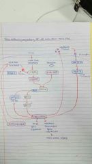
Draw the cGAMP pathway |
|
|
|
What are nuclear hormone receptors? |
Transcription factors that are also receptors |
|
|
What is an example of a homodimeric nuclear hormone receptor? |
Heat shock proteins can act as an inhibitor to the nuclear receptors in the cytosol. Ligand binding kicks off the HSP, allowing it to translocate to the nucleus and bind to the response element section of DNA |
|
|
Where are heterodimeric nuclear receptors often found? |
In the nucleus, often already bound to DNA |
|
|
How do heterodimeric nuclear receptors change with ligand binding? |
No ligand = bound to co repressors --> gene expression OFF |
|
|
What does ligand binding do to the helix 12 position of nuclear receptors? |
Ligand binding causes retraction of H12 and space for a co activator to join |
|
|
What is APO-LBD? |
Nuclear hormone receptor with extended H12 and attached co repressors |
|
|
What is HOLO-LBD? |
Nuclear hormone receptor with H12 retracted with a small enough space that only co activators can bind |
|
|
What enzyme do co repressors recruit and what does it do? |
HDAC (histone deacetylase) which removes acetyl groups from chromatin, closing them off and inhibiting transcription
|
|
|
What enzyme do co activators recruit and what does it do? |
recruit HAT (histone acetyl transferase) which acetylate chromatin, opening it up and allowing transcription to occur |
|
|
What are the two broad classes of caspases? |
Apoptotic and inflammatory |
|
|
Caspases are what type of protease? |
Cytesine |
|
|
What are the two types of apoptotic caspases and how do they differ? |
Initiator caspases: require clustering on a signalling hub in order to dimerise and activate Executioner caspases: already dimeric, require cleavage to activate |
|
|
What is apoptosis? |
A 'neat and tidy' form of programmed cell death that involves the budding off of 'apoptotic blebs'- little bits of cell that can be easily phagocytosed by macrophages |
|
|
What are the two types of apoptosis? |
Intrinsic- signal coming from within e.g. cytochrome C found in the cytosol |
|
|
How does the apoptosome form and how does it lead to cell death? |
The apoptosome forms an oligomeric complex made up of APAF-1 after it binds to its ligand, cytochrome C. This apoptosome has a CARD (caspase activation and recruitment domain) to attract and activate caspase 9. Caspase 9 can then activate caspase 3 which goes on to cleave substrates and initiate apoptosis. |
|
|
What is pyroptosis? |
A form of planned necrosis that involves the rupture of the plasma membrane and spilling of cellular contents (as well as the release of cytokines) leading to an inflammatory response.
|
|
|
What mediates apoptosis? |
Apoptotic caspases which are activated via the intrinsic or extrinsic pathway |
|
|
What mediates pyroptosis? |
Inflammatory caspases activated by the inflammasome |
|
|
What is the ASC protein and what does it do? |
It is an adaptor protein involved in the activation of capases by the inflammasome. It has a PYD domain to attach to the inflammasome nucleating protein and a CARD domain to attach to the caspase, serving as the connector between the two. |
|
|
How is the ASC protein prion-like? What is the ASC speck? |
The ASC protein is prion like because it can exist in two states. When it is in its prion like state, the ASC protein can induce other ASC proteins to adopt this same state. |
|
|
Explain runaway inflammasome signalling |
Receptor activation in response to infection --> ASC speck formation --> cytokine release and cell death (pyroptosis) --> ASC release --> extracellular activation of caspase 1 and IL-1B --> phagocytosis of ASC speck --> perpetuation of signalling in neighbouring cell |