![]()
![]()
![]()
Use LEFT and RIGHT arrow keys to navigate between flashcards;
Use UP and DOWN arrow keys to flip the card;
H to show hint;
A reads text to speech;
34 Cards in this Set
- Front
- Back
|
define magnification and give the equation to calculate it |
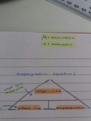
= how many times larger the image is compared to the actual object |
|
|
define resolution |
ability to distinguish between two separate points -depends on the wavelength of radiation you're using, if two points are closer than half a wavelength, they will be seen as one point
e.g wavelength of visible light = 400-700nm, so light microscopes can resolve points 200nm apart
|
|
|
explain transmission electron microscopy (TEM) |
- beam of electrons passed through the specimen and focussed using electromagnets on a screen - magnification = x500,000 - resolution = 0.2nm -gives high resolution image, but you can only view dead specimens |
|
|
explain scanning electron microscopy (SEM) |
-electrons are bounced off the surface of the specimen (which is coated in gold) - magnification = x100,000 - resolution = 5-20nm - gives a surface image wih 3D appearance, but is very expensive and requires great skill to use |
|
|
name the components of eukaryotic cells (animal / plant) |
•mitochondria •ribosomes •nucleus •nucleolus •nuclear pore • nuclear envelope •cell surface membrane • lysosome (animal only) •gogli apparatus •rough endoplasmic reticulum •smooth endoplasmic reticulum • cell wall • chloroplasts/chlorophyll •vacuole •tonoplast
|
|
|
describe the difference between eukaryotic cells and prokaryotic cells |
eukaryotic (animal, plant, fungal) cells are more complicated, have more organelles, and have a true nucleus, unlike prokaryotic (bacterial) cells, which are smaller and simpler |
|
|
explain the function of the rough endoplasmic reticulum (RER) and the smooth endoplasmic reticulum (SER) |
RER - a membrane bound organelle - surface covered in ribosomes - a transport system for products of protein synthesis - linked to nuclear envelope SER - similar to RER but without ribosomes - had cisternae - synthesises, stores and process lipids and carbohydrates |
|
|
explain the function of the golgi apparatus/vesicle |
apparatus - fluid filled flattened sacs - processes lipids/proteins - modifying and packaging the products of RER and SER (glycoproteins) vesicle - small fluid filled sac produced by gogli apparatus - membrane bound - stores lipids/proteins processed by the gogli apparatus - transports them out of the cell via cell surface membrane |
|
|
describe the function of the lysosome |
round membrane-bound organelle - contains hydrolytic digestive enzymes called lysozymes - used to digest invading cells and break down worn out cell components (apoptosis - controlled cell death) |
|
|
explain the function/structure of mitochondria and chloroplasts |
mitochondria - oval shaped with double membrane - inner one is folded to make cristae - contains circular DNA and is self replicating - site of aerobic respiration where ATP is produced chloroplasts - found in plant/algal cells - has double membrane - thylakoid membranes stack up to form grana - held together by lamellea - site of photosynthesis - most chlorophyll found in grana and stroma (thick fluid in chloroplasts) - also contains starch grains, lipid droplets, cirular DNA and 70S ribosomes |
|
|
briefly outline the stages of cell fractionation |
chop up fresh tissue into ice cold buffer solution - reduce rate of hydrolytic enzyme controlled reactions which may break down other organelles - buffer keeps ph constant so protein tertiary structure is not affected - isotonic too (cells not damaged by osmosis)
blend tissue in a homogeniser - to break open cell, releasing organelles
filter mixture - removing debris such as connective animal tissue or plant cellulose
pour mixture into tube, put into ultracentrifuge - denser organelles sink to the bottom of the tube first (e.g nuclei) forming a pelet called the sediment
the liquid layer on top (supernatant) is poured into a fresh tube - this separates it from the sediment to be spun at a faster speed, separating out less dense organelles
SMALLER ORGANELLES - LESS DENSE - NEED TO BE SPUN FASTER TO SEPARATE |
|
|
how fast does the ultracentriguge need to spin to separate out all the organelles |
nuclei - 1000×g mitochondria - 3500×g lysosomes - 16,500×g ribsosomes/membranes - 20,000×g |
|
|
describe in detail prokaryotic cells |
• smaller, simpler cells - bacteria • large range of tolerance • genetic material exists as cicular DNA (plasmids) • have 70S ribosomes • flagella for movement • food stores - glycogen + oil droplets • has a cell surface membrane • has a cell wall made of glycoproteins (murien) • has mucilagenous slime capsule outside cell wall for protection from immune system |
|
|
why do we need mitosis? |
- growth and repair - replacement of worm out cells - asexual reproduction |
|
|
describe/explain the cell cycle |
has three stages: • interphase (cell growth/DNA replication) • nuclear division (mitosis) • cytokinesis - this process occurs in cells with the ability to divide - the cell cycle starts when a cell is produced by cell division and ends with that cell dividing to produce two identical daughter cells |
|
|
describe the three stages of the interphase |
G1 PHASE - cell grows - new organelles and proteins are made SYNTHESIS PHASE - DNA replication to double cells genetic content G2 PHASE - preparation for mitosis - continues growth - proteins needed for cell division are made - ATP content increased providing energy for cell division |
|
|
explain stage 1 of mitosis |
PROPHASE - chromosomes condense getting shorter/thicker - protein bundles called centrioles move to opposite poles of the cell - this forms a network of protein fibres called the spindle - nuclear envelope breaks down, releasing chromosomes into the cytoplasm |
|
|
explain stage 2 of mitosis |
METAPHASE - chromosomes line up across the centre of the cell - they become attached to the spindle by their centromeres |
|
|
describe stage 3 of mitosis |
ANAPHASE - centromeres divide separating each pair of sister chromatids - the spindle contracts pulling chromatids to opposite poles of the spindle
|
|
|
explain stage 4 in mitosis |
TELOPHASE - chromatids reach opposite poles of the spindle - they unccoil and become long/thin, now called chromosomes - nuclear envelope forms around each group of chromosomes, forming two nuclei - cytoplasm divides (cytokinesis) - producing two genetically identical daughter cells which will begin the interphase again |
|
|
how does mitosis cause cancer |
cancer is a result of mutations to the genes which control the cell cycle/mitosis, and leads to uncontrolled growth/division of cells, this forms a tumour |
|
|
how can we treat cancer with our knowledge of mitosis |
- some are designed to control cell division by disrupting the cell cycle - some targets of the cell cycle include:
• G1 PHASE - some chemical drugs prevent the production of enzymes required for DNA replication meaning the cell cannot enter the synthesis stage
• SYNTHESIS STAGE - radiation/some drugs damage DNA - at points in the cell cycle DNA is checked for sevre damage, and if damage is detected the cell will kill itself (apoptosis) which prevents further growth of the tumour |
|
|
briefly explain viral replication |
- first they attach to receptor proteins on the host cell with their attachment proteins - they inject their DNA/RNA into the host cell - this provides instructions for the host cell to produce viral components like nucleic acids, enzymes, and structural proteins - these components assemble into new viruses which break out of the host cell and begin infecting other cells |
|
|
how can cancer genes be caused by proto-oncogenes? |
normally proto-onco genes induce cells to divide in a normal way but when mutated, they are called oncogenes and lead to uncontrolled cell divison |
|
|
how can tumour suppressor genes cause cancer? |
normally these genes stop the cell cycle if genes are mutated, but if the tumour suppressor gene itself is mutated, it allows the cell cycle to continue even in the presence of mutated DNA |
|
|
what do plasma (cell-surface) membranes consist of? |
phospholipids, cholesterol, proteins, glycolipids, and glycoproteins |
|
|
describe the function of phospholipids in cell surface membranes |
• they form the membrane bilayer • they allow transport of lipid soluble substances, but not water soluble substances due to hydrophobic tails • they give membrane flexibility |
|
|
describe the function of proteins in cell surface membranes |
• extrinsic proteins - on surface • intrinsic proteins - consist of channel proteins (which transport water soluble substances) and carrier proteins (which allow for active transport) • provide structural support • form cell surface receptors for cell recognition • form receptor sites e.g for hormones |
|
|
explain the role of cholesterol in cell surface (plasma) membranes |
• provides strength/stability and reduces lateral movement • reduces fluidity at higher temps • prevents leakage of dissolved ions and water from the cell |
|
|
explain the function of glycolipids/glycoproteins in the cell surface membrane |
• glycoproteins form receptors for hormones and neurotransmitters • they act as recognition sites • they maintain stability of the membrane and help cells attach to form tissues |
|
|
define diffusion |
the net movement of a substance from an area of high concentration to an area of low concentration - a passive process |
|
|
what is the difference between (simple) diffusion and facilitated diffusion |
simple diffusion involves the movement of very small molecules like oxygen, carbon dioxide and lipid soluble molecules across the membrane ...... while facilitated diffusion occurs through a hydrophilic protein channel or a carrier protein, and can transport water soluble substances |
|
|
describe a carrier protein |
• molecules bind to these proteins, which causes them to change shape • they're highly specific • these proteins pumps are also ATPase enzymes as they catalyse the splitting of ATP into ADP + Pi |
|
|
define active transport |
the movement of molecules from an area of lower concentration to an area of higher concentration (against the concentration gradient) using ATP and carrier proteins |