![]()
![]()
![]()
Use LEFT and RIGHT arrow keys to navigate between flashcards;
Use UP and DOWN arrow keys to flip the card;
H to show hint;
A reads text to speech;
12 Cards in this Set
- Front
- Back
|
Describe the normal position of the heart within the chest cavity, relating it particularly to the ribcage and sternum. |
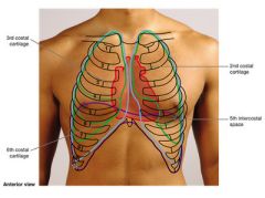
Lies mainly behind sternum. 4 corners of heart: Right: 3rd costal cartilage Between 5th and 6th costal cartilage Left: 3rd costal cartilage 5th intercostal space (further L) |
|
|
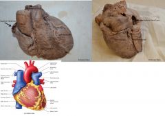
Identify the apex and base of the heart and the chambers that form them. |
|
|
|
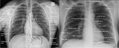
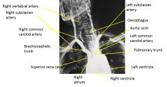
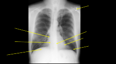
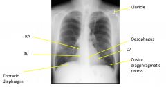
Recognise the main features of the heart on a plain posterior/anterior x-ray. |
|
|
|
|
|
|
|
|
|
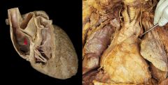
Recognise and describe the 4 chambers of the heart and the markings evident on the inside surfaces of these chambers |
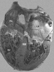
2 atria that have auricular appendages attached Vena cava>R atrium Pulmonary veins>L atrium 2 ventricles containing papillary muscle and chordae tendonae R ventricle>pulmonary artery L ventricle>aorta |
|
|
Recognise and describe the 4 main valves of the heart and appreciate when, and by what means, they open and close. |
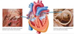
Tricuspid: RA/RV Pulmonary: RV/pulmonary trunk Mitral: LA/LV (2 flaps) Aortic: LV/ ascending aorta LV relaxes: aortic closes, mitral opens Blood LA->LV LA contracts LV contracts: mitral closes, aortic opens Blood: LV->aorta |
|
|
Describe the location, arrangement, and function of the chordae tendonae and papillary muscle |
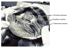
Chordae tendonae: bundles of collagen fibres that arise form papillary muscle to limit movement of cusps and prevent backflow of blood. On mitral and tricuspid valves in ventricles. |
|
|
Locate the fossa ovalis and the ligamentum arteriosum and appreciate their significance. |
Fossa ovalis is a depression in RA, remnant of thin fibrous sheet that covered foramen ovale during fetal development (closes at first breath) Ligamentum arteriosum is remnant of ductus arteriosus (fetal structure that shunts blood from pulmonary arteries to aorta (bypass lungs)) |
|
|
Locate and trace the 2 main coronary arteries and their major branches and how what regions of the heart each one supplies |
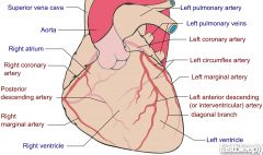
Arises from ascending aorta Right coronary artery Supplies: RA, RV Branches: Marginal artery, posterior interventricular artery Left coronary artery Supplies: LA, LV Branches: Anterior interventricular artery, Circumflex artery |
|
|
Identify the interventricular septum and appreciate its significance. |
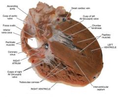
Seperates sides of the heart. Stops mixing of blood. |
|
|
Know the location of the SAN and AVN and know the location of fast conducting fibres of the heart. |
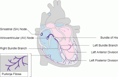
SAN - RA near entrance of superior vena cava AVN - floor of RA between A and V Fast conducting fibres (Purkinje) - inner ventricular walls |