![]()
![]()
![]()
Use LEFT and RIGHT arrow keys to navigate between flashcards;
Use UP and DOWN arrow keys to flip the card;
H to show hint;
A reads text to speech;
127 Cards in this Set
- Front
- Back
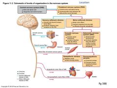
|
CNS |
-Brain and Spinal Cord. -Integration and control center. -Interprets sensory input and dictates motor output.
|
|
|
PNS
|
Everything else: Cranial nerve, spinal nerves, ganglia, enteric plexuses in small intestine, sensory receptors in skin. |
|
|
PNS system includes
|
-Somatic nervous system -Autonomic nervous system -Enteric nervous system |
|
|
Sensory (afferent) division
|
-Somatic and viscera sensory nerves fibers. -Conducts impulses from receptions to the CNS. |
|
|
Motor (efferent) division
|
Transmits impulses from CNS to effector organs: Muscles and glands. Two divisions: Somatic and Autonomic Nervous System. |
|
|
Somatic nervous system = Body
|
Conduct impulses from CNS to skeletal muscles Voluntary nervous system (Counscios control of skeletal muscles)
|
|
|
Autonomic nervous system = Automatic
|
Consist of visceral motor nerve fibers. Regulates smooth and cardiac muscles and glands. Involuntary. Two functional subdivision: Sympathetic and Parasympathetic. |
|
|
Sympathetic = Fight or flight
|
Mobilitizes body system during activity. |
|
|
Parasympathetic = Rest and digest
|
Conserves energy. Salivation, Lacrimation, Urination, Digestion, Defecation. |
|
|
Nervous System
|
Is master controling and communicating system body, via electrical and chemical signals. |
|
|
N.S has three overlapping functions
|
-Sensory input -Integration -Motor output |
|
|
Sensory Input
|
Information gathered by sensory receptos about internal and external changes
|
|
|
Integration
|
Processing and interpretation of sensory input
|
|
|
Motor output
|
Activation of effector organs (muscles and glands) produces a response. |
|
|
|
|
|
Neurons (nerve cells)
|
-Are structural units of nervous system. -Extreme longevity - 100 + years. -Amitotic (except olfactory epithelium and hippocampus). -High metabolic rate - require continuous and abundant oxygen and glucose. -All have a cell body and one or more processes. |
|
|
Neuron Cell Body (perikaryon or soma)
|
Contains nucleus, cytoplasm, typical organells and Nissl boddies.
|
|
|
Nuclei
|
Cluster of neurons cell bodies in the CNS |
|
|
Ganglia
|
Cluster of neurons cell bodies in PNS
|
|
|
Dendrites (Afferent)
|
Receiving/ input portions. Graded potentials. |
|
|
Axon (Efferent) |
Propagates nerve impulses. Action potentials. |
|
|
Axon Hillock
|
Action Potential generated here Cone-shape part at cell body No protein synthesis here. |
|
|
Axon Collateral
|
Branches of the axon
|
|
|
Axon Terminal
|
The end of an axon.
|
|
|
Multipolar Neuron
|
-Many processes extend fron the cell body -All are dendrites except for a single axon -Most abundant in body -Major neuron type in the CNS |
|
|
Bipolar Neuron
|
-Two processes extend from the cell -One is a fused dendrite, the other is an axon -Rare -Found in some special sensory organs like olfactory mucosa, eye and ear. |
|
|
Unipolar Neuron |
-One process extends from the cell body and forms cental and peripheral processes, which together comprise an axon. -Found mainly in PNS -Common only in dorsal root ganglia of the spinal cord and sensory ganglia of cranial nerves |
|
|
Neuroglia (Glia cells)
|
Small cells that surround and wrap delicate neurons
|
|
|
Neurons (Nerve cells)
|
Excitable cells that transmit electrical signals.
|
|
|
Neuroglia of CNS Neurons
|
-Astrocites -Microglial cells -Ependymal cells -Oligodendrocytes |
|
|
Astrocites |
-Star shape -Large and most abundant in CNS Functions: Support, Nutrition and Blood Brain Barrier -Support neurons with microfilaments and nutrition. -Secrete chimicals to regulate growth, migration and interconnection among neurons in the embryo. -Maintain a good chemical enviroment for generation Action Potentials. |
|
|
Glial Scars |
Are proliferation of astrocytes when CNS axons are cut and won't regrow. |
|
|
Microglia Cells
|
-Inmune like cells: Phagocytosis -Found in CNS -Functions as phagocytes to remove cellular debris and microbes. |
|
|
Ependymal Cells
|
-Found in CNS: CSF -Line the ventricales of the brain and central canal of spinal cord. -Secrete CSF (cerebrospinal fluit) which protects and nourishes the brain/spinal cord. -Form blood-cerebrospinal fluit barrier. |
|
|
Neuroglia of PNS-
|
-Satellite cells -Schwann cells |
|
|
Satellite cells
|
-Found in PNS: Support, Nutrition -Surround neuronal cell bodies to provide structural support. -Regulates exchange of materials between neuronal cell bodies and interstitial fluid |
|
|
Schwann Cells (Neurolemmocytes)
|
-Found in the PNS: Myelin formation -Form and maintain myelin sheath arounf PNS axons -1 cell myelinates 1 axon -Participate in axon regeneration |
|
|
Myelin Sheath
|
-Are a mulpitple lipoprotein covering -It electrically insulates the axon and increases the speed of nerve impulses (AP) conduction. -Nodes of Ranvier |
|
|
Nodes of Ranvier
|
Gaps in the myelin sheath. |
|
|
Myelinated fibers
|
Segmented sheath surrounds most long or large diamenter axons
|
|
|
Nonmyelinated fibers
|
Do not contain sheath Conduct impulses more slowly |
|
|
Demyelination
|
-Loss / destruction of myelin sheaths due to disorder or radiation/chemotherapy -May cause deterioration of affected nerves. |
|
|
Steps of myelination of a nerve fiber
|
1. A schwann cell envelops an axon 2. The schwann cell then rotates around the axon, wrapping its plasma membrane loosely around it in successive layers. 3. The schwann vell cytoplasm is force between the membranes. The right membrane wrapping surroundings the axon from the myelin sheath. |
|
|
In Nonmyelinated Axons (continuous conduction) |
Conduction is slow because it takes time for ions and for gates of channel proteins to move, and this must occur before voltage can be regenerated. |
|
|
In Myelinated Axons (Saltatory conduction)
|
Conduct is fast. Myelin keeps current in axons (voltage doesn't decay much). APs are generated only in the myelin sheath gaps and appear to jump rapidly from gap to gap.
|
|
|
Chemically gated ion channels (Ligand channels) |
Opens in response to binding of the appropriate neurotrasmitter
|
|
|
Voltage-gated ions channels
|
Open in response to changes in membrane potential. |
|
|
Mechanically gated channels
|
Open and close in response to phisical deformation of receptos, as sensory receptors. |
|
|
Leakage (nongated) channels
|
Channels, which are always open |
|
|
Gated channels
|
In which part of the protein changes shape to open/close to channel
|
|
|
Multiple Sclerosis (MS)
|
-Autoimmune demyelinating disease. -Demyelinates CNS axons -Turns them into ninfunctioning lesions called Scleroses. -Impulses conduction ceases, but axons are nor damaged -Cycles of remission and relapse are typical -Symptoms: Visial & speech distrubances, muscle weakness/ tingling/ paralysis, incontinence. |
|
|
Hydrocephalus
|
Pathology in which the production of CSF exceeds reabsorption. An increase in cranial pressure expands the unfused skull.
|
|
|
1.Lateral Ventricles
|
There are two, one in each hemisphere. Horseshoe in appearance. Seperated by the Septum Pellucidum-thin membrane.
|
|
|
a. Interventricular Foramen
|
Connects the lateral ventricles to the 3rd ventricle
|
|
|
2. 3rd Ventricle
|
b. Cerebral Aquaduct connects the 3er ventricle to the 4th ventricle
|
|
|
3. 4th Ventricle
|
CSF reabsorbed by the Arachnoid granulation-project into veins of the dural sinuses.
|
|
|
Meninges
|
3 connective tissue membranes that lie external to the CNS organs. -Cover and protects the CNS, blood vessels and enclose venoues senuses. -Contains CSF |
|
|
A. Epidural Space. 1. Dura Mater |
"Tough Mother" The outer meningeal covering. Two layers: -Endosteal layer -Meningeal layer |
|
|
-Endosteal layer
|
Fused with the periosteum in the cranial bones. a.Dura sinus: Large blood sinus between the two laters. Drain blood from cranial venis into jugular veins. |
|
|
-Meningeal layer
|
Faces the arachnoid mater. The inner dural layer.
|
|
|
B. Subsural Space 2.Arachnoid mater |
Much "Thinner" covering. Forms a "Smooth" covering the brain. Weblike connecting with the next layer - The Pia Mater.
|
|
|
C. Subarachnoid Space (CSF floods through this layer). 3. Pia Mater |
"Gentle Mother" This layer has direct contact with the surface of the brain. COntains blood vessels supplying the brain.
|
|
|
CSF Circulation
|
1.The choroid plaxus of each ventricles produces CSF. 2.CSF flows the ventricles and into the subarachnoid space via the median and lateral apertures. 3.CSF flows through the subarachnoid space. 4.CSF is absorbed into the dural venous sinuses via the arachnoid granulations. |
|
|
Where is spinal anesthesia injected?
|
Subarachnoid
|
|
|
Cerebrum
|
Cerebral hemisphere are paired (left and right) superior parts of the brain. -Includes more than half of the brain mass. -The surface is made of ridges (gyri) and grooves (sulci). |
|
|
3 main region of the cerebral hemisphere
|
1. Cortex (gray matter) 2. White matter 3. Basal nuclei (deep pockets of gray matter) |
|
|
Lobes
|
Fissures (deep grooves) divide the cerebrum into lobes. Frontal, Parietal, Occipital, and Temporal Lobe. |
|
|
Visual Areas
|
Occipital lobe
|
|
|
Audiroty Area
|
Temporal lobe |
|
|
Olfactory Cortex (Smell) |
Temporal lobe and small area of frontal lobe.
|
|
|
Gustatory Cortex
|
Temporal lobe (involved in the perception of taste). Visceral Sensory Area: Posterior to gustatory cortex. |
|
|
Vestibular Cortex (Equilibrium)
|
Temporal lobe
|
|
|
Grey Matter in the Brain (outer)
|
-Outer layer in the cerebral cortex; composed mostly of neuron cell bodies.
|
|
|
White Matter in the Brain (Inner) |
-Fibers tracts deeps to the grey matter. -Corpus callosum connects hemisphere.
|
|
|
Grey Matter in the Spinal Cord (Inner) |
-Surround central canal. -Forms butterfly or H shape -Dominated by cell bodies neurons neuroglia. and unmyelinated axons. -Great amount in spinal cord segments serving limbs
|
|
|
White Matter in the Spinal Cord (Outer) |
-Superficial -Contains large numbers of myelinated and unmyelinated axons.
|
|
|
Longitudinal Fissure
|
Separetes the cerebral hemisphere
|
|
|
Transverse Cerebral Fissure
|
Separetes the cerebral hemisphere and the cerebelum.
|
|
|
Central Sulcus
|
Separetes frontal lobe from the parietal lobe
|
|
|
Parieto-occipital sulcus
|
Separetes the occipital from the parietal lobe
|
|
|
Lateral Sulcus
|
Outlines temporal lobe. |
|
|
Insular lobe (Gyri of Insula) |
Is burries under portions of the temporal , parietal and frontal lobe.
|
|
|
Part of the hemisphere removed and the brain being abe to adapt. What is it called?
|
Adaptation after injury
|
|
|
Thalamus
|
-Location: walls of the 3rd ventricle. -Makes up to 80% of diencephalon. -The sensory relay center on the body. -Olfactory doesn't relay here. -Relays most sensational to the cerebrum. -Ascending fibers synapse here before entering to Cereberum. -Many important nuclei are found here. Mostly grey matter. -Nuclei cell bodies, for hearing, vision, taste. -Key role in meditaion, motor activities, cortical arousal, learning and memory. |
|
|
Hypothalamus
|
-Location: below the Thalamus. -Control center of the ANS. -Synthesizes, store and releases hormones. -Regulates secretion from pituitary gland. The infundibulum connects Hypo. and Pituitary -Temperature center. -Coordinates heart rate, blood pressure, respiratory rate from Pons and Medulla. -Regulates fear, anger, hunger, sexual feelings. |
|
|
Mamillary Body
|
-Two visible bumps on the posterior of the Hypothalamus. -Memory and emotional response to odors. |
|
|
Epithalamus
|
-Location: Above the Thalamus. -Includes the Pineal gland. -Control of the Circadian Rhythm (biological clock) -Releases Melatonin (Hormone). -Induces sleep. High levels during darkness. |
|
|
Limbic System
|
-"Emotional Brain" -Links conscious to the unconscious. -Emotions, memory, mood, behavior, olfaction.
|
|
|
Limbic System Components
|
-Cingulate gyrus -Hypothalamus -Hippocampus -Amygdala -Olfactory bulbs -Fornix |
|
|
Cingulate Gyrus
|
(Limbic lobe) Integration of sensory input, adds emotional content. |
|
|
Hypothalamus
|
Contol center of ANS |
|
|
Hippocampus
|
Long term memory, creating new memories
|
|
|
Amygdala
|
Control emotions, fear, interprets facial expressions, new social situations, identifyng danger. |
|
|
Olfactory Bulbs
|
Olfaction
|
|
|
Fornix
|
Connects parts of the limbic system together.
|
|
|
Brain Stem
|
-Midbrain, Pons, Medulla Oblongata. -Different form spinal cord due to having nuclei of gray matter embedded in the white. -Produces programmed, automatic behaviors necessary for survival -Positioned between cerebrum and spinal cord. |
|
|
Midbrain
|
-Upper part of the brain stem. -Origin for cranial nerves 3 and 4. |
|
|
Pons
|
-"Bridge", relay station for descending and ascending fibers. -Responsible for respiratory rhythm, inhalation and exhalation. -Origin for cranial nerves 5 and 7. |
|
|
Medulla Oblongata
|
-Becomes the spinal cord at the foramen magnum. -Heart and respiratory rates. -Origin of CN's IX-XII. -Vasomotor center- smooth muscle tone of blood vessels. -Autonomic center for: .-Emesis: vomiting. .-Deglutiton: swallowing. Coughing, hiccupping, and sneezing. |
|
|
Cerebellum
|
- 10% volume of brain and 50% of the brain's neurons. -Arbor Vitae (tree of life) -Coordination of movements and spatial reception. -Balance and equilibrium. -Time keeper, distinguishing pitch and similar sounding words, planing and scheduling tasks.
|
|
|
Broca's Area
|
-Involves in our ability to speak. -Patients with lesions in Broca's understand words, but cannot speak. |
|
|
Wernicke's Area
|
-Involved in understanding spoken and writen words. -Patients with lesions in Wernicle's can speak, but words are nonsensible. |
|
|
NREM Sleep (Non Rapid Eye Movement)
|
Four stage, Stages 3 and 4 are thought to be restorative stages
|
|
|
REM Sleep (Rapid Eye Movement) |
Occurs at approximately 90 min of sleep, after the fourth stage of NREM. -Temporary paralysis: Skeletal muscles are actively inhibited, except the ocular muscles and the diaphragm. -EEG, heart rate, respiratory rate, BP,and GI motility change. -Deprevation of REM patient can become moody and depressed. |
|
|
Memory
|
The storage and retrieval of information. Two stages: -Short Term Memory (STM) -Long Tern Memory (LTM)
|
|
|
Short Term Memory
|
-AKA. Working memory. -Temporary holding of information. -Limit to 7 or 8 pieces of info. |
|
|
Long Term Memory
|
Limitless capacity |
|
|
Declaritive Memory
|
-Specific information. -Related to conscious thoughts and languages ability. -Stored in the LTM with context in which learned. |
|
|
Non Declarative Memory
|
-Less conscious or unconscious. -Acquired through experience and repetition. -Best remembered by doing. Hard to unclear. -Includes: Procedural memory Motor memory Emotional memory |
|
|
Procedural Memory
|
-Basal nuclei relay to Thalamus and pre-motor cortex. -Dopamine from Substantia Nigra needed. -Skills: ex. Playing piano
|
|
|
Motor Memory
|
Cerebellum.
|
|
|
Emotional Memory
|
-Amygdala. -The hippocampus and surrounding temporal lobes function in consolidation and access of Memory. -Acetylcholine needed for memory formation and retrieval. |
|
|
Cranial Nerves
|
-12 pairs of cranial nerves are associated with brain. -Most are mixed nerves, but two pairs purely sensory |
|
|
Cranial Nerves I - XII 1. Olfactory Nerves |
Sensory F / Motor F / PS* Fibers/ -Sensory nerve of smell Yes (smell) / No / No |
|
|
2. Optic Nerve
|
-Purely sensory (visual) function Yes (vision) / No / No |
|
|
3. Oculomotor Nerves
|
-Raising eyelid, directing eyeball, constricting iris (parasympathetic), and controlling lens shape. No / Yes / No |
|
|
4. Trochlear Nerves
|
-Motor nerve that directs eyeball No / Yes / No |
|
|
5. Trigeminal Nerves
|
-Three divisions: Ophthamic, Maxillary, Mandibular. -Convey sensory impulses from various areas of face (V1 and V2). -Supply motor fibers (V3) for mastication Yes (general sensation) / Yes / No |
|
|
6. Abdunces Nerves
|
-Motor, innervating lateral rectus muscle No / Yes / No |
|
|
7. Facial Nerves
|
-Motor: nerves of face. Facial expression. -Sensory:(taste) from anterior two-thirds of tongue Yes (taste) / Yes / Yes |
|
|
8. Vestibulocochlear Nerves
|
-sensory function: hearing and balance; Yes (hearing and balance) / Some / No |
|
|
9. Glossopharyngeal Nerves
|
-Motor functions: tongue and pharynx for swallowing. -Sensory functions: taste and general sensory impulses from pharynx and posterior tongue. Yes (taste) / Yes / Yes |
|
|
10. Vagus Nerves
|
-Most motor fibers are parasympathetic fibers that help regulate activities of heart, lungs, and abdominal viscera. -Sensory: taste buds of posterior tongue and pharynx Yes (taste) / Yes / Yes |
|
|
11. Accessory
|
-innervate trapezius and sternocleidomastoid muscles No / Yes / No |
|
|
12. Hypoglossal Nerves
|
-Innervate extrinsic and intrinsic muscles of tongue that contribute to swallowing and speech No / Yes / No |
|
|
Trigeminal Neuralgia (Tic Douloureux)=(Twitch-Pain) |
Cause by inflammation of trigeminal nerve, is widely considered to produce most excruciating pain known.
|
|
|
|
|