Reading...
![]()
Play button
![]()
Play button
![]()
Use LEFT and RIGHT arrow keys to navigate between flashcards;
Use UP and DOWN arrow keys to flip the card;
H to show hint;
A reads text to speech;
140 Cards in this Set
- Front
- Back
|
Central Nervous System
|
Composed of the brain and spinal cord
|
|
|
Surface anatomy of the brain
|
-cerebral hemispheres, cerebellum, and brain stem
|
|
|
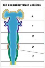
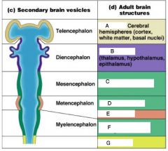
A) Telencephalon
B) Diencephalon C) Mesencephalon D) Metencephalon E) Myelencephalon |
|
|
A) Cerebrum
B) Diencephalon C) Brain Stem; midbrain D) Brain Stem; pons E) Cerebellum F) Brain Stem; Medulla onlongata H) Spinal Cord |
|
|
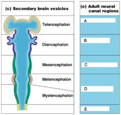
Telencephalon
|
Lateral Ventricles
|
|
|
Diencephalon
|
Third Ventricles
|
|
|
Mesenecephalon
|
Cerebral aqueduct
|
|
|
A) Lateral Ventricles
B) Third Ventricles C) Cerberal Ventricles D) Fourth Ventricles E) Central Canal |
|
|
Know
|
|
|
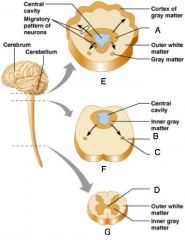
Spinal Cord
|
External to which is white matter composed of myelinated fiber tracts
|
|
|
Brain
|
-Similar to spinal cord but with more grey matter
-Cerebellum has gray matter in nuclei |
|
|
A) Inner gray matter
B) Outer white matter C) Gray Matter D) Central Cavity E) Region of cerebellum F) Brain Stem G) Spinal Cord |
|
|
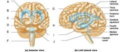
Ventricles of the Brain
|
Arise from the expansion of the lumen of the neural tube
|
|
|
The ventricles are (3)
|
-The paired C-shaped lateral ventricles
-The third ventricles found in the diencephalon -The fourth ventricle found in the hindbrain dorsal to the pons |
|
|
A) Lateral ventricle
B) Septum pellucidum C) Third ventricle D) Cerebral aqueduct E) Fouth Ventricle F) Central Canal G) Anterior Horn H) Interventricular foramen I) Inferior horn J) Lateral Seperture |
|
|
The cerebral hemipsheres contain ______ and shallow ______
|
Ridges (gyri)
Grooves (sulci) |
|
|
Fissures
|
Deep groves in the cerberal hemispheres
|
|
|
Cerebral hemipsheres are seperatted by the
|
longitudinal fissure
|
|
|
The Cerebral hemipsheres have three basic regions
|
Cortex, white matter and basal nuclei
|
|
|
Deep sulci divide the hemipsheres into five lobes
|
-Frontal
-Parietal -Temporal -Occipital -Insula |
|
|
Central Suculus
|
Seperates the frontal and parietal lobes
|
|
|
Parieto-occipital suculus
|
Seperates the parietal and occipital lobes
|
|
|
Lateral sulcus
|
seperates the parietal and temporal lobes
|
|
|
What two structures border the central sulcus
|
precentral and postcentral gyri
|
|
|
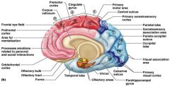
Cerebral cortex
|
superficial gray matter, that accounts for 40% of the mass of the brain. It enables sensation, communication, memory, understanding and voluntary movements
|
|
|
Each hemisphere in the cerebral cortex acts
|
contralaterally: controls opposite sides of the body
|
|
|
What is different about the hemispheres of the cerebral cortex
|
they are not equal in function
|
|
|
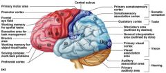
Motor Areas
|
control voluntary movement
|
|
|
Sensory areas
|
conscious awareness of sensation
|
|
|
Association areas
|
integrate diverse information
|
|

|
Know
|
|
|
Know
|
|
|
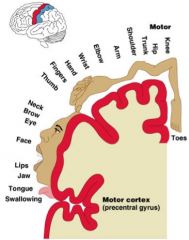
Primary motor cortex
|
loacted in the precentral gyrun
|
|
|
What is the primary motor cortex composed of
|
pyramidal cells whos axons make up the corticospinal tracts
|
|
|
What does the primary motor complex allow
|
conscious control of precise, skilles, voluntary movements
|
|
|
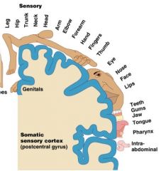
Motor homunclulus
|
Caricature of relative amounts of cortical tissue devoted to each motor function
|
|
|
Know
|
|
|
premotor cortex
|
located anterior to the precentral gyrus
|
|
|
What does premotor cortex control
|
learned, repititous, and patterned motor skills
|
|
|
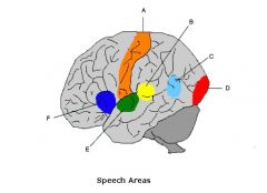
Brocas Area
|
-Located anterior to the inferior region of the premotor area
-present in one hemisphere |
|
|
What role does brocas area play
|
-motor speech area that directs muscles of the tongue
-activates as one prepares to speak |
|
|
Frontal eye field
|
Located anterior to the premotor cortex and superior to brocas area
|
|
|
What does the frontal eye field control
|
voluntary eye movement
|
|
|
What are the sensory areas of the brain
|
-Primary somatosensory cortex
-Somatosensory association cortex -Visual and auditory areas -Olifactory, gustatory, and vestibular cortices |
|
|
Primary somatosensory cortex
|
Located in the postcentral gyrus
|
|
|
What role does the primary somatosensory cortex play
|
-recieves information from the skin and skeletal muscles
-exhibits spatial discrimination |
|
|
Somatosensory homunculus
|
-caricature of relative amounts of cortical tissue devoted to each sensory function
|
|
|
Know
|
|
|
Know
|
|
|
Somatosensory association cortex
|
located posterior to the primary somatosensory cortex
|
|
|
What is the role of the somatosensory association cortex
|
Intergrates sensory information
|
|
|
Primary visual (striate) cortex
-Location -Function |
-seen on the extreme posterior tip of the occipital lobe
-Recieves visual information from the retinas |
|
|
Visual association area
-Location -Function |
-Surrounds the primary visual cortex
-Interprets visual stimuli (color, form, and movement) |
|
|
Primary Auditory cortex
-Location -Function |
-Located at the superior margin of the temporal lobe
-Revieves information related to pitch, rythm, and loudness |
|
|
Auditory association area
-Location -Function |
-Located posterior to the primary auditory cortex
-Stores memories of sounds and permits preception of sounds |
|
|
Where is wernickes area located
|
Auditory association area
|
|
|
What did carl wernicke describe about the brain
|
That people who suffer neurophysiological damage to this area are unable to understand the content word while listening, and unable to produce meaningful sentence; there speech has grammatical structures but no meaning
|
|
|
What are the association areas (4)
|
-prefrontal cortex
-Language areas -General interpretation area -Visceral association |
|

|
Know
|
|
|
Prefrontal cortex
-Location -Function |
-in the anterior portion of the frontal lobe
-Involved with intellect, cognition, recall, and personality |
|
|
The prefrontal cortex is necessary for
|
judgement, reasoning, persistance, and conscience
|
|
|
Which cortex is linked to the limbic system
|
Prefrontal cortex
|
|
|
Where are the language areas located
|
-large areas surrounding the left lateral sulcus
|
|
|
Wernickes area (language area)
|
involved in sounding out unfamilary words
|
|
|
Brocas area (language area)
|
Speech preperation and production
|
|
|
Lateral prefrontal cortex (language area)
|
Language comprehension and word analysis
|
|
|
Lateral and ventral temporal lobe (language area)
|
coordinate auditory and visual aspects of language
|
|
|
General Interpretation Area
-Location -Function |
-Found in one hemisphere, usually the left
-Integrates incoming signals into a single though -Involved in processing spatial relationships |
|
|
A) Motor Cortex
B) Wernickes area C) Angular gyrus D) Visual cortex E) Auditory Cortex F) Brocas Area |
|
|
Lateralization
|
each hemisphere has the abilities not shared with its partner
|
|
|
Cerebral dominance
|
-designates the hemisphere dominant for language
|
|
|
Left Hemisphere
|
-Controls language, math and logic
|
|
|
Right Hemisphere
|
-controls visual-spatial skills, emotion, and artistic skills
|
|
|
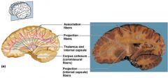
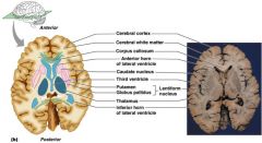
Cerebral White Matter
|
consists of deep myelinated fibers and their tracts
|
|
|
Cerebral white matter is responsible for the communication between
|
The cerebral cortex and lower CNS center, and areas of the cerebrum
|
|
|
Commissures
|
Connect corresponding gray areas of the two hemispheres
|
|
|
TEST QUESTION!!!!
|
|
|
TEST QUESTION!!!
|
|
|
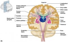
Basal Nuclei/ganglia
|
Masses of gray matter found deep within the cortical white matter
|
|
|
The corpus stratum is composed of three parts
|
-Caudate nucleus
-Lentiform nucleus -Fibers of internal capsule |
|
|
Lentiform Nucleus
|
Composed of the putamen and the globus pallidus
|
|
|
Know
|
|
|
What are the functions of Basal Nuclei (4)
|
-Influence muscualr activity
-Regulate attention and cognition -Regulare intensity of slow or sterotyped movements -Inhibit antagonistic and unnecessary movement |
|
|
Diencephalon
|
-Central core of the forebrain
|
|
|
What three paired structures does the diencephalon consists of
|
-Thalamus
-Hypothalamus -Epithalamus |
|
|
What structure enlcoses the third ventricle
|
-Dienchephalon
|
|
|
Know
|
|
|
Thalamus
|
Paired, egg-shaped masses that form the superolateral walls of the third ventricle
|
|
|
The thalamus is connected to the midline by the
|
intermediate mass
|
|
|
All inputs ascending to the cerebral cortex pass through the (TQ)
|
Thalamus
|
|
|
Thalamus plays a key role in (5)
|
mediating sensation, motor activities, cortical arousal, learning, and memory
|
|
|
Hypothalamus
|
Located below the thalamus, it caps the brainsetm and forms the inferolateral walls of the third ventricle
|
|
|
Mammillary Bodies
|
small, paired nuclei bulging anteriorly from the hypothalamus
|
|
|
The mammillary bodies are the relay station for (TQ)
|
Olfactory pathways
|
|
|
Infundibulum
|
Stalk of the hypothalamus; connects to the pituitary gland
|
|
|
What is the functions of the hypothalamic function
|
-blood pressue
-rate and force of heartbeat -digestive tract motility, -rate and depoth of breathing, and many other visceral activities |
|
|
Hypothalamic is involved with perception of (3)
|
-pleasure
-fear -rage |
|
|
The hypothalamic controls mechanisms needed to maintain
|
normal body temperature
|
|
|
The hypothalamic regulates feelings of
|
hunger and satiety
|
|
|
The hypothalamic regulates
|
sleep and the sleep cycle
|
|
|
Endocrine functions of the hypothalamus
|
Releasing hormones control secreation of hormones by the anterior pituitary
|
|
|
The supraoptic and paraventricular nuclei produce
|
ADH and oxytocin
|
|
|
Epithalamus
|
Most dorsal portion of the diencephalon; forms roof of the third ventricle
|
|
|
Pienal gland
|
extends from the posterior border and secreats melatonin
|
|
|
Melatonin
|
a hormone involved with sleep regulation, sleep-wake cycles, and mood
|
|
|
Choroid plexus
|
a structure that secreats cerebral spinal fluid
|
|

|
Know
|
|
|
The brain stem consists of three regions
|
-midbrain
-pons -medulla oblongata |
|
|
The brain stem is similar to the spinal cord by contains
|
embedded nuclei
|
|
|
The brain stem controls (TQ)
|
automatic behaviors necessary for survival
|
|
|
The brain stem is associated with (TQ)
|
10 of the 12 pairs of cranial nerves
|
|
|
Know
|
|
|
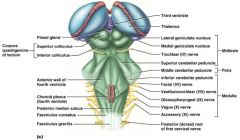
Midbrain
|
located between the diencephalon and the pons
|
|
|
Cerebral aqueduct
|
hollow tube that connects the third and fourth ventricles
|
|
|
Midbrain nuclei control cranial nerves _____ and _____
|
III(oculomotor) IV(trochlear)
|
|
|
Corpora quadrigemina
|
four domelike protrusions of the dorsal midbrain
|
|
|
Superior colliculi (TQ)
|
Visual reflex centers
|
|
|
Inferior colliculi (TQ)
|
Auditory replay centers
|
|
|
Substantia nigra
|
functionally linked to basal nuclei
|
|
|
Pons
|
Forms part of the anterior wall of the fourth ventricle
|
|
|
The fibers of the pons relay
|
impulses between the motor cortex and the cerebellum
|
|
|
pons are the origin of the cranial nerves ___, _____ and ____
|
V (trigeminal)
VI (abducens) VII (facial) |
|
|
pons contain nuclei of the
|
reticular formation
|
|
|
Cardivascular control center
|
adjust force and rate of heart contractions
|
|
|
Respiratory centers
|
control rate and depth of breathing
|
|
|
The cerebellum provides
|
precise timing and appropriate patterns of skeletal muscle contraction
|
|
|
The cerebellar activity occurs
|
subconciously
|
|
|
Folia
|
transversely oriented gyri
|
|
|
Arbor vitae
|
distinctive treelike pattern of the cerebellar white matter
|
|
|
Cerebellum Anatomy
|
two bilaterally symmetrical hemispheres connected medially by the vermis
|
|
|
cerebellum recieves impulses of the intent
|
to initiate voluntary muscle contraction
|
|
|
Proprioceptors and visual signals inform
|
the cerebellum of the bodys condition
|
|
|
the cerebellum cortex calculates
|
the best way to perfrom a movement
|
|
|
A "blueprint" of coordinated movement is sent
|
to the cerebral motor cortex
|
|
|
Two functional brain systems
|
-Limbic system
-Reticular formation |
|
|
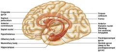
Limbic system
|
structures on the medial acpects of cerebral hemispheres and diencephalon
|
|
|
Limbic system includes (3)
|
-rhinencephalon
-amygadala -hypothalamus |
|
|
Amygadala
|
deals with anger, danger, and fear responses
|
|
|
Which system puts emotional responses to odors
|
Limbic System
|
|
|
Know
|