![]()
![]()
![]()
Use LEFT and RIGHT arrow keys to navigate between flashcards;
Use UP and DOWN arrow keys to flip the card;
H to show hint;
A reads text to speech;
44 Cards in this Set
- Front
- Back
|
LECTURE4LECTURE4 Chemical Kinetics What does it study? What does rate of reaction depend on? |
- the study of how fast a reaction occurs The rate of a reaction depends on:- Chemical species concentrations (including catalyst) - Kinetic constants (kcat, kf, kb, Km) - T, P, pH, viscosity, phase, … |
|
|
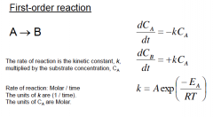
What describes the Kinetics of a First Order reaction A-->B What is the unit for k? |
|
|
|
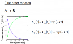
What does a First order graph look like? |
|
|
|
What is Allosteric Activation? |
2nd substrate / product binds; reaction rate increases |
|
|
What is Allosteric Inhibition? |
2nd substrate / product binds; reaction rate decreases |
|
|
How does Substrate & product inhibition regulates cellular metabolism |
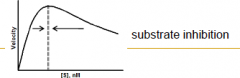
A part of a negative-feedback loop to achieve a desired set-point |
|
|
What is an Assay and what are the different types of Assay? How do you evaluate the quality of assay? |
- Assay is measuring a concentration of something, in this case [PRODUCT] - There are 2 main types: Direct vs. Indirect: Absorbance, Fluorescence, Luminescence, Light Scattering, Heat, Radiation, Mass - Quality: dynamic range, sensitivity / background, dependence on pH / temperature / solvent cost, throughput, generalizable |
|
|
Indirect Assay of Cofactor Usage: how does it work? |
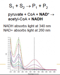
Measurement of Change in Absorbance at Two Wavelengths, since the 2 cofactors absorb diff wavelengths. |
|
|
Direct Assay: list of Assays e.g. separating by size/shape, Measuring mass, UV Visualizations or Fluorescence |
Mass Spec: counts every single molecule, HPLC: size shape charge, UV-Vis Spectrometry: measures light absorbance over wavelength, Fluorescence Detector: excites sample (at lambda1) & measures emission (at lambda2), Refractive Index: measures light refractionOthers: pH, conductivity, redox, |
|
|
What does Mass Spec do? what are the steps? what does it measure? |
Measures size, shape and charge of the molecules1. Ionization 2. Separation (magnetic or electrostatic) 3. Counting |
|
|
MASS SPEC STEP1: What are the types for ionization? |
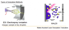
IONIZATION METHODS: 1. ESI(Electrospray ionization) 2.MALDI (putmolecules on a surface and shoot laser to charge) |
|
|
MASS SPEC STEP2&3: What are the types for Mass analyzer? What do they measure? |
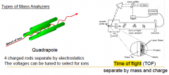
Separation and Counting They measure mass and charge ratio m/z 1.Quadrapole: 4 charged rods separate by electrostaticsThe voltages can be tuned to select for ions. Highly charged molecules will off quickly, lightly charged will slant. You can filter out molecules you don't need. 2. Time of flight (TOF): separate by mass and charge. Measures how long it takes to reach the detector very precisely. |
|
|
What are the extra steps in Mass Spec |
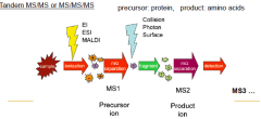
repetition of Fragmentation into smaller molecules. Repeating separation, collision Photon surface, separation ... detection. |
|
|
WHat are the steps in Liquid Chromatography Mass Spec? (LC-MS) |
1. Separated by size/hydrophobicity using chromatography column.
2. Detect the retention time (when the samples elute/appears) 3. Sample enters MS for identification. |
|
|
LECTURE5LECTURE5: Protein folding, Structure prediction and Design. |
LECTURE5LECTURE5: Protein folding, Structure prediction and Design. |
|
|
What enzyme characteristics control its catalysis? How can you change these characteristics? |
Changing enzyme's AA will alter these characteristics 1. Substrate specificity, kcat, Km: Increase kcat, decrease Km 2. Temperature-dependence: Increase or decrease optimal temperature 3. Solvent-dependence: eliminate sensitivity to solvent 4. Allosteric inhibition / activation: eliminate inhibition 5. Protein half-life: increase t1/2 |
|
|
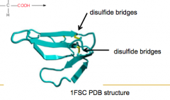
Which AA have Sulfur? What is special about them? |
Cysteine and Methionine because they can form disulfide bridges (covalent bonds) which makes the protein much stronger. |
|
|
What is the Protein Engineering examples given in class with Lipitor? |
Originally produced through inorganic cat with extreme conditions 80C, pH9. Also includes forming highly reactive epoxides as an intermediate before making Lipitor. Enzymatic conditions: 40C, pH7.3 (ambient). rate increases by 1000 fold, after mutating 35 AA's. (very complicated pathway) |
|
|
What are the Protein Engineering strategies? |
-Nature’s Toolbox: find a natural enzyme that works well - Directed Evolution: iterative mutagenesis + selection - Rational Design: use computational modeling to identify mutants |
|
|
Q: Can we predict the structure and function of a protein from its amino acid sequence? Q: Can we predict the amino acid mutations that will increase an enzyme’s kcat, decrease its Km, increase thermostability, change its cofactor requirement (e.g. NADPH to NADH)? |
These are the unanswered questions... been studied for 30 years++ |
|
Q: What happens if we change the Serine at 237, the Glutamic acid at 459, or the Threonine at 186? |
Will have drastic changes on the reaction rate |
|
|
What are the 2 most common types of protein folding? are there others? |
Alpha-Helix and Beta-Sheet There are currently ~1393 types of protein folds http://cathdb.infoProtein Folding |
|
|
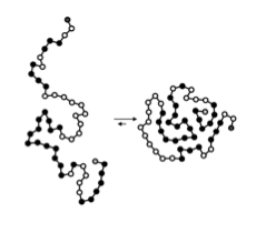
How does a protein find its native structure? |
Protein folding is hierarchical - Spatially local interactions form first, followed byglobal interactions - H2O-Protein interactions play an important role |
|
|
What are the hierarchy of Protein folding? |
1. Nucleation 2. Growth and coalescence: to form 2ndary structures 3. Readjustment for maximum overall stability 4. Quaternary association |
|
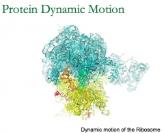
Molecular Simulation of a Ribosome (EQ State): Still always jiggling. Do we want drug that binds to part that move a lot or a little? |
We want to bind to the parts that only move a little. Lots of movement = high entropy, and if bound means entropy of that part decreases . Therefore, large Energy of binding. |
|
|
How does Classical theory (2 states model) apply to protein folding? |
The result is WRONG: If look at the Activation Energy, will need many years to fold, however in nature - proteins fold within micro-secs |
|
|
What is the real mechanism of protein folding? Why does it work that way? |
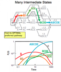
Unlike the classical theory, actual folding goes through many intermediate states from disorder to order. - There are many different unique pathways to fold so doesn't have to search long to find the next step/. |
|
|
What are the 4 examples for free energy landscapes for protein folding? How would you describe numbers of high-E, low-E, and minimum-E states that a protein has? |

1. Smooth 2. Rugged 3. Flat (diffusion controlled) 4. Rate-limiting intermediate Protein has millions high-E, hundreds of low-E and few/one minimum free energy state |
|
|
What are the challenges of predicting protein's structure from it's AA? |
1. S=klogZ=klnW if we have many degrees of freedom, Z= # of ways molecule can rotate, vibrate and move. 2. Our best models for calculating attractive & repulsive atomic energies/enthalpy are approximate. |
|
|
What are the 2 approaches used in predicting protein structure. |
1. homology prediction 2. physics-based prediction |
|
|
What is Homology Prediction? |
Predicting protein structure by comparing similar AA seq may have similar native structures. (evolution) |
|
|
What is Physics-Based Predictions? What are Force Fields and Free Energies? What is the important assumption? |
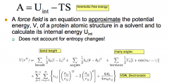
Physics-Based Predictions is trying to minimize the free energy G(fix T,P), minimize A(fix T,V). A=U-TS (easier to fix V&T) A force field is an equation to approximate the potential energy, V, of a protein atomic structure in a solvent and to calculate its internal energy. Key Assumption: If U (internal E, characterizes enthalpic interactions) is greatly minimized, A is minimized. Even if +dS |
|
|
How do we calculate force on an atom? |
F is calculated in the x,y and z direction as the derivative of potential E (V) function. |
|
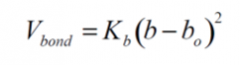
Potential of a bond, what does it depend on? |
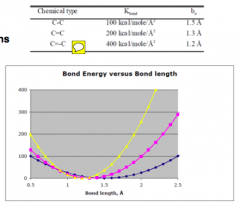
Interaction energies depend on atomic positions, as seen each bond have different preferred length. |
|
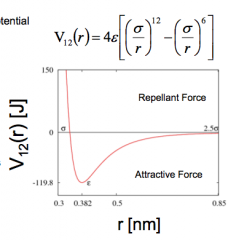
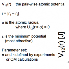
FORCES: Van Der Waals. What do each term means? |
|
|
|
FORCES: Electrostatic Interactions. What are the equations for Force and potential. What is the example used in class? |
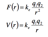
Example: T-Box TSC factor. Elec attraction between +AA and -Pi-DNA backbone. |
|
|
FORCES: H-Bonding. Occurs between which functional groups? Contributes largely to which type of protein folding? Parallel or anti-parallel AA are more stable? |
Attraction by (dipole-dipole electrostatics) between (NH+)-(N-), (NH+)-(O-), OH(-)---H(+) ANTI-PARALLEL BETA-SHEET |
|
|
Hydrophobic Interactions. What do they form, why are form?Any special examples? |
Hydrophobic cores and hydrophillic surfaces are formed. H2O maximize H-Bond. Example: Transmembrane Proteins. |
|
|
Covalent Forces: Disulfide Bonds |
|
|
|
What is the Computational Design Workflow for Designing and enzyme that will catalyze new reactions? |
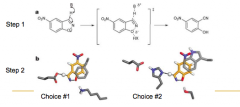
1. Identify transition state (TS) structure of reaction-of-interest 2. Identify AA residues & their configuration that will bind & stabilize TSThis is called the ‘binding pocket’ or ‘active site’ of the protein 3. Starting with a scaffold protein with an existing binding pocket,mutate AA residues and change their configuration untilit matches the target identified in Step 2 4. Calculate protein structure – substrate interaction energies. Negative enough? |
|
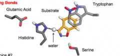
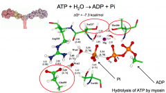
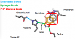
Q: What are the molecular interactions between the substrate and amino acids inside this target substrate binding pocket? |
|
|
|
How accurate is computational prediction to the experimental measurements? |
Root mean square Difference = 0.95 (smaller than C-C length) |
|

Same [S]0, same [E]tot. which enzyme design is the best? What happens if one of the Key AA is mutated? DATA: KE70 is the first designed molecule. key AA's: W110, S211, E231 |
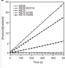
Best: KE59, it was mutated further and works even better than KE70. When the "Key catalytic AA residues" is mutated (KE59 E231Q) - It doesn't catalyze the reaction just as well. Very evident in the graph. |
|
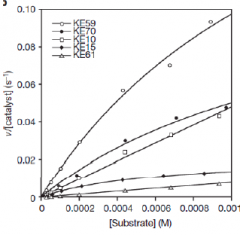
Diff [S]0, same [E]tot, measure [P] => Plot Rate vs [S]0 (initial S) Which enzyme design has the lowest Km, highest Km? |

Realize this is the reciprocal of Lineweaver-Burke Plot. Lowest Km: KE59 [E]strongbind Highest Km: KE61 [E]weakbind |