![]()
![]()
![]()
Use LEFT and RIGHT arrow keys to navigate between flashcards;
Use UP and DOWN arrow keys to flip the card;
H to show hint;
A reads text to speech;
39 Cards in this Set
- Front
- Back
|
Rib and Vertebrae Articulations |
For T2-T10 Rib 2 head articulates with demifacets T1 and T2 Rib 2 tubercule articulates with T2 transverse process costal facet T1 3 costal facets: 2 on body, 1 on transverse process 1st rib articulates only with T1 T11, T12 ribs 11,12 don't have tubercules so no articulation at transverse process |
|
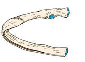
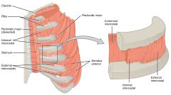
Label the Rib structures |
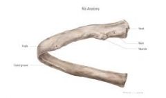
Typical Rib 2-10 blue at end away from head=cup for costal cartilage |
|
|
What's different about the 1st, 2nd, 11th, and 12th rib? |
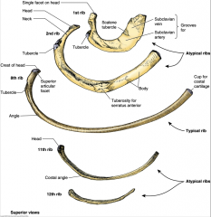
Rib 1: grooves for subclavian A,V and a tubercule for scalene muscle attachment Rib 2: Tuberosity for serratus anterior. R11, 12-no tubercules (no attachments to transverse processes), no costal cartilage (no sternal articulations) |
|
|
Whats the relation between costal cartilage and the sternum? |
True ribs (1-7): costal cartilage attach directly to sternum. For rib 1-fibrocartiligenous joint. For ribs 2-7=synovial False Ribs (8-10): costal cartilage attach to costal cartilage of rib above Floating Ribs (11,12)-no CC, attach only to vert. |
|
|
What muscles are in the intercostal space? |
External intercostal mm, internal intecostal mm., innermost intercostal mm, subcostal mm, transversus thoracis. |
|
|
External intercostal mm. where, innervation, action |
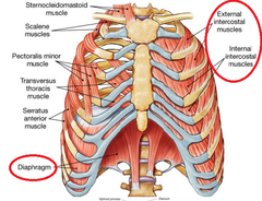
Span 1 rib. go "hands in pocket" Nerve: intercostal nerve action: elevate ribs for inspiration |
|
|
Internal intercostal mm. Where, what, nerve, action |
Deep to external intercostals span 1 rib. go out like you're going to party (opp of external intercostals) nerve: intercostal nerve action: depress ribs for forced exhalation |
|
|
Innermost intercostal mm. where, action, nerve |
deepest part of muscles sandwiching the intercostal vessels (internal intercostals are the superficial "bread") nerve: intercostal n. action: depresses ribs for forced expiration, esp in lateral side
|
|
|
Transversus thoracis mm. OINA |
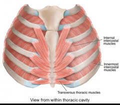
Nerve: intercostal nerve action: depresses costal cartilage near bottom of rib cage |
|
|
Subcostal mm. |
nerve: intercostal nerve action: depresses ribs for forced expiration. span about 2 ribs where: deepest, seen only on posterior thoracic cavity wall |
|
|
What nerves, vessels are in the intercostal space? |
Sandwich between internal intercostals and innermost intecostals in each intercostal space Superior to inferior: Intercostal Vein-->intercostal A.-->intercostal N. (VAN) |
|
|
Where do the intercosal veins drain to? |
superior-subclavian Inferior R side-azygous Inferior L side-hemiazygous, accessory hemiazygous |
|
|
Intercostal aa are branches of... |
superior intercostal artery-comes from subclavian A.=1st 2 intercostal nn. other intercostal aa from internal thoracic A. |
|
|
Where do intercostal Nn come from? |
Ventral rami of spinal nerves |
|
|
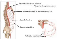
What are the branches of the internal thoracic arteries? |
4 branches: pericardiophrenic aa (run down lateral sides of mediastinum to the pericardial sac) anterior intercostal aa (anastamose with posterior intercostal arteries from the aorta) musculophrenic A (right branch of the split of the internal thoracic) superior epigastric A (left branch off of the split of the internal thoracic A.) |
|
|
Where do the internal thoracic Aa. come from? |
Subclavian Aa |
|
|
Is inspiration active or passive? What causes it? |
Inspiration is active. Takes energy for external intercostals and scalenes to expand ribcage and for diaphragm to depress abdominal viscera |
|
|
Is expiration active or passive? what causes it? |
passive expiration=passive. due to elastic recoil of thorax and abdomen and lungs forecful expiration=active. ribcage needs to contract with help from internal and innermost intercostals, transversus thoracis, subcostals |
|
|
What type of movement do the upper ribs have in respiration? Explain it. |
Pump handle movement. more anteroposterior expansion than transverse. done by external intercostals (moves sternum out and in) |
|
|
What type of movement do the lower ribs have in respiration. Explain it. |
Bucket handle movement. Lateral expansion as opposed to anteroposterior. |
|
|
How does the motion of the ribs and sternum play a role in respiration? |
when the ribs expand (upper=anteroposteriorly, lower=laterally) that creates a vaccum and air rushes into the lungs to fill that vaccuum |
|
|
Draw out the interal thoracic artery and branches |
|
|
|
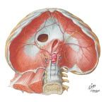
Describe the Diaphragm. What happens to it during inspiration, expiration? |
musculotendinous structure with two domes with a central tendon. It flattens out during inspiration,pushing viscera down. during expiration it passively reaches 5th rib (R dome) or 5th intercostal space (L dome) |
|
|
What is the diaphgragm's sensory and motor innervation? |
Motor: phrenic sensory: phrenic (central) and intercostal NN (periphery) |
|
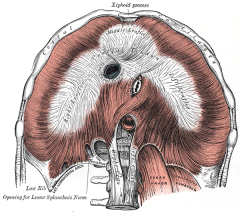
Label the 5 arcuate ligaments |
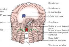
Median: over aortic hiatus, bound by crura Medial (2): L1 vert body-->transverse process. psoas major pass uner lateral (2): transverse process L1-12th rib quadratus lumborum pass under |
|

What does the caval hiatus contain? Locate it on diagram |

T8 Level Inferior vena cava, sometimes branches of right phrenic N. |
|

What does the esophogeal haitus conatain? Locate it on a diagram |

At T10 level Contains esophogus and anter and posterior vagal trunks (used to be vagus N.). its encircled by the crura |
|
What does the aortic hiatus contain? Locate it on a diagram. |
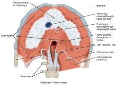
behind median arcuate ligament At T12 level Contains aorta, azygous and hemiazygous veins, and the thoracic duct |
|
|
What arteries supply the diaphragm? |
Superior phrenic AA (from aorta), pericardiophrenic and musculophrenic (from internal thoracic), lower intercostal AA, and inferior phrenic AA (from lumbar aorta) |
|
|
clinical: Hiatal Hernia |
when weakness at esophogeal hiatus allows part of the stomach or intestines to go up that hiatus Type 1/sliding-stomach pushed into thorax below esophagus Type 2-4-paraesophageal hernia-stomach/intestines come up through hiatus next to esophagus which may become strangled (no bl. Supply) |
|
|
Describe the angle between spinous processes from thoracic to lumbar region |
Angle between spinousprocess and angle of vertebral body starts at around 45 degrees and slowlyapproaches perpendicular in lumbar region Also T12 has a mammilary process |
|
What are the ligaments associated with the rib cage? Label them. |
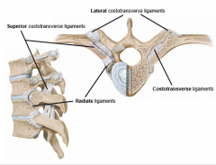
costotransverse ligament — neck of rib to transverse process superior costotransverse ligament — rib neck to transverse process of next superior vert. lateral costotransverse ligament — tubercle to transverse processof same vertebra radiate ligaments — vertebral body to rib neck=looks like donutC |
|
|
Sternocostal Joints |
Rib 1+manubrium-cartaligineous Rib 2+manubrium+body of sternum=synovial 3-7 + sternum-synovial joints |
|
|
What do intercostal nn do? |
motor innervation to all innercostal mm. cutaneous innervation to lateral sides of trunk and front of chest/abdomen |
|
|
What is the top opening of the rib cage called? The bottom opening of the rib cage? |
Top: superior thoracic aperture bottom: inferior thoracic aperture |
|
|
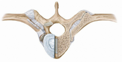
Costovertebral joints synovial or cartiligenous? |
2 synovial cavities (1/bone) each rib seperated from vert. by intra-articular ligament |
|
|
What type of respiratory movement does contraction of the diaphragm allow? |
Vertical expansion of rib cage |
|
|
What type of respiratory movement do ribs 11-12 have? |
Caliper or pincer movement-like theyre coming to clap together in the front |
|
|
What's the mneumonic to remember diaphragm hiatuses? |
I=IVC ATE=8 (T8) TEN=T10 EGGS= esophogeal AT=aortic NOON=T12 |