![]()
![]()
![]()
Use LEFT and RIGHT arrow keys to navigate between flashcards;
Use UP and DOWN arrow keys to flip the card;
H to show hint;
A reads text to speech;
80 Cards in this Set
- Front
- Back
|
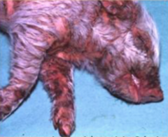
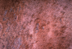
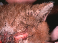
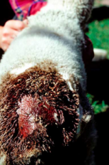
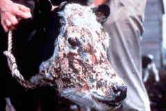
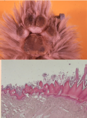
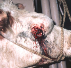
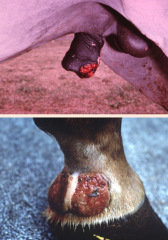
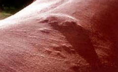
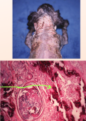
Suppurative epidermitis from S. hyicus |
|
|
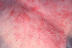
Staph. aureus |
|
|
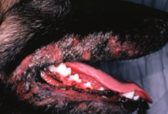
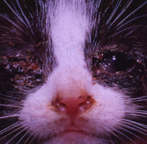
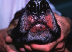
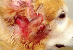
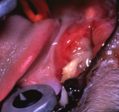
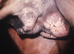
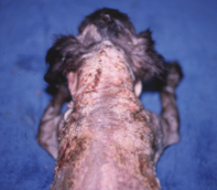
Mucocutaneous pyoderma Bacterial infection where the floppy lips are chronically wet, since this is the place susceptible to trauma and therefore bacteria can enter |
|
|
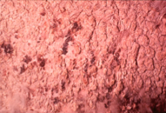
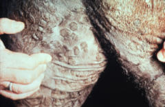
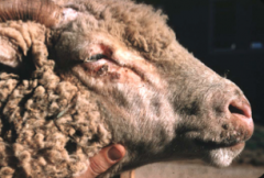
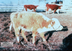
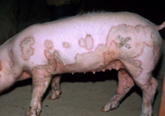
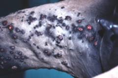
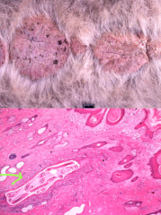
Dermatophillus dermatitis due to D. congolensis bacteria. Affects cattle, sheep, and horses. Aka. rain scald. |
|
|
Trichophyton equinum in a horse. Fungal infection of tissue causing dermatophytosis. Recall: dermatophytosis is due to either Microsporum or Trichophyton species |
|
|
Trichophyton verrucosum |
|
|
Malassezia pachydermatis. A fungal infection that affect the external ear canal and skin |
|
|
Candidiasis in a horse. A normal fungus in the GI and skin that can overgrow when the animal is immunocompromised. Targets mucous membranes and mucocutaneous junctions |
|
|
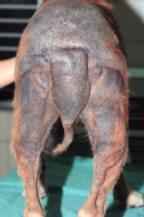
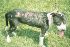
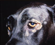
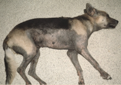
Generalized demodecosis due to Demodex canis. Occurs in young or immunosuppressed animals |
|
|
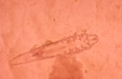
Demodex canis |
|
|
Sarcoptic mange |
|
|
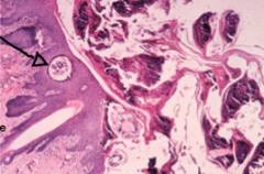
Sarcoptes scabei. This is a highly contagious mite that burrows deep in the skin (stratum corneum) and causes intense pruritic excoriations (hypersensitivity) |
|
|
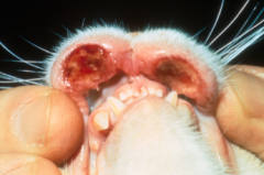
Notoedres cati. Affects cats and rabbits |
|
|
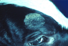
Bovine chorioptic mange |
|
|
Flea hypersensitivity. Caused by Ctenophalides felis or C. canis. Causes types 1 and 4 hypersensitivity |
|
|
Queensland itch. Type 1 and 4 hypersensitivity to Culicoides (sandflies) Other biting flies include Haematobia irritans (hornfly), Stomoxys calcitrans (stable fly), horse flies, and black flies |
|
|
Myiasis. An infection from the larvae of dipterous flies. |
|
|
Mosquito bite hypersensitivity |
|
|
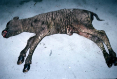
Lumpy skin disease due to capripoxvirus |
|
|
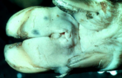
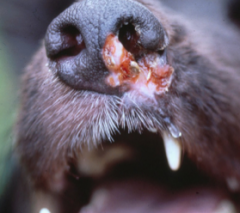
Contagious pustular dermatitis (orf). Due to parapoxvirus |
|
|
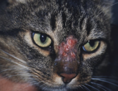
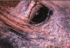
Feline herpesviral conjunctivitis and rhinitis. (FHV1) |
|
|
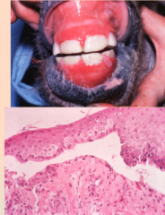
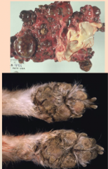
Picornovirus causing swine vesicular disease |
|
|
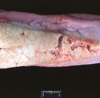
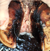
Picornovirus causing foot and mouth disease |
|
|
Thermal burn from heating pad |
|
|
Irritant contact dermatitis |
|
|
Skin infarction from Erysipelas |
|
|
Pemphigus foliaceus. Common and mild. Superficial vesicles that become pustules |
|
|
Bullous pemphigus. Separation of basal layer from basement membrane, thereby creating vesicles and ulcers |
|
|
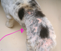
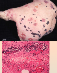
Leukotrichosis due to copper deficiency |
|
|
Photosensitization. 3 types: 1. Photodynamic substance (from plants or drugs) 2. Abnormal porphyrin metabolism 3. Hepatogenous (impaired liver can't properly excrete phylloerythrin) |
|
|
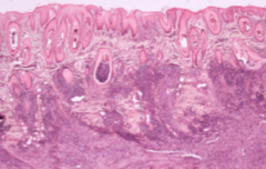
Ichthyosis (congenital increased adherence of keratinocytes) |
|
|
Nutritional disease causing epidermal growth disorder in the stratum corneum |
|
|
Canine distemper causing "hard pad" |
|
|
Superficial necrolytic dermatopathy |
|
|
Diffuse seborrhea |
|
|
Pityriasis rosea |
|
|
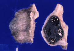
Squamous cell carcinoma |
|
|
Epitheliotropic lymphoma |
|
|
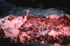
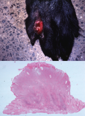
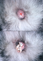
Pyotraumatic dermatitis (hot spot) |
|
|
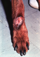
Acral lick dermatitis |
|
|
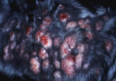
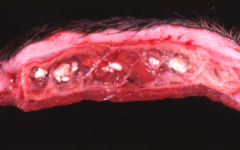
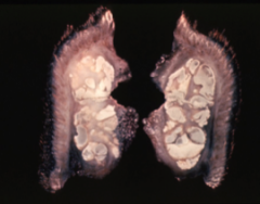
Mycobacterial granuloma |
|
|
Sporothrix schenckii (Sporotrichosis). Fungal. |
|
|
Pythium insidiosum. causes water mold (fungal) |
|
|
Leishmania (protozoal) |
|
|
Habronemiasis (parasitic). Caused by the larvae of Habronema or Draschia |
|
|
Onchocerciasis (biting midges - parasitic) |
|
|
Stephanofilariasis (eosinophilic caused - parasitic) |
|
|
Eosinophilic granuloma (immune mediated) |
|
|
Eosinophilic granuloma (immune mediated) |
|
|
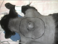
Vaccine-associated sarcoma |
|
|
Injection site reaction |
|
|
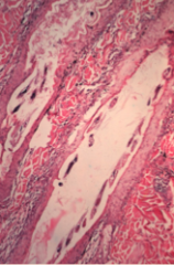
Ehlers Danlos syndrome (collagen dysplasia -- alteration in dermal collagen) |
|
|
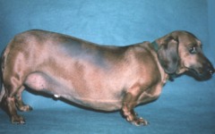
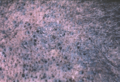
Calcinosis cutis (dermal deposits from hyperadrenocorticism) |
|
|
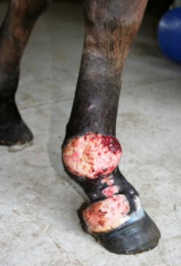
Calcinosis circumscripta (dermal deposits at pressure points). Common in German shepherds. |
|
|
Lymphoma |
|

|
Mast cell tumour |
|
|
Cutaneous histiocytoma |
|
|
lipoma (mesenchymal tumor) |
|
|
SubQ fibroma (mesenchymal tumor) |
|
|
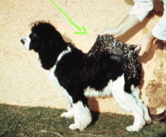
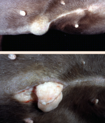
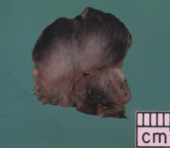
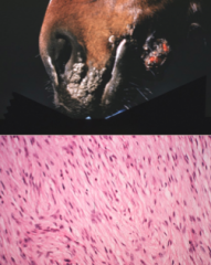
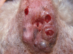
equine sarcoids (bovine papillomavirus induces fibroblast proliferation) |
|
|
perianal gland adenoma (epithelial neoplasia) |
|
|
Follicular (basal cell) tumor. aka Trichoblastoma |
|
|
Follicular cyst |
|
|
Name 3 mesenchymal tumors (5 possible answers) |
Lipoma Fibroma/Fibrosarcoma Equine sarcoid Hemangioma/Hemangiosarcoma Hemangiopericytoma |
|
|
Name 3 round cell tumors (5 possible answers) |
lymphoma mast cell tumor cutaneous histiocytoma cutaneous and systemic histiocytosis extramedullary plasmacytoma |
|
|
3 types of dermal dermal deposits |
calcinosis cutis calcinosis circumscripta mucin (Sharpeis) |
|
|
Causes for dermatitis |
immune mediated/idiopathic parasitic fungal mycobacteria trauma |
|
|
4 responses of the dermis to injury |
dermal inflammation alter dermal collagen dermal deposits dermal neoplasia |
|
|
6 responses of the epidermis to injury |
inflammation necrosis altered epidermal adhesion (bullous diseases) altered pigmentation altered growth and keratinization epidermal neoplasia |
|
|
What's a pustule |
aggregates of neutrophils in the epidermis |
|
|
Names some things that can cause superficial epidermal inflammatory disease |
same as dermal: bacteria fungi parasites (fleas, flies, mites) viral (pox, herpes, calici, picorno) |
|
|
What can cause epidermal necrosis? |
physical injury (burns, lacerations) chemical injury ischemia and infarction |
|
|
3 things that cause altered epidermal adhesion (bullar diseases) |
Pemphigus foliaceus Pemphigus vulgaris Bullous pemphigus |
|
|
Name 3 neoplasias of the epidermis (out of 5) |
Papilloma Squamous cell carcinoma in situ (Bowen's disease) Squamous cell carcinoma Epitheliotropic lymphoma Melanoma |
|
|
bilateral alopecia due to hypothyroidism - increased number of catagen follicles - lack of hair shaft in follices - coat is dull and hyperpigmented |
|
|
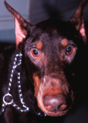
comedomes from hyperadrenocorticism |
|
|
3 presentations of skin with hyperadrenocorticism |
1. epidermal thinning 2. comedomes + increased bruising 3. calcinosis cutis |
|
|
Calcinosis cutis (hyperadrenocorticism) |
|
|
folliculitis due to Demodex canis |
|
|
Folliculitis furunculosis |