![]()
![]()
![]()
Use LEFT and RIGHT arrow keys to navigate between flashcards;
Use UP and DOWN arrow keys to flip the card;
H to show hint;
A reads text to speech;
95 Cards in this Set
- Front
- Back
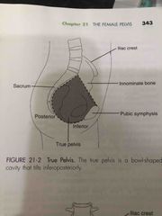
True pelvis = region deep to the pelvis inlet (linea terminalis) |
- lesser/minor -situated in the true pelvis includes the bladder, loops of small bowel, genital tract, and ovaries - tilts inferoposteriorly |
|
|
Normal measurements? Vaginal cavity ? Cervical canal ? Premenarchal uterus ? Multiparous Uterus ? Uterine tubes? Adult ovary? Ovarian volumes ? Premenarche (3-15years)? Menstruating? Premenopausal? Postmenopausal (1-5)? (10-15)?
|
Back (Definition) |
|
|
Key words |
Back (Definition) |
|
Front (Term) |
— |
|
Front (Term) |
— |
|
Front (Term) |
Anatomy |
|
Front (Term) |
— |
|
Front (Term) |
- |
|
Front (Term) |
Sono |
|
Front (Term) |
— |
|

Front (Term) |
Back (Definition) |
|
Front (Term) |
Back (Definition) |
|
|
How can you tell if the bladder is appropriately full enough for a pelvicultrasound? |
It will extend superior to the fundus of the uterus. |
|
|
Why do we have the patient fill their bladder before a scan? |
To provide an accoustic window for better visibility of the pelvicorgans. |
|
|
What structures are seen posterior to a distended bladder? |
Uterus, vagina, posterior cul de sac, rectum and ovaries (dependingtheir location) |
|
|
re arrange in the correct order, anterior to posterior Skin Uterus/Vagina Rectus Abdominus Muscles Bladder Spine Uterus/Vagina Rectum Skin Spine |
Skin Rectus Abdominus Muscles Bladder Uterus/Vagina Rectum Spine |
|
|
What structures are always seen midline? |
Rectus abdominus muscles,
bladder, urethra, vagina, rectum andcervix (usually) |
|
|
What structures are seen in the adnexa? |
Ovaries, broad ligament, pelvic wall muscles, pelvic bones |
|
|
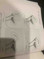
If the uterus is not midline, what are the names of the 2 other positions itcould be? |
Levorotated
Dextrorotated |
|
|
What are the names of the different uterine positions referring to itsflexion? |
Anteverted Retroverted Retroflexed Anteflexed |
|
|
What is the average size of a nulliparous uterus? |
6-8cm |
|
|
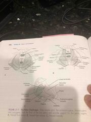
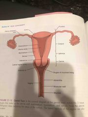
4 parts of the uterus ? and the 3 uterine layers? |
4 parts = fundus, body, isthmus, cervix 3 uterine layers = myometrium endometrium perimetrium |
|
|
Name the 2 layers of the endometrium?
Which layer is mostly sloughedoff during menstruation? |
Zona Basalis and Zona Functionalis. Zona functionalis is sloughed off menstruation. |
|
|
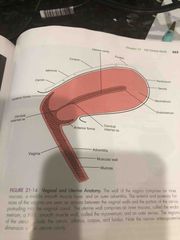
Describe the structure of the vagina, where it’s located and its U/Sappearance. |
Collapsed muscular tube with a thin mucosa. Located posterior andinferior to the urinary bladder sitting at a 90 degree angle to thecervix. |
|
|
Name the 4 parts of the oviducts? |
Interstitial/Intramural isthmus, ampulla and infundibulum |
|
|
Describe the structure, location and the ultrasound appearance of the Fallopian tubes? |
Muscular tubes lined with mucosa attached laterally to the uterus at the cornua.Not visualized on ultrasound unless abnormnal.
|
|
|
What is another name for the ovarian fossa?
|
Fossa of Waldeyer. |
|
|
Name the 2 tissue layers that make up the ovary and what structures they contain? |
Cortex – outer layer; contains the primary follicles
Medulla – middle layer; contains blood vessels, nerves and lymphatics |
|
|
What is the tunica albuginea?
|
Connective tissue covering surrounding the ovary.
|
|
|
What is the ultrasound appearance of an ovary?
|
They are hypoechoic to the surrounding tissue with round, anechoic areaswithin them that represent the growing follicles
|
|
|
During a routine scan of a 2 year old girl, the ovary size calculated to be 5 cc. Is this anormal value for this patient? What is the normal range for this age?
|
This is larger than normal. Normal pediatric range = < 3
|
|
|
How is ovarian volume calculated?
|
Length x width x height (depth) x 0.52
|
|
|
What is considered the normal size of an ovary?
|
Pediatric years = < 3 cc
Reproductive female = 10 + 6 cc Postmenopausal female = < 5 cc |
|
|
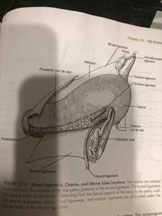
Suspensory ligament? |
attaches ovary to lateral pelvic wall |
|
|
Ovarian ligament? |
– attaches ovary to uterus
|
|
|
Broad ligament?
|
attaches uterus to lateral pelvic wall |
|
|
Round ligament?
|
attaches cornua of uterus to labia majora |
|
|
Cardinal ligament? |
lateral cervix to lateral pelvic wall |
|
|
Uterosacral ligament ? |
-posterior cervix to sacrum |
|
|
Mesovarium ? |
- fold in the broad ligament (anterior to the ovary) |
|
|
Mesosalpinx? |
- fold in the broad ligament (envelopes the fallopian tube) |
|
|
What is the alternate name for the ligament--- Suspensory ligament ? (attaches ovary to lateral pelvic wall) |
Infundibulopelvic ligament |
|
|
What is the alternate names for the ligament--- Cardinal Ligament? (2) -– attaches lateral cervix to lateral pelvic wall |
Mackenrodt ligament and Transverse cervical ligament |
|
|
What 3 muscles compose the levator ani muscle group?
|
Puborectalis Pubococcygeus Iliococcygeus |
|
|
What muscles are not usually demonstrated with ultrasound? |
Coccygeus Piriformis |
|
|
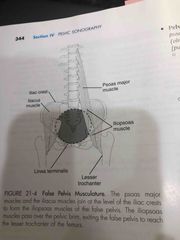
Which muscles lie in the false pelvis? |
-Psoas - iliopsoas |
|
|
Which muscle lies in the true and false pelvis? |
Rectus abdominus |
|
|
Which muscles lie in the true pelvis? |
-Obturator internus -Piriformis -Coccygeus X - Levator ani (3) |
|
|
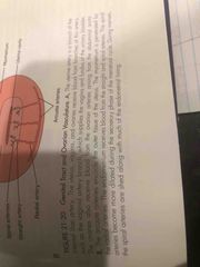
What is the linea terminalis? |
The imaginary line that separates the true and false pelvis located at the pelvicbrim/pelvic inlet.
|
|
|
Is the shape of the pelvic space the same from the superior edge of the false pelvis tothe inferior edge of the true pelvis?
|
No, the false is more round and the true is more oval.
|
|
|
Normally, is the uterus and ovaries found in the true pelvis or false pelvis?
|
-True pelvis |
|
|
Are the pelvic muscles located intraperitoneal or extraperitoneal (outside the peritonealcavity)? |
-Extraperitoneal |
|
|
What four bone make up the pelvis?
|
Sacrum,
Coccyx, Right and Left innominate bone |
|
|
What are the three parts of the innominate bone? |
- Ilium, Ischium, and Pubis |
|
|
List another 2 name(s) for the Rectouterine pouch ? |
-Pouch of Douglas -Posterior cul de sac |
|
|
4 Parts of fallopian tube/oviduct? |
4 Parts = -Interstitial/Intramural -Isthmus -Ampulla -Infundibulum with fimbriae |
|
|
Interstitial/intramural ?
|
- attaches at cornea - continuous (egg travels into uterus) (shoulder)
|
|
|
Isthmus? |
Isthmus- second part of uterine tube (upper arm)
|
|
|
Ampulla? |
- largest longest section, least straight, usually where fertilized, outside the cornea (lower arm)
|
|
|
Infundibulum with Fimbriae ? |
- (fingers) or fimbriae tubae, are the finger-like projections located at the ends of the fallopian tubes, closest to the ovaries. The majority of the fimbriae do not touch the ovary but rather hover very close by, activated by hormones to catch a released egg and move it down into the fallopian tube. |
|
|
Do we see fallopian tubes on u/s?
|
-Usually dont see fallop tubes unless it is infected or has pathology |
|
|
mesovarium?
|
The mesovarium is the portion of the broad ligament of the uterus that suspends the ovaries. The ovary is not covered by the mesovarium; rather, it is covered by germinal epithelium.
|
|
|
mesosalpinx ?
|
The mesosalpinx is part of the lining of the abdominal cavity in higher vertebrates, specifically the portion of the broad ligament that stretches from the ovary to the level of the fallopian tube.
|
|
|
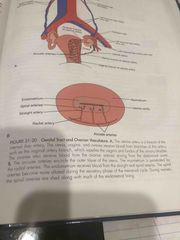
Vasculature of the pelvis to the uterus
(vessel diameter?) large to smallest? outside uterus? inside uterus? |
outside uterus--- AO, common illiac artery, internal illiac artery uterine artery inside uterus tissue--- Arcuate Artery, Radial Artery, Spiralarterioles, Straight arterioles |
|
|
Arterial Uterine Blood flow? |
Aorta Common iliac arteries Internal Iliac arteries Uterine arteries (Hypogastric arteries) Arcuate arteries Radial arteries Spiral arterioles + Straight arterioles |
|
|
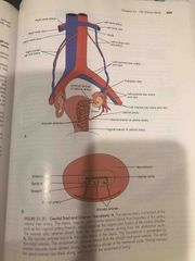
Venous Uterine Blood flow? |
Uterine veins
Internal iliac veins Common iliac veins IVC |
|
|
Ovarian Arterial Blood Flow/Supply? |
The ovaries have 2 blood supplies 1) Aorta----> Rt + Lt ovarian artery --->ovary 2) Uterine arteries (2) ---> ovaries (2) |
|
|
Ovarian venous blood flow / return? |
Venous flow =(back to the heart via IVC)
RT Ovary---->Rt ovarian vein--->IVC (highway path) Lt Ovary---->Lt ovarian vein--->Lt renal vein--->IVC (detour) |
|
|
the ovary sits ________to the vessels and ureter. |
-the ovary sits anterior to the vessels and ureter. |
|
|
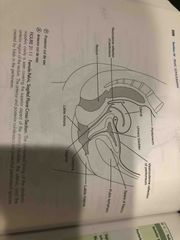
Space of Retzius?
|
Space of Retzius - fat filled, rarely seen unless full of fluid- located posterior to the symphysis pubis and anterior to the bladder
|
|
|
Anterior cul-de-sac / Vesicouterine pouch?
where is it located? |
Anterior cul-de-sac / Vesicouterine pouch
vesico = bladder -between the bladder and uterus (vesico - uterine)more anterior than posterior |
|
|
Posterior cul-de-sac / Rectouterine pouch / Pouch of Douglas ?
|
3 names for one meaning = Posterior cul-de-sac / Rectouterine pouch / Pouch of Douglas
-more posterior between the rectum and the uterus (recto - uterine) -when patient is on their back, fluid will collect here ** spaces may have free fluid depending on gravity/patient position - we need to look for it** |
|
|
The ureters ?
|
The ureters themselves are usually too small to visualize (unless they are dilated). We can determine if they are functional, however, by observing the ureteric jets These can be demonstrated using the colour doppler mode (and are sometimes visible even without colour) Usually two ureters/jets, a Rt and a Lt, are seen unless the patient has duplications of the ureter(s) The ureters insert into the inferior posterior part of the urinary bladder so to visualize the jets the probe must be angled inferiorly (towards the patients feet) The direction the jets of urine are seen is often opposite to the side of their location |
|
|
the bladder? appearance? |
What are we looking for:
-The bladder wall should appear smooth and thin. Look for thickening, diffuse or focal -Clear anechoic fluid in the lumen; no stones and/or debris seen |
|
|
how do we calculate bladder volume pre and post void? |
Measure bladder volume before and after urination
Pre-void volume : SAG x TRN x AP x 0.52 = volume in cc/ml Post-void volume : SAG x TRN x AP x 0.52 = volume in cc/ml |
|
|
urethra? |
The escape route.
Voiding the urinary bladder occurs through the urethra which is located more inferior than the ureters. The urethra (refered to as the urethral hump) is often seen as a small hypoechoic “area” at the midline inferior as aspect of the bladder and anterior to the vagina |
|
|
What’s behind the bladder?
|
THE UTERUS Once the sonographer has assured adequate bladder fullness has been achieved and completed a thorough scan for urinary pathologies, the sonographer will evaluate the female reproductive organs. |
|
|
the different uterus positions seen with flexions towards the right or left sides? |
left = levorotated right = Dextrorotated A patient may have variations of both anterior to posterior flexion and side to side flexion. |
|
|
the cervix? |
-The cervix is the most inferior part of the uterus and is cylindrical in shape. -The endocervical canal is the passage way that runs through the inner part of the cylinder. It is lined with mucosa (appears echogenic on ultrasound). -In the transverse plane, shadowing will be seen from the edges of the cervix. (vampire teeth) The shadowing comes from the fornices…tiny recesses (spaces) where the inferior part of cervix projects into the superior part of the vagina |
|
|
vag? |
The vagina is a long, collapsed muscular tube located inferior to the cervix. The inner part of the “tube” (the vaginal canal) is lined with mucosa (appears echogenic on ultrasound) |
|
|
the ovaries? -location? appearance on u/s? |
The ovaries are paired, small, ovoid-shaped organs located in the adnexa of the female pelvis.They usually that contain a varying number of small, round, fluid-filled follicles. Structures that are fluid-filled often appear anechoic (black) on ultrasound.
|
|
|
u/s appearance of ovaries? |
Sonographic Appearance:Hypoechoic compared to surrounding structuresVarying number of small, anechoic follicles |
|
|
Ovarian Position?
|
-Variable (but will not be anterior to the uterus)
-Lie in ovarian fossa or Fossa of Waldeyer -Within the peritoneal cavity (intraperitoneal organs) -Use iliac arteries as guide. -MID SAGITAL to see canal top to bottom |
|
|
Each ovary has______ arterial blood supplies
|
Each ovary has two arterial blood supplies:
1- Ovarian artery – branch directly off abdominal aorta 2- Branch from uterine artery |
|
|
Each ovary has ____ venous drainage system via:
Lt Ovarian vein > empties into left renal vein Rt ovarian vein > empties into IVC |
Each ovary has one venous drainage system via:
Lt Ovarian vein > empties into left renal vein Rt ovarian vein > empties into IVC |
|
|
Ovarian volume calculation? |
Length x Width x Height x 0.523 =
ie: Sag x Trn x AP x 0.523 = ____ |
|
|
the ligament that suspends the ovary from the pelvis wall ? |
Infundibulopelvic ligament aka suspensory ligament |
|
|
the hammock shaped group of muscles that supports the pelvic organs? |
levator ani |
|
|
the portion of the uterus which undergoes the most change under the influence of hormones? |
the zona functionalis layer |
|
|
which ligament is responsible for anteversion/flexion? |
round ligament |
|
|
the longest portion of the uterine tube ? aka fallopian tube |
the ampulla |
|
|
the ovaries are located on a shallow depression of the broad ligament called the ? |
fossa of Waldeyer |
|
|
the age group which would have a 1:1 uterus to cervix ratio ? |
post menopausal |
|
|
the age group which would have a 1:2 uterus to cervix ratio ?
|
newborn 1 uterus 2 cervix |
|
|
what structure produces the liner shadowing on a transverse image of the cervix? |
the fornices |