Reading...
![]()
Play button
![]()
Play button
![]()
Use LEFT and RIGHT arrow keys to navigate between flashcards;
Use UP and DOWN arrow keys to flip the card;
H to show hint;
A reads text to speech;
75 Cards in this Set
- Front
- Back
|
Typical symptoms a patient may have with an abscess formation include all of the following EXCEPT:
A) fever. B) decreased white blood count C) pain D) increased white blood count |
B) decreased white blood count
|
|
|
The splenic artery is considered to be the:
A) superior border of the pancreas B) lateral border of the pancreas C) anterior border of the pancreas D) inferior posterior border of the pancreas |
A) superior border of the pancreas
|
|
|
The splenic vein is considered to be the:
A) superior posterior border of the pancreas B) superior border of the pancreas C) medial posterior border of the pancreas D) inferior posterior border of the pancreas |
C) medial posterior border of the pancreas
|
|
|
Fusion of the lower pole of the kidneys is called a:
A) cross-renal ectopia B) pelvic kidney C) supernumerary kidney D) horseshoe kidney |
D) horseshoe kidney
|
|
|
The most echogenic portion of the kidney is/are the:
A) cortex B) sinus C) medullary pyramids D) parenchyma |
B) sinus
|
|
|
A cortical bulge in the lateral border of the kidney is called a/an:
A) junctional parenchymal defect B) dromedary hump C) extrarenal pelvis D) column of Bertin |
B) dromedary hump
|
|
|
Dilatation of the renal pelvis with thinning of the renal cortex is characteristic of:
A) duplex collection system B) column of Bertin C) hydronephrosis D) extrarenal pelvis |
C) hydronephrosis
|
|
|
The kidneys are located in the:
A) peritoneal cavity B) retroperitoneal cavity C) perirenal cavity D) perirenal space |
B) retroperitoneal cavity
|
|
|
A triangular-shaped lesion on the peripheral border of the kidney most likely represents a(n):
A) renal tumor B) artifact from rib C) IVC compression D) junctional parenchymal defect |
D) junctional parenchymal defect
|
|
|
The normal sonographic texture of the spleen is:
A) homogeneous with internal echoes equal to or less echogenic than those of the liver. B) hypoechoic C) isoechoic but more echogenic than the liver D) hyperechoic |
A) homogeneous with internal echoes equal to or less echogenic than those of the liver.
|
|
|
A potential space located between the liver edge and right kidney is:
A) Morison’s pouch B) Douglas’ pouch C) cul-de-sac D) Winhauer’s space |
A) Morison’s pouch
|
|
|
Renal cell carcinoma commonly invades the IVC via the:
A) renal vein B) renal artery C) portal vein D) splenic vein |
A) renal vein
|
|
|
Which of the following is a benign fatty tumor of the kidney?
A) angiomyolipoma B) hypernephroma C) neuroblastoma D) lymphoma |
A) angiomyolipoma
|
|
|
The most common benign neoplasms in the spleen include all of the following EXCEPT:
A) cavernous hemangiomas B) infarctions C) cystic lymphangiomas D) hamartomas |
B) infarctions
|
|
|
The left renal vein courses:
A) posterior to the IVC B) anterior to the IVC C) anterior to the aorta D) anterior to the superior mesenteric artery |
C) anterior to the aorta
|
|
|
A pelvic kidney has a(n):
A) abnormal appearance in a normal location B) normal appearance in an abnormal location C) normal appearance in a normal location D) abnormal renal pelvis |
B) normal appearance in an abnormal location
|
|
|
Splenomegaly may result from all except which of the following?
A) an inflammatory process B) a left subphrenic abscess C) a metastatic disease to the spleen D) polycythemia vera |
B) a left subphrenic abscess
|
|
|
In a _____ hematoma of the spleen, the splenic capsule remains intact.
A) interparenchymal B) subcapsular C) intraperitoneal D) interperitoneal |
B) subcapsular
|
|
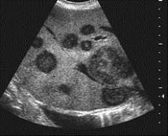
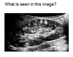
See image. What is the most likely diagnosis?
A) multiple hemangiomas B) multiple metestatic lesions C) multiple cyst D) multiple adenomas |

B) multiple metestatic lesions
|
|
|
A renal sonogram is performed. The finding of hypoechoic areas adjacent to the renal sinus is most consistent with
A) bifid renal pelvis B) renal pyramid C) column of Bertin D) junctional parenchymal defect |
B) renal pyramid
|
|
|
A triangular-shaped lesion on the peripheral border of the kidney most likely represents a(n)
A) renal tumor B) artifact from rib C) IVC compression D) junctional parenchymal defect |
D) junctional parenchymal defect
|
|
|
The vessel seen posterior to the IVC on the sagittal scan represents the:
A) right adrenal artery B) right renal artery C) left renal artery D) left renal vein |
B) right renal artery
|
|
|
Which statement describes the correct anatomic location of structures adjacent to the spleen?
A) the diaphragm is anterior, lateral and inferior to the spleen B) the fundus of the stomach and lesser sac are medial and posterior to the splenic hilum C) the left kidney lies inferior and medial to the spleen D) the right adrenal and kidney lie superior to the spleen |
C) the left kidney lies inferior and medial to the spleen
|
|
|
When accessory spleens are present, they usually are located:
A) at the inferior margin of the spleen B) on the posterior aspect of the spleen C) near the hilum of the spleen D) near the kidney |
C) near the hilum of the spleen
|
|
|
Which of the following statements about the spleen is FALSE?
A) A prominent bulge along the medial surface of the spleen can be seen in normal patients. B) The normal-sized spleen should not extend caudal to the mid portion of the left kidney. C) The spleen is a retroperitoneal organ. D) the sonographic texture of the normal spleen is homogeneous. |
C) The spleen is a retroperitoneal organ.
|
|
|
The normal sonographic texture of the spleen is:
A) homogeneous with internal echoes equal to or less echogenic than those of the liver B) hypoechoic C) isoechoic but more echogenic than the liver D) hyperechoic |
A) homogeneous with internal echoes equal to or less echogenic than those of the liver
|
|
|
The splenic artery is a branch of which of the following vascular structures?
A) abdominal aorta B) gastric artery C) celiac axis D) superior mesenteric artery |
C) celiac axis
|
|
|
The most common solid renal mass found in childhood is:
A) renal cell carcinoma B) angiomyolipoma C) Wilms’ tumor D) Von Hippel-Lindau tumor |
C) Wilms’ tumor
|
|
|
A sign of transplant rejection is:
A) Resistive Index (RI) greater than 0.7 B) RI greater than 0.4 C) RI less than 0.4 D) No RI |
A) Resistive Index (RI) greater than 0.7
|
|
|
Which of the following describes the sonographic appearance of chronic pyelonephritis?
A) echogenic cortex B) hypoechoic enlarged kidney C) inability to distinguish the cortex from the medullary regions D) echogenic foci in the medullary regions |
C) inability to distinguish the cortex from the medullary regions
|
|
|
A post-transplant perinephric fluid collection can present as all of the following except:
A) ureterocele B) hematoma C) urinoma D) lymphocele |
A) ureterocele
|
|
|
Pyonephrosis refers to the presence of:
A) blood in a dilated collecting system B) pus in a dilated collected system C) urine in a dilated collecting system D) a perinephric abscess |
B) pus in a dilated collected system
|
|
|
Adult polycystic disease may be characterized by all except which of the following?
A) It is a latent disease until the third or fourth decade of life. B) It is an autosomal-dominant disease. C) It may be associated with cysts in the liver, pancreas, and spleen. D) The involved kidneys are small and extremely echogenic. |
D) The involved kidneys are small and extremely echogenic.
|
|
|
The normal bladder wall should be smooth and thin, and measure:
A) 2–4 mm B) 3–6 mm C) 5–7 mm D) 5–10 mm |
B) 3–6 mm
|
|
|
The adrenal glands are separated from the kidneys by the:
A) perinephric fascia B) perinephric capsule C) crus of the diaphragm D) perinephric fat |
D) perinephric fat
|
|
|
Adrenal hypofunction occurs in:
A) adrenogenital syndrome B) Addison’s syndrome C) Conn’s syndrome D) Cushing’s syndrome |
B) Addison’s syndrome
|
|
|
Excessive secretion of aldosterone occurs in:
A) Addison’s syndrome B) Cushing’s syndrome C) Waterhouse-Friderichsen syndrome D) Conn’s syndrome |
D) Conn’s syndrome
|
|
|
Adrenal insufficiency typically is caused by:
A) adrenal adenoma B) adrenal cyst C) lymphadenopathy D) metastases |
D) metastases
|
|
|

1. Superior medial and inferior lateral
2. Removes foreign material from the blood; initiates an immune reaction, resulting in a production of antibodies and lymphocytes, major destruction site of RBC 3. Erythrocyte, leukocyte, hematocrit, and hemoglobin 4. Splenule (accessory spleen), aplasia (absent spleen), polysplenia (multiple small spleens, wandering spleen) |
|
|

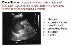
B) an incidental isoechoic “mass” is identified near the splenic hilum. Most suspicious for a splenule or accessory spleen.
|
|
|

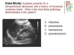
D. Granulomatosis demonstrates hyperechoic foci dispersed within the splenic parenchyma ; may demonstrate shadowing
Chronic infarction would demonstrate as an echogenic mass with well-defined margins; Harmartoma looks similar to hemangioma |
|
|

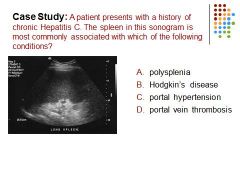
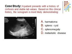
C. The key words are most commonly. While portal vein thrombosis and Hodgkin’s disease may be associated with splenomegaly, portal hypertension is most commonly associated with splenomegaly.
|
|
|

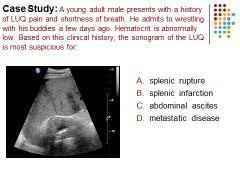
A) Hypoechoic masses within the spleen after a history of trauma and a decrease in the patient’s hematocrit is most suspicious for splenic rupture
|
|

|
B) Splenic cysts are rare finding and can be congenital, infective, neoplastic, parasitic, or previous hx of trauma in etiology. Pts are typically asymptomatic
|
|
|

1. Inner medulla and outer cortex
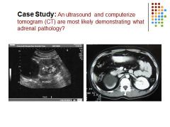
2. Located in Gerota’s fascia within the perinephric fat; located anterior, medial and superior to each kidney. Lie lateral to the diaphragmatic crura. Rt lies posterior and lateral to the IVC. Lt lies lateral to the aorta and posterior to the spleen. 3. Addison disease; Cushing disease, hyperaldosteronism |
|

|
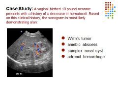
C) Adrenal Hemorrhage: A 10lb neonate delivered vaginally is at risk for adrenal hemorrhage. With a decrease in hematocrit and an avascular complex mass superior to the left kidney is most suspicious for an adrenal hemorrhage. Adrenal Hemorrhage is seen more often in full term infants and large-for-gestational-age babies; bilateral in 10%, more often on the right
|
|

|
Adrenal cyst – an anechoic structure is identified superior to the right kidney, most suspicious for an adrenal cyst. Differential: renal or hepatic cyst
|
|

|
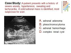
B - Adrenal pheochromcytoma: this is a rare vascular tumor of the adrenal medulla. A small percentage are malignant. Clinical findings include hypertension, sweating, tachycardia, chest or epigastric pain, headache, palpitations, severe anxiety and an increase in epinephrine and norepinephrine. Sono findings include a solid homogeneous adrenal mass. May appear complex (necrosis) or calcify (chronic necrosis)
|
|

|
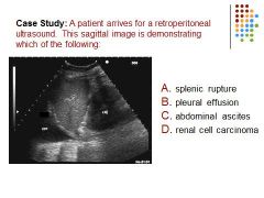
B) Anechoic fluid is identified superior to the diaphragm suspicious for a pleural effusion
|
|
|

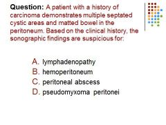
D - Think this one out!
1. Patient with a history of cancer = possible metastatic disease 2. Lymphadenopathy is possible but doesn’t typically appear cystic or causes matting of the bowel 3. Hemoperitoneum – not likely…no evidence of recent trauma or hx portal hypertension 4. Abscess – not as likely without a history of fever 5. Pseudomyxoma peritonei can be a metastatic disease, appearing on US are multiple septated cystic areas and has a tendency to cause matting of the bowel 6. Even if we don’t remember the sonographic appearance of pseudomyxoma peritonei, we can deduce the same result by working with the patient hx |
|

|
C – 13 cm; keyword is once…a 17cm spleen is enlarged also
|
|

|
B) anechoic
|
|

|
A) Pelvis
|
|
|

moderate hydronephrosis
|
|
|

simple renal cyst
|
|
|

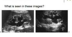
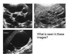
AUTOSOMAL DOMINANT POLYCYSTIC KIDNEY DISEASE - Image A: When the disease is advanced, the renal parenchyma is replaced by numerous non-communicating cysts of varying size (arrows). Image B: When the disease is early, the kidneys are of normal size and only a few cysts (arrows) are present
|
|

|
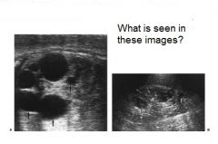
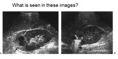
AUTOSOMAL RECESSIVE POLYCYSTIC KIDNEY DISEASE (ARPKD) Image A: a longitudinal image of a kidney in a newborn shows enlargement with marked increase in central echogenicity with compressed cortex. Image B: kidney of a 5 year old boy with ARPKD is mildly enlarged and heterogeneous with visible small cysts
|
|
|

Multicystic Dysplastic Kidney
|
|

|
Pyonephrosis
|
|

|
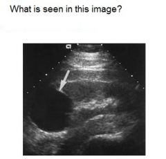
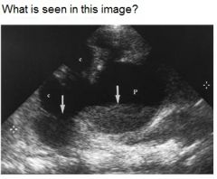
RENAL ABSCESS – Image A: intrarenal abscess produces thick-walled mass containing echogenic fluid within the kidney. The perirenal spacecontains mostly normal echogenic fat and an area of inflammatory infiltration (curved arrow). Image B: this renal abscess contains a large pocket of air (white arrow) that produces intense reverb artifact
|
|

|
Echogenic End Stage Kidney
|
|

|
Multiple Angiomyolipomata in a child
|
|

|
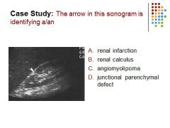
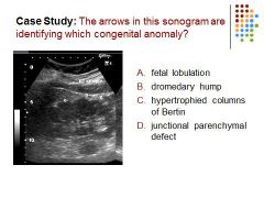
D) Junctional Parenchymal Defect
|
|
|

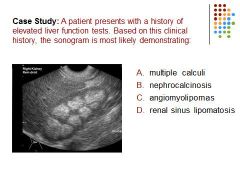
B) Nephrocalcinosis
|
|

|

B) Nephrocalcinosis
|
|
|

B) angiomyolipoma
|
|

|
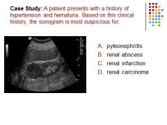
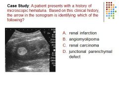
D) renal carcinoma
|
|

|
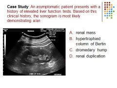
B) Hypertrophied column of Bertin – an isoechoic “mass” extends into the renal sinus
|
|

|
A) Fetal lobulation
|
|
|

B) Hydronephrosis
|
|

|
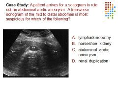
B – Horseshoe kidney
|
|
|

D – Thoracentesis (also known as pleural tap is an invasive procedure to remove fluid or air from the pleural space) is optimally performed with the patient in a sitting position bending at the waist slightly forward typically resting on the table
|
|
|

C – portal vein thrombosis
|
|
|

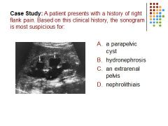
B – nephrolithiasis (kidney stones)
|
|
|

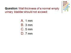
C – 5 mm / the thickness of the normal bladder is 5 mm when empty and 3 mm when distended
|