Reading...
![]()
Play button
![]()
Play button
![]()
Use LEFT and RIGHT arrow keys to navigate between flashcards;
Use UP and DOWN arrow keys to flip the card;
H to show hint;
A reads text to speech;
10 Cards in this Set
- Front
- Back
- 3rd side (hint)
|
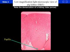
H & E stain of a pig kidney showing the demarcations of the different lobes. There are approximately 18 lobes in an adult human kidney but they are only demarcated in the fetus. Know which regions are associated with higher osmolarity (medulla vs. cortex) and where the papillae are (at the base).
|
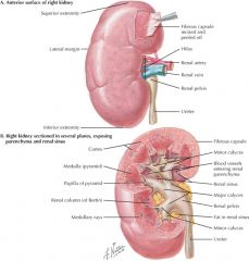
The kidneys are situated retroperitoneally on either side of the posterior abdominal wall. In an adult, the kidneys are reddish-brown, bean-shaped organs, each weighing approximately 150 grams. Each kidney is surrounded by a dense connective tissue capsule and has a hilar indentation on one side. At the hilar region, renal vessels, lymphatics, and nerves enter and leave the kidney, and the ureter exits the kidney.
|
|
|
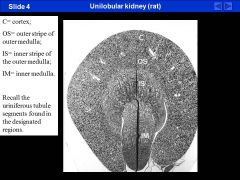
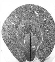
Low magnification view of a unilobular rat kidney. See the difference between the cortex (C),
outer medulla (OS and IS), and the inner medulla (IM). Again, which parts of the nephron are found in each location? |
Netter drawings showing the difference between a juxtamedullary nephron and a superficial nephron.
Know what segments are found in each location: CORTEX 1. renal corpuscles 2. convoluted tubules 3. collecting ducts 4. medullary rays OUTER MEDULLA 1. thick segments of LOH 2. part of the descending thin segments 3. collecting ducts INNER MEDULLA 1. thin segments of LOH 2. collecting ducts 3. vasa recta The outer medulla has an inner and outer stripe but you are NOT responsible for knowing the difference between them. Note, medullary rays are made of thick ascending and thick descending segments as well as collecting ducts. |
|
|
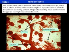
This slides shows the renal vasculature. The balls are glomeruli (remember there are 1 million in each kidney). This is the location of filtration. You can also see the interlobular artery branching to
intralobular branches and then afferent and efferent arterioles. Also, note the peritubular capillary plexus. |
Glomerular filtrate passes across the glomerular wall -->
capsular space of Bowman --> opening of urinary pole of the renal corpuscle --> uriniferous tubules. The first segment of the uriniferous tubule is termed the PROXIMAL TUBULE. It is within the proximal tubule that most of the glomerular filtrate is reabsorbed back into a rich PERITIBULAR CAPPILARY PLEXUS which surrounds these tubules. The first portion of the proximal tubule follows a highly convoluted course within the cortical substance of the kidney, and has therefore been termed the PROXIMAL CONVOLUTED TUBULE/ (PARS CONVOLUTA). The portion of the proximal tubule that descends from the cortex into the outer stripe of the outer medulla has been termed the DESCENDING PROXIMAL TUBULE (AKA PARS RECTA/ DESCENDING THICK SEGMENT OF THE LOOP OF HENLE/ STRAIGHT SEGMENT OF THE PROXIMAL TUBULE (S3). |
|
|
S1-S3 all have microvillus brush border.
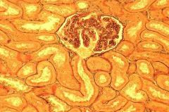
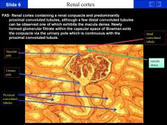
macula densa parietal epithelial proximal tubules -contain debris in lumen urinary pole A periodic acid shift stain of the renal cortex containing a renal corpuscle. This basically shows the carbohydrate-rich glycocalyx of the brush boarder (pinkish) and debris in the lumen of the proximal tubule (due to ruptured cells and cellular debris that result from the fixation process). The urinary pole is at the bottom of the renal corpuscle and the vascular pole is approximately 180 degrees away. Also, note the afferent arteriole contains renin granules and marks the macula densa a component of the Juxtaglomerular apparatus, while the efferent arteriole does not (this can help distinguish them). This is seen as a cluster of nuclei at the top of the slide. |
The cuboidal cells that make up the wall of the PROXIMAL CONVOLUTED tubule have:
~elaborate basolateral folds, ~numerous mitochondria, ~an extensive lysosomal system, ~and a microvillous brush border coated with a rich glycocalyx. Based on differences in fine structure, histochemistry and function, the proximal convoluted tubules have been further subdivided into two segments. Comparing FIRST SEGMENT OF THE PROXIMAL CONVOLUTED TUBULE (S1) to the the SECOND SEGMENT OF THE PROXIMAL TUBULE (S2), we see that S1 has more: ~TALLER cells ~TALLER microvillous BRUSH BORDER ~MORE elaborate intercellular INTERDIGITATION, ~MORE MITOCHONDRIA, ~LESS prominent LYSOSOMES |
|
|
Dr. Andrews said during his lecture: DON’T WORRY ABOUT THIS SLIDE
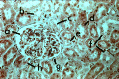
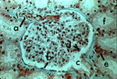
A view of the renal cortex containing a renal corpuscle. A = Nucleus of a podocyte on the inner part of Bowman’s capsule. B = Distal Tubule C = Macula Densa (this is really not clear) D = Proximal tubule E = Ascending Segment (difficult to determine this on this slide) F = Distal tubules G =parietal epithelium of Bowman’s capsule H = Urinary pole |
|
|
|
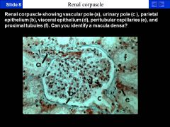
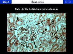
This slide is much clearer and could show up on a test.
A = Vascular pole. B = Parietal epithelium (cells are flat). C = Urinary Pole. D = Visceral Epithelium (Nucleus of podocyte) E = Peritubular Capillaries. F = Proximal Tubules. The space between the parietal and visceral epithelium is called Bowman’s space or Urinary space. The macula densa is near A at the top of the slide. |
|
|
|
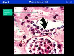
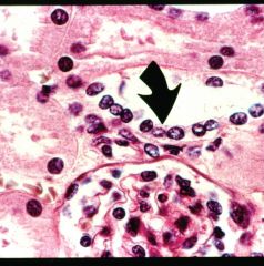
to left= proximal tubles
bottom= renal corpuscule A close up H&E stain of the macula densa. It means dark spot and it stains darkly because of the nuclei. It is adjacent to the glomerulus. |
|
|
|
~~IMPORTANT SLIDE ~~
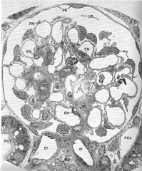
(bottom is vascular pole, can’t see urinary pole ) PE – parietal epithelium US- urinary space Ef – efferent arteriole Af – afferent arteriole – can see renin granules in walls of afferent arteriole En – endothelial cell Po- podocytes make up visceral layer MC – mesangial cells Pca – peritubular capillaries Fm – filtration membrane MD – macula densa RED ARROWS: indicating the place at the vascular pole where simple squamous cells termed the parietal epithelial layer of bowman’s capsule, reflect to form the visceral layer of bowman’s capsule composed of podocytes. |
Glomerular capillaries are unique in being lined by a GLOMERULAR ENDOTHELIUM perforated by numerous open fenestrations.
These endothelial loops are surrounded by a thick basal lamina termed the GLOMERULAR BASEMENT MEMBRANE (i.e. 240 to 340 nm thick in humans), and an epithelium termed the VISCERAL LAYER OF BOWMAN'S CAPSULE. The specialized cells comprising this visceral layer are termed PODOCYTES (podo= greek for foot) |
|
|
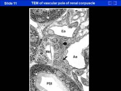
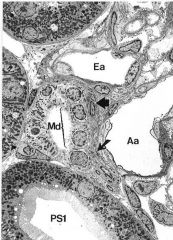
PS1: proximal tube S1 – can see microvillous brush border and abundance of mitochondria.
LITTLE ARROW: RENIN granules BIG ARROW: Extra-glomerular mesangial cells Md: macula densa Aa: Afferent a. Ea: Efferent a. |
|
|
|
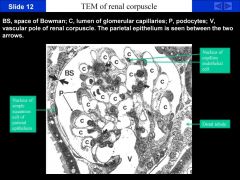
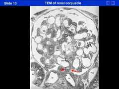
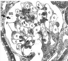
Another TEM of the renal corpuscle.
BS = Bowman’s space. P = Podocytes. C = capillary lumen. V = Vascular pole. Between the two large arrows is the thin parietal epithelium. Between the capillaries are mesangial cells. You can also see the macula densa on the bottom right. MASCULA DENSA IS ONLY ON THE VASCULAR POLE!! neVER SEE IT ON URINARY POLE |
THE JUXTAGLOMERULAR APPARATUS
As the STRAIGHT DISTAL ASCENDING SEGMENT goes back into the cortex, it passes between the afferent and efferent arterioles of the renal corpuscle from which it arose. The EXTRAGLOMERULAR MESANGIUM. (or LACIS CELL because they are surrounded by a lot of extracellular matrix… "lacis" or network.) is the compact cushion of small cells that separates the distal tubule from the renal corpuscle at this site. They look kinda like the mesangial cells in the glomerulus. The cells in the wall of the DISTAL TUBULE in this region become thinner, sometimes taller, and the DARK STAINGING nuclei appear clumped to form the MACULA DENSA (Greek= dense spot). Smooth muscle cells in the wall of the AFFERENT ARTERIOLE adjacent to the macula densa are modified into cells that synthesize, store (in membrane-bound GRANULES), and secrete the hormone RENIN. JUXTAGLOMERULAR CELLS= RENIN PRODUCING CELLS. juxtaglomerular cells + macula densa = JUXTAGLOMERULAR APPARATUS. |