Reading...
![]()
Play button
![]()
Play button
![]()
Use LEFT and RIGHT arrow keys to navigate between flashcards;
Use UP and DOWN arrow keys to flip the card;
H to show hint;
A reads text to speech;
145 Cards in this Set
- Front
- Back
- 3rd side (hint)
|
nares
|
anterior openings of nasal cavity (nostril)
|
|
|
|
choanae
|
the paired openings between the nasal cavity and the nasopharynx;
called also posterior nasal apertures |
|
|
|
bones of the head
|
Frontal
Parietal Temporal Occipital Sphenoid Maxilla Mandible Palatine Zygoma Lacrimal Ethmoid Vomer Inferior Concha |
13 of them from his list
|
|
|
foramen of the head
|
Inferior alveolar foramen
Foramen Lacerum Foramen Magnum Mental foramen Internal Acoustic Meatus Carotid canal Foramen Ovale Pterygomaxillary Fissure Foramen Rotundum Foramen Spinosum Superior Orbital Fissure Optic Canal Stylomastoid Foramen Inferior Orbital Fissure Zygomatico-facial foramen Zygomatico-temporal foramen Nasolacrimal canal Infraorbital foramen Frontonasal Duct Hiatus Semilunaris Auditory Tube |
21 of them from his list
|
|
|
name and locate the sinuses
|
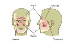
paranasal sinuses: the mucosa-lined air cavities in the cranial bones which communicate with the nasal cavity, including the ethmoidal, frontal, maxillary, and sphenoidal sinuses
|
|
|
|
The four extrinsic muscles in each half of the tongue
-extrinsic alter position -intrinsic (4 of these also)alter shape |
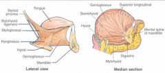
genioglossus (dorsum from XII)
hyoglossus (side and under XII) styloglossus(side and under XII) palatoglossus (side XI) |
|
|
|
name the three scetions of the V craniofacial nerve, and locate landmarks around it
|
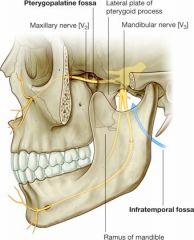
look at picture
|
|
|
|
pterygopalatine fossa
|
on each side is just posterior to the upper jaw. This small fossa communicates with the cranial cavity, the infratemporal fossa, the orbit, the nasal cavity, and the oral cavity. A major structure passing through the pterygopalatine fossa is the (maxillary division of the trigeminal nerve) maxillary nerve [V2]
|
|
|
|
infratemporal fossa
|
an area between the posterior aspect (ramus) of the mandible and a flat region of bone (lateral plate of the pterygoid process) just posterior to the upper jaw (maxilla)
conduit for one of the major cranial nerves—the mandibular nerve (the mandibular division of the trigeminal nerve [V3]), which passes between the cranial and oral cavities |
|
|
|
pharyngeal isthmus
|
a space between the posterior wall of the pharynx and the free border of the soft palate
-where the nasal pharynx communicates with the oral pharynx during respiration |
|
|
|
the auditory tube
|
-the opening is at the lateral wall of nasal pharynx
-it is located inferoposterior to the inferior nasal concha - |
|
|
|
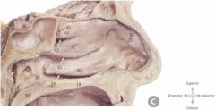
1
|
scalp
|
|
|
|
2
|
calvaria of skull
|
|
|
|
3
|
diploe (spongy marrow of cranial bones)
|
|
|
|
4
|
superior sagittal sinus
|
|
|
|
5
|
falx cerebi (a midline fold of dura mater between the two cerebrral hemispheres)
-attaches to crista galli of ethmoid bone |
|
|
|
6
|
inferior sagittal sinus
|
|
|
|
7
|
crista galli
|
|
|
|
8
|
straight sinus
|
|
|
|
9
|
great cerebral vein
|
|
|
|
10
|
confluence of sinuses
|
|
|
|
11
|
opening of the right tranverse sinus
|
|
|
|
12
|
occipital sinus
|
|
|
|
13
|
falx cerebelli (midline fold to seperate cerebelli hemispheres)
|
|
|
|
14
|
frontal sinus
|
|
|
|
15
|
corpus callosum
|
|
|
|
16
|
anterior cerebral artery
|
|
|
|
17
|
septum pellucidum
|
|
|
|
18
|
fornix
|
|
|
|
19
|
thalamus and interthalamic adhesion
|
|
|
|
20
|
optic chiasm
|
|
|
|
21
|
pituitary gland
|
|
|
|
22
|
sphenoidal sinus
|
|
|
|
23
|
mamillary body
|
|
|
|
24
|
midbrain
|
|
|
|
25
|
mesencephalic tectum
|
|
|
|
26
|
pineal gland
|
|
|
|
27
|
cerebraal aqueduct
|
|
|
|
28
|
cerebellum
|
|
|
|
29
|
fourth ventricle
|
|
|
|
30
|
cisterna magna
|
|
|
|
31
|
basilar artery
|
|
|
|
32
|
left vertebral artery
|
|
|
|
33
|
pons
|
|
|
|
34
|
medulla oblongata
|
|
|
|
35
|
spinal cord
|
|
|
|
36
|
anterior arch atlas
|
|
|
|
37
|
body and dens of atlas
|
|
|
|
38
|
pharyngeal tonsil
|
|
|
|
39
|
ostium (opening) of auditory tube
|
|
|
|
40
|
nasal pharynx
|
|
|
|
41
|
middle nasal concha
|
|
|
|
42
|
inferior nasal concha
|
|
|
|
43
|
hard palate
|
|
|
|
44
|
soft palate and uvula
|
|
|
|
45
|
mandible
|
|
|
|
46
|
hyoid bone
|
|
|
|
47
|
genioglossus muscle
|
|
|
|
48
|
geniohyoid muscle
|
|
|
|
49
|
mylohyoid muscle
|
|
|
|
50
|
epiglottis
|
|
|
|
51
|
thyroid cartilage
|
|
|
|
52
|
cricoid cartilage
|
|
|
|
53
|
trachea
|
|
|
|
54
|
oral pharynx
|
|
|
|
55
|
laryngeal pharynx
|
|
|
|
56
|
esophagus
|
|
|
|
pterygopalatine fossa
card 1 basic info |
-communicates with the cranial cavity, the infratemporal fossa, the orbit, the nasal cavity, and the oral cavity
-a major structure passing through is (maxillary division of the trigeminal nerve) maxillary nerve [V2] -The fossa is seen through a narrow cleft, the pterygomaxillary fissure, between the pterygoid process of the sphenoid bone and the posterior wall of the maxilla. The medial wall of the fossa is the vertical plate of the palatine bone -parasympathetic fibers from the facial nerve [VII] and sympathetic fibers originating from the T1 spinal cord level join branches of the maxillary nerve [V2] in the pterygopalatine fossa |
|
|
|
seven exits and entrances of the pterygopalatine fossa
|
1. The foramen rotundum
2. The pterygoid canal 3. The sphenopalatine foramen 4. pharygeal foramen 5. pterygomaxillary fissure 6. The greater foramen 7. lesser palatine foramen |
|
|
|
The foramen rotundum
|
-enters the superoposterior aspect of the pterygopalatine fossa
-large hole in top portion of landers drawing -is a canal that runs through -trigeminal nerve branch the (maxillary nerve [V2] passes through) |
|
|
|
The opening pterygoid (vidian) canal
|
-inferior and medial to the foramen rotundum
-just below the rotundum in landers drawing -sympathetic fibers from the internal carotid plexus join to form the nerve running through the pterygoid canal |
|
|
|
The sphenopalatine foramen
|
-The foramen opens medially into the lateral wall of the nasal cavity
-nasal nerve travels through this -situated on the superior aspect of the medial wall of the fossa |
|
|
|
bones of the head
|
all
|
|
|
|
Arterial Branches of head
|
External Carotid Branches
Lingual (floor of mouth) Facial (floor of mouth) Maxillary (maxilla) Superficial Temporal |
|
|
|
Venous Tributaries
|
Jugular Venous System
Superficial Temporal Maxillary Facial Lingual Retromandibular External Jugular Internal Jugular |
|
|
|
Cranial Nerves
just name them |
V, trigeminal
VII, facial nerve IX, glossopharyngeal XII= hypoglossal nerve, |
|
|
|
hypoglossal nerve
|
it supplies motor fibres to all of the muscles of the tongue, except the palatoglossus muscle
-hyoglossus -styloglossus -genioglossus |
|
|
|
glossopharyngeal nerve
|
- It receives sensory fibres from the posterior one-third of the tongue, the tonsils, the pharynx, the middle ear and the carotid body.
-It supplies parasympathetic fibres to the parotid gland via the otic ganglion. - It supplies motor fibres to stylopharyngeus muscle - It contributes to the pharyngeal plexus |
|
|
|
facial nerve
|
-controls the muscles of facial expression, and
-taste to the anterior two-thirds of the tongue. -It also supplies preganglionic parasympathetic fibers to several head and neck ganglia |
|
|
|
facial nerve branches
All Two Zebras Bit My Cousin |
-auricular (ear)
-temporal (top of head) -zygomatic (eye) -buccal (cheek) -mandibular (chin) -cervical (jaw) |
|
|
|
trigeminal nerve
|
-The fifth nerve is primarily a sensory nerve, but it also has certain motor functions (biting, chewing and swallowing
|
|
|
|
three branches of trigeminal nerve
|
-V1 opthalmic (sensory)carries sensory information from the scalp and forehead, the upper eyelid, the conjunctiva and cornea of the eye, the nose (including the tip of the nose), the nasal mucosa, the frontal sinuses and parts of the meninges (the dura and blood vessels).
-V2 maxillary (sensory) caries sensory information from the lower eyelid and cheek, the nares and upper lip, the upper teeth and gums, the nasal mucosa, the palate and roof of the pharynx, the maxillary, ethmoid and sphenoid sinuses, and parts of the meninges. -V3 mandibular (sensory and motor) carries sensory information from the lower lip, the lower teeth and gums, the floor of the mouth, the anterior ⅔ of the tongue, the chin and jaw (except the angle of the jaw, which is supplied by C2-C3), parts of the external ear, and parts of the meninges |
|
|
|
Inferior alveolar foramen
(mandibluar foramen) |
the opening on the medial surface of the ramus of the mandible, leading into the mandibular canal.
|
|
|
|
1 Frontal sinus
7 Sphenoidal sinus 8 Pituitary gland 13 Pharyngeal recess 14 Salpingopharyngeal old 15 Tubal elevation 16 Opening of auditory tube 18 Soft palate 19 Hard palate 34 Atrium 37 Levator elevation 38 Salpingopalatal fold 39 Inferior meatus 40 Inferior nasal concha 41 Middle meatus 42 Middle nasal concha 43 Superior meatus 44 Superior nasal concha 45 Spheno-ethmoidal recess |
name these structures
|
|
|
|
Foramen Lacerum
what passes through |
-triangular hole in the base of the skull located at the base of the medial pterygoid plate.
-the artery of pterygoid canal, -the nerve of pterygoid canal, and some -venous drainage. |
|
|
|
Foramen Magnum
what passes through it |
-the great hole at the bottom of the skull
the transmission of the medulla oblongata and its membranes, the foramen magnum transmits the accessory nerve (the eleventh of the twelve cranial nerves), the vertebral arteries, the anterior and posterior spinal arteries, the membrana tectoria and alar ligaments. |
|
|
|
Mental foramen
|
-located at the front of the mandible on both sides
the mental nerve passes through this |
|
|
|
Internal Acoustic Meatus
what passes through |
a canal in the temporal bone of the skull that carries nerves from inside the cranium towards the middle and inner ear compartments.
-transmits the facial and vestibulocochlear nerves and the internal auditory branch of the basilar artery |
|
|
|
Carotid canal
|
-On the interior surface of the temporal bone
-transmits into the cranium the internal carotid artery, and the carotid plexus of nerves. -Sympathetics to the head also pass through the carotid canal. They have several motor functions: raise the eyelid (superior tarsal muscle), dilate pupil, innervate sweat glands of face and scalp and constricts blood vessels in head. |
|
|
|
Foramen Ovale
|
foramen ovale is situated in the anterior part of the sphenoid bone, posteriolateral to the foramen rotundum
-Several nerves, arteries and veins pass through the foramen ovale. They are as follows: * Mandibular nerve (the third branch (V3) of the trigeminal nerve) * Accessory meningeal artery (small meningeal or parvidural branch, sometimes derived from the middle meningeal artery) * Lesser superficial petrosal nerve (note: the lesser superficial petrosal nerve sometimes passes through a special canal (canaliculus innominatus of Arnold), situated medial to the foramen spinosum) * Emissary veins (from the cavernous sinus to the pterygoid plexus) The contents of this foramen neatly form the acronym 'MALE'. |
|
|
|
Pterygomaxillary Fissure
|
transmits the terminal part of the internal maxillary artery.
-is the large hole on the left side of landers drawing -a cleft just posterior to the inferior orbital fissure between the lateral pterygoid plate and the maxilla |
|
|
|
Foramen Spinosum
what passes through |
located in the base of the skull, on the sphenoid bone, situated lateral to the foramen ovale, in a posterior angle
- * the middle meningeal artery * a recurrent branch, the nervus spinosus, from the mandibular nerve (the mandibular nerve is the third branch (V3) of the trigeminal nerve) |
|
|
|
Optic Canal
|
transmits the optic nerve and ophthalmic artery (with accompanying sympathetic nerve fibres) into the orbital cavity
-superior suface of sphenoid bone |
|
|
|
how many bones in the skull
|
28
|
|
|
|
unpaired bones of the skull
|
Occipital
Sphenoid Frontal Vomer Ethmoid Mandible |
|
|
|
paired bones of the skull
|
Parietal
Temporal Zygoma Palatine Maxilla Lacrimal Inferior Concha Nasal (3) Ear Ossicles |
|
|
|
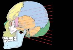
The cranium can be subdivided into
|
•an upper part (the calvaria), which surrounds the cranial cavity containing the brain;
•a lower anterior part—the facial skeleton (viscerocranium) |
|
|
|
The bones forming the calvaria are the
|
paired temporal and parietal bones, and the unpaired frontal, sphenoid, ethmoid, and occipital bones.
|
|
|
|
The bones forming the facial skeleton are the
|
paired nasal bones, palatine bones, lacrimal bones, zygomatic bones, maxillae, inferior nasal conchae, and the unpaired vomer.
|
|
|
|
Anterior Ethmoidal Foramen
|
anterior ethmoidal nerves and vessels.
|
|
|
|
Carotid Canal
|
internal carotid artery, deep petrosal nerve and other postganglionic
sympathetics (carotid plexus). |
|
|
|
Cribriform Plate
|
olfactory nerves
|
|
|
|
Facial Canal
|
facial nerve proper
|
|
|
|
Foramen Cecum
|
emissary vein from nasal cavity to superior sagital sinus.
|
|
|
|
Foramen Lacerum
|
meningeal br. of ascending pharyngeal a., emissary v. from pterygoid
venous |
|
|
|
Foramen Magnum
|
junction of spinal cord and brainstem, anterior and posterior spinal arteries
and veins, alar ligaments, tectorial membrane, apical ligament, spinal accessory nerve. |
|
|
|
Foramen Ovale
|
mandibular br. of trigeminal nerve, accessory meningeal a., lesser petrosal n.
|
|
|
|
Foramen Spinosum
|
middle meningeal a., reccurent br. of mandibular n.
|
|
|
|
Foramen Vesalii
|
emissary vein from pterygoid venous plexus.
|
|
|
|
Greater Palatine Foramen
|
anterior (greater) palatine nerves and vessels, posterior (lesser)
palatine nerves and vessels. These are branches of pterygopalatine or descending palatine nerves and vessels. |
|
|
|
Hiatus of the Facial(greater superficial petrosal) canal
|
greater superficial petrosal n., and
petrosal branches of middle meningeal artery. |
|
|
|
Hypoglossal Canal
|
hypoglossal nerve and meningeal branch of ascending pharyngeal a.
|
|
|
|
Incisive (Nasopalatine) Canal
|
nasopalatine branches of descending palatine nerves and
vessels |
|
|
|
Inferior Orbital (Sphenomaxillary) Fissure
|
maxillary nerve branches zygomatic
infraorbital,lacrimal br. of greater petrosal n., Infraorbital vessels. |
|
|
|
(Inferior) Tympanic Canaliculus
|
tympanic branch of glossopharyngeal nerve (will become
lesser petrosal) , tympanic br. of ascending pharyngeal a.. |
|
|
|
Infraorbital foramen
|
infraorbital br. of maxillary n., infraorbital vessels.
|
|
|
|
Internal Acoustic Meatus
|
vestibulocochlear n., facial n., internal auditory br. of basilar a.
|
|
|
|
Jugular Foramen
|
internal jugular v., glossopharyngeal n., vagus n., spinal accessory n.
|
|
|
|
Mandibular (Inferior Alveolar) Foramen
|
inferior alveolar n. and vessels.
|
|
|
|
Mental Foramen
|
mental n. and vessels.
|
|
|
|
Mastoid Canaliculus (Tympanomastoid Fissure)
Mastoid Foramen |
auricular branch of vagus n.
mastoid branch of occipital a., emissary v. |
|
|
|
Nasolacrimal Canal
|
tear duct
|
|
|
|
Optic Foramen
|
optic n., ophthalmic a., central retinal a.
|
|
|
|
Petrotympanic (Squamotympanic) Fissure
|
chorda tympanic branch of the facial n.
|
|
|
|
Pharyngeal Canals (along with vomerovaginal canals)
|
pharyngeal br. of maxillary a.,
pharyngeal brs. of greater petrosal n. |
|
|
|
Posterior Superior Alveolar (Maxillary) Foramen
|
posterior superior alveolar n. and vessels
|
|
|
|
Pterygoid (Vidian) Canal
|
nerve of pterygoid canal (combined sympathetic postganglionics of
deep petrosal nerve and parasympathetic preganglionics from greater petrosal nerve.) |
|
|
|
Pterygomaxillary Fissure
|
maxillary a., posterior superior alveolar n.
|
|
|
|
Pterygopalatine (Descending Palatine) Canal
|
descending palatine nerves and vessels.
|
|
|
|
Pterygopalatine (Sphenopalatine) Foramen
|
pterygopalatine vessels, nasopalatine n., posterior
nasal nerves., pharyngeal nerves and vessels. |
|
|
|
Stylomastoid Foramen
|
facial nerve proper
|
|
|
|
Superior Orbital Fissure
|
occulomotor, ophthalmic br. of trigeminal n., trochlear n.,abducent n..
ophthalamic v. |
|
|
|
Supraorbital Fissure
|
supraorbital n. and vessels
|
|
|
|
Zygomaticofacial Foramen
|
zygomaticofacial br. of zygomatic n.of zygomatic br. of maxillary
|
|
|
|
Zygomatico-orbital Foramen
|
zygomatic n. of V2
|
|
|
|
Zygomaticotemporal Foramen
|
zygomaticotemporal br. of zygomatic br. of maxillary
|
|
|
|
superciliary arches
|
superior to the rim of the orbit on each side
-Between these arches is a small depression (the glabella) the unibrow area |
|
|
|
coronal suture
|
In upper regions, the frontal bone articulates with the parietal bone
|
|
|
|
lambdoid suture
|
parietal bone then articulates with the occipital bone
-is in a upside down V shape |
|
|
|
sphenoparietal suture
|
frontal bone articulates with the greater wing of the sphenoid bone and articulates with the parietal bone
|
|
|
|
sphenosquamous suture
|
frontal bone articulates with the anterior edge of the temporal bone
|
|
|
|
sutural bones or wormian bones
|
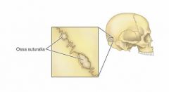
at the lamboid suture
-between the parietal bones and the occipital bone |
|
|
|
zygomatic arch is made of what two bones
|
-zygomatic process of temporal bone
-temporal process of zygomatic bone |
|
|
|
Three parts of the sphenoid bone
|
-a centrally placed body, -paired greater and lesser wings projecting laterally from the body,
-and two downward projecting pterygoid processes immediately lateral to each choana |
|
|
|
pharyngeal tubercle
|
Prominent on the basilar part (next to the sphenoid) of the occipital bone
-a bony protuberance for the attachment of parts of the pharynx to the base of the skull |
|