![]()
![]()
![]()
Use LEFT and RIGHT arrow keys to navigate between flashcards;
Use UP and DOWN arrow keys to flip the card;
H to show hint;
A reads text to speech;
215 Cards in this Set
- Front
- Back
|
What is the Clavaria? What is the Diploe? |
The calvaria or skullcap (feminine Latin noun with plural calvariae; however, many medical texts list the word as calvarium, neuter Latin noun with plural calvaria) is the upper part of the neurocranium and covers the cranial cavity containing the brain. \ The Diploe is composed of cancellous bone between the upper and lower table components that form the Clavaria, it contains red bone marrow inside. |
|
|
Where is the Epidural& subdural located? |
Epidural ("Extradural") is located between the Lower table and the Dura matter. The Subdural layer is located between the dura matter and the Subarachnoid space. |
|
|
Where are the Dural/ Venous Sinuses located in the skull?What do they do? Where do they drain? |
Located between/within Dura matter. They receive blood from internal and external veins of the brain, receive cerebrospinal fluid (CSF) from the subarachnoid space, and ultimately empty into the internal jugular vein. |
|
|
What are the 5 layers of the scalp? |
Skin Dense C/T Aponeurosis Loose C/T Pericranium |
|
|
What are the two layers the Dura Matter is composed of? |
Periosteal layer: Periosteum consists of dense irregular connective tissue, which attaches the Dura Sinues and the Calvaria to ensure they don't collapse. The meninges are three layers of protective tissue called the dura mater, arachnoid mater, and pia mater that surround the neuraxis. |
|
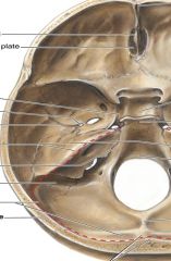
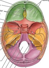
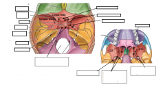
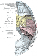
Label the Sinus's in this image. |
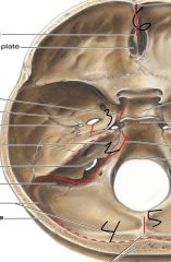
1.) Superior Petrosal 2.) Inferior Petrosal 3.) Carotid groove for Cavernous Sinus 4.) Transverse Sinus 5.) Occipital sinus 6.) Frontal crest for Sup. Sagital Signus. |
|
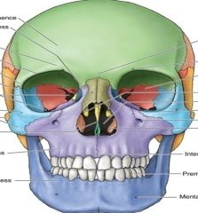
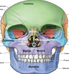
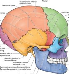
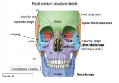
Label the colored regions. Frontal bone Zygomaic Maxilla Mandible Inferior Nasal Concha (INC) Ethmoid Vomer |
|
|
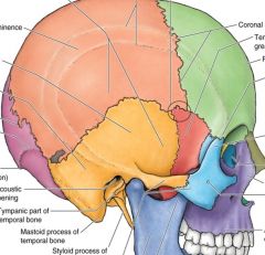
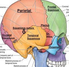
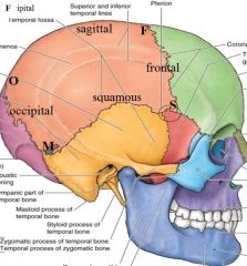
Label the colored regions Parital Temporal Squamous Pterion Gr. Wing Frontal Squamous Occiptal Squamous Zygomatic |
|
|
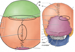
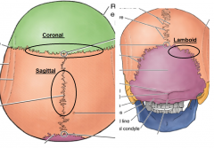
Label the following Sutures. Coronal Sagital Lamboid |
|
|
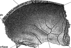
Label the margins and angles of the Partial bone (Orange) |
Angles: S- Sphenoid M- mastoid F- Frontal O-Occiptal angle |
|
Where is the superior sagital sinus located? |
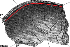
From the frontal margin to the occiptal margin. |
|
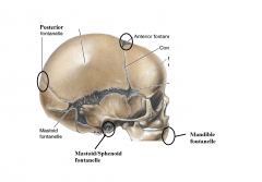
At what age are these Fontanelle's fused in the skull? |
Posterior Fontanelle - 1st year Anterior Fontanelle - 18 months Mastoid/Sphenoid Fontanelle - 28 dyas Mandible Fontanelle - 2nd year |
|
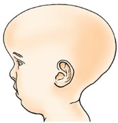
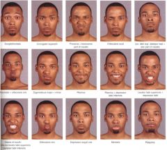
What condition is? Scaphoncephaly, Oxycephaly? Or Plagiocephaly? Which suture closes prematurely to cause this deformaty? |
Scaphoncephaly. Premature closure of the sagital Sutures will cause this deformity |
|
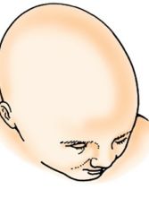
What condition is? Scaphoncephaly, Oxycephaly? Or Plagiocephaly? What sutures in the skull would have had to prematurely close to get this deformity? |
Oxycephaly This caused by premature closure of the Coronal Sutures. |
|
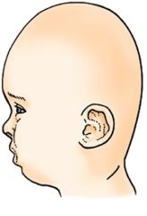
What condition is? Scaphoncephaly, Oxycephaly? Or Plagiocephaly? What sutures in the skull would have had to prematurely close to get this deformity? |
Plagiocephaly. Caused by premature closer of the lamdboid sutures unilaterally. |
|
|
When does "Obliteration"/full fusion of the sutures in the Clavaria happen? |
Age 40-50 years old. |
|
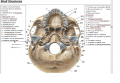
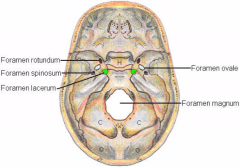
Anterior cranial fossa1.Foramen cecum2. Frontal crest &sulcus of sup.sagittal sinus3.Cribriform plate/lamina cribrosa: 4. Ant.+post.ethmoidal foramina: ant.+post. ethmoidal nerves & vessels 5. Lesser wings of sphenoidMiddlecranial fossa6.Sella turcica: hypophysealfossa,dorsum, tuberculum: forpituitary gl.7. Optic canal: CN II, ophthalmicartery8. Prechiasmaticsulcus: optic chiasma9. Sup.orbital fissura:CN’s: III, IV, V1,VI, sup. + inf. ophthalmic veins; sympathetic postg. Nn.10.Foramen rotundum: CNV2 11.Foramen ovale: CNV3,lesser petrosal n (CNIX), accessory meningeal a.12. Foramen spinosum: Middlemeningeal a/v,meningeal br. of mandibular n.13.Internal opening of carotid canal&Foramen lacerum:InternalCarotid a.& Sympathetic carotid nerve plexus; Greater petrosal n.(CNVII); Deep petrosal sympathetic n.14. Hiatus & groove for Greater petrosal n & Hiatus & groove for Lesser petrosal n 16. Internal acoustic opening&meatus: CNVII, CN VIII, labyrinthine a/v17. Jugular foramen: CNIX, X, XI, Int. Jug. V., mening.br.ofascending pharyngeal &occipital aa.18.Hypglossal canal: CNXII19. Foramen magnum: brainstem- spinalcord, Spinal accessory n, Vertebral aa/vv, Symp. Posg. N. |
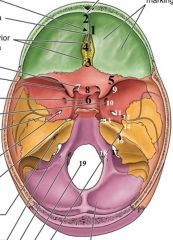
Anterior cranial fossa 1.Foramen cecum 2. Frontal crest &sulcus of sup. sagittal sinus 3.Cribriform plate/lamina cribrosa: CN I 4. Ant.+post. ethmoidal foramina: ant.+post. ethmoidal nerves & vessels 5. Lesser wings of sphenoid Middle cranial fossa 6.Sella turcica: hypophyseal fossa, dorsum, tuberculum: for pituitary gl. 7. Optic canal: CN II, ophthalmic artery 8. Prechiasmatic sulcus: optic chiasma 9. Sup.orbital fissura: CN’s: III, IV, V1,VI, sup. + inf. ophthalmic veins; sympathetic postg. Nn. 10.Foramen rotundum: CNV2 11.Foramen ovale: CNV3, lesser petrosal n (CNIX), accessory meningeal a. 12. Foramen spinosum: Middle meningeal a/v, meningeal br. of mandibular n. 13.Internal opening of carotid canal & Foramen lacerum: Internal Carotid a. & Sympathetic carotid nerve plexus; Greater petrosal n.(CNVII); Deep petrosal sympathetic n. 14. Hiatus & groove for Greater petrosal n & Hiatus & groove for Lesser petrosal n 16. Internal acoustic opening&meatus: CNVII, CN VIII, labyrinthine a/v 17. Jugular foramen: CNIX, X, XI, Int. Jug. V., mening.br.of ascending pharyngeal &occipital aa. 18.Hypglossal canal: CNXII 19. Foramen magnum: brainstem- spinal cord, Spinal accessory n, Vertebral aa/vv, Symp. Posg. N. |
|
|
In the Neurocranium: Base: Internal surface of the skull, what plate is a continuation of the cribiform plate? |
The cribiform plate and the Cristae Galli is seen in the internal neurocranial base and is part of the ethmoid. |
|
|
What does the Sella Turcica/Hypphseal Fossa contain? What boney region in the Internal Neurocraniual base does the Sella Turcica reside |
Pituitary Gland. The sphenoid bone. The Hypophseal fossa sits within the Sella Turcica |
|
|
What vessels leave through the Sup.orbital fissure? |
CN’s: III (Occulomotor), IV (Trochlear), V1 (Opthalmic),VI (Abducent), sup. + inf. ophthalmic veins; sympathetic postg. Nn. |
|
|
What nerve leaves through the Foramen Rotundum |
Foramen rotundum: CNV2 (Maxillary) |
|
|
What nerve exits Foramen Ovule? |
CNV3 (Mandibular), Lesser Petrosal nn (CNIX), accessory Meningeal a. The lesser petrosal nerve is the General visceral efferent (GVE) component of the glossopharyngeal nerv |
|
|
What vessels exit the Foramen Spinosum? |
Middle Meningeal a/v and the Meningeal branch of the mandibular n. |
|
|
What vessels exit the Carotid opening and the Foramen Lacerum? |
Internal Carotid a. & Sympathetic carotid nerve plexus; Greater petrosal n.(CNVII); Deep petrosal sympathetic n. The greater (superficial) petrosal nerve originates at the geniculate ganglion, where the nervus intermedius and facial nerve join. It contains mainly preganglionic parasympathetic fibers and some sensory taste afferent from the soft palate |
|
|
What vessels exit the External Auditory Meatius? |
CNVII (Fasical) , CN VIII (Auditoy/Vestibulochochlear), labyrinthine a/v |
|
|
What vessels exit the Jugular foramen? |
CNIX (Glossopharyngeal), X (Vagus), XI (Accessory), Int. Jug. V., mening.br.of ascending pharyngeal &occipital aa. |
|
|
What vessels exit the Hypglossal canal? |
CN XII hypoglossal nerve |
|
|
What exits the Foramen Magnum? |
Foramen magnum: brainstem- spinal cord, Spinal accessory n, Vertebral aa/vv, Symp. Posg. N. |
|
|
What exists the Cribiform |
CN I (Olfactory) |
|
|
What exits the Foramen Cecum? |
Emissary Vien. The emissary veins connect the extracranial venous system with the intracranial venous sinuses. They connect the veins outside the cranium to the venous sinuses inside the cranium. They drain from the scalp, through the skull, into the larger meningeal veins and dural venous sinuses. |
|
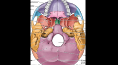
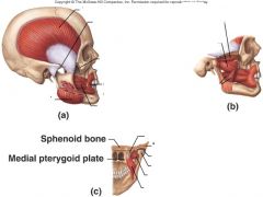
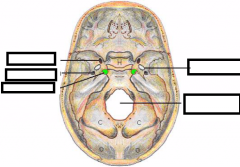
Locate the Sphenoid bone and the Temporal bone on this external base skull diagram. For the Sphenoid, located the following: - Greater Wing - Ptergoid's plates (Medial/Lateral) For the Temporal, locate the following: -Sqamous -Tempanic -Mastoid -Petrous |
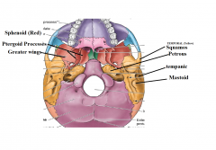
Sqaumous part is the top portion of the tempral region. Tempanic is near the ear Mastoid part is at the mastoid process Petrous is inferior portion of the Temporal bone. |
|
|
Where is the Pyterygoid canal located? What does it contain? |
Contains the Greater Peterosal N. of CN VII (Fascial) and the Deep Petrosal N symp. n |
|
|
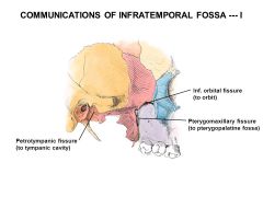
Where is the Pterygopalatine fossa? What does it contain? |
btwn the pterygoid process &infratemporal surface of maxillary body |
|
|
What are the functional components of the Olfactory nerve? (CNI) |
Special viceral sensory for SMELL |
|
|
Functional Components of the Optic nerve? (CNII) |
Special Somatic sensory for VISION (Balance, vision, |
|
|
Functional Components of the oculomotor nerve? (CNIII) |
Somatic motor to all the Extraocular muscles (Except the superior oblique & lateral rectus) General Visceral Motor = Parasympathetic preganglionic to cilary parasympathetic ganglion, then from it, via postganglionic parasympathetic to cilary smooth muscle for reflex and to pupillae smooth muscle for pupillary reflex. Preganglionic autonomicneurons originate from the brain or spinal cord; postganglionic neuronsoriginate from ganglia located outside the CNS. |
|
|
Functional Components of the oculomotor nerve? (CNIV) |
Somatic Motor to superior oblique muscle |
|
|
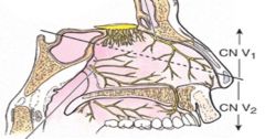
Functional Components of the Trigomental nerve? (CNV) |
General Somatic sensory from face, anterior scalp, eyes, sinues, nasal &oral cavities, anterior two thirds of the tongue, external surface of tempanic membrane Special Visceral Motor = Mastication, tensor tempani, tensor veli palatatini, mylhyooid & anterior belly of digasric muscle. |
|
|
Functional Components of the Abducent nerve? (CNVI) |
Somatic motor to lateral rectus occular muscle. |
|
|
Functional Components of the fascial nerve? (CNVII) |
Special visceral (Brachial) motor to muscles of fascial expression, stapedius, stylohyoid & posterior belly of diagasric. General Visceral motor = parasympathetic preganglionic to a.) pterygopalatine parasympathetic ganglia. Than to lacrimal gland and glands of nasal mucosa and paranasal sinuses. B.) to submandibular parasympathetic ganglion to submandibular, sublingual, and intralingual salviary glands and the glands of the oral mucosa. Special visceral sensory for taste from the anterior two-thirds of the tongue General somatic sensory from skin of concha of auricle, & external surface of tempanic membrane. |
|
|
Functional Components of the Vestibulocochlear nerve? (CNVIII) |
Vestibular division - Special somatic sensory for BALANCE Cochlear division" Special somatic sensory for HEARING |
|
|
Functional Components of the Glossopharyngeal nerve (CNIX) |
General somatic sensory from: posterior 1/3rd of tongue, tonsils, pharynx, and tympanic cavity including the internal surface of tympanic membrane. General Visceral sensory from: carotid sinus (Baroreceptors internal carotid artery), and carotid body (chemoreceptors from bifurcation of common carotid artery). Special visceral sensory for taste of posterior 1/3rd of tongue Special visceral motor (Brachial) to stylopharyngeus muscle General visceral motor preganglionic to otic ganglion and then to parotid gland. |
|

What nerve innervates the stylopharyngeus muscle? This nerve also innervates what gland via the otic ganglion? |
CN Nine glossopharyngeal Parotid gland |
|
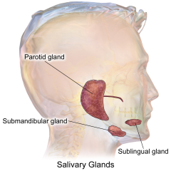
Which CN innervate these glands? |
Parotid is innervated by the Glossopharyngeal (CN IX) Sunlingual and Submandibular glands are innervated by the facial nerve. |
|
|
Functional Components of the Vagus nerve (CNX) |
General Somatic sensory = External ear, external tympanic membrane, dura mater of posterior cranial fossa and root of tongue. General Visceral sensory = Pharynx, Larynx, thoracic and abdnomnal viscera, and aortic body chemoreceptor. Special visceral sensory for TASTE from root of tongue and epiglottis Special visceral motor (Brachial) to muscles; soft palate, pharyngeal and laryngeal muscles. General visceral motor = preganglionic parasympathetic to parasympathetic ganglia to smooth muscles and glands of the pharynx, larynx, thoracic and abdominal viscera and cardiac muscle. |
|
|
Functional Components of the Accessory nerve (CNXI) |
Spinal root: Somatic motor and visceral motor to SCM and traps. (Dual origins from Brachial arch IV and occipital myotomes/CN XI) Cranial Root of Accessory CN joins vagus nerve to intrinsic muscles of larynx and soft palate. |
|
|
Functional Components of the Hpoglossal nerve (CNXII) |
Somatic motor to intrinic and extrinic muscles of tongue except platoglossus (CNX) and Geniohyoid (C1N) |
|
|
What structure is formed from the 1st pharyngeal arch? What nerve emerges from this arch? What mj muscles/bones/ligaments are innervated by it? |
Mandible CNV (Trigemental) Mastication muscles - MEDIAL/Lateral pterygoid - Masster - Temporalis. Mylohyoid Anterior belly of Diastric Tensor Tympani Tensor Veli Palatini Malleus (Bone) Incus (Bone) Sphenomandibular lig. |
|
|
What structure is formed from the 2nd pharyngeal arch? What nerve emerges from this arch? What mj muscles/bones/ligaments are innervated by it? |
Hyoid Fasical CNVII Stapedius sTYLOHYOID Posterior belly of digastric Fascial expression muscles - Buccinator -Auricularis - Occipitalfrontalis - Platysma Stapes (bone) Styloid Process (bone) Lesser conu of Hyoid (bone) Upper body of hyoid Stylohyoid ligament |
|
|
What structure is formed from the 3rd pharyngeal arch? What nerve emerges from this arch? What mj muscles/bones/ligaments are innervated by it? |
forms part of Hyoid Glossopharyngeal CNIX Styolopharyngeus Greater conu of Hyoid Lower body of Hyoid, |
|
|
What structure is formed from the 4th - 6th pharyngeal arch? What nerve emerges from this arch? What mj muscles/bones/ligaments are innervated by it? |
Superior and Recurrent laryngeal branch of Trigemental nerve (CNV) Cricothyroid Levator Veli Palatini Palatoglossus Palatpharyngeus - Striated muscle of Esophagus - Contriction muscle of Pharynx - Intrinisic muscle of Larynx Stylohyoid ligament (Same with Arch 3/Glossopharyngeal) |
|
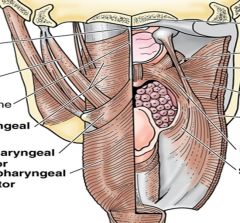
Label the muscles in this diagram |

|
|
|
Where is the Inion? (Point to it) |
the projecting part of the occipital bone at the base of the skull. |
|
|
Where is the Nasion? (Point to it) |
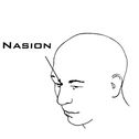
he point where the bridge of the nose meets the forehead |
|
|
Where is the Glabella (point to it)? |
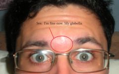
the smooth part of the forehead above and between the eyebrows. |
|
|
Where is the Lambda? |
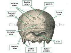
Where the Lamboid and sagittal suture meet |
|
|
Where is the Bregma? |
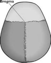
point on the skull at which the coronal suture is intersected perpendicularly by the sagittal suture |
|
|
Where is the Asterion? |
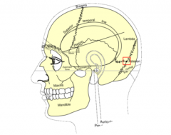
n human anatomy, the asterion is a visible, so-called craniometric, point on the exposed skull, just behind the ear, where three cranial suturesmeet:the lambdoid,parieto-mastoid, andoccipito-mastoid sutures, |
|
|
What lies under the Pterion? |
Anterior branches of the Middle Menigeal Artery |
|
|
What are "Sensory Ganglion"? List the sensory ganglion of the brain. |
They are unipolar neurons outside the brain and skull cavity. Trigeminal Ganglion: Trigeminal CN. Ophthalmic, mandibular, maxillary NN. Geniculate Ganglion: Fascial NN. Vestibular Ganglion: Vestibulocochlear Spiral Ganglion: Vestibulocochlear Superior/Inferior CN IX Ganglion: Glossopharyngeal Superior/Inferior CN X Ganglion: Vagus The signal passes from the sensory ganglion (in the skull cavity) to the sensory nuclei (in the brain). |
|
|
Where is the spiral Ganglia located? What ganglion is located in the internal acoustic meatus? |
Located inside the cochlea. The vestibular ganglion in vestibular. |
|

What is located inside the Petrous part of the temporal bone? (Regions of the ear) What is the Eustachian tube? |
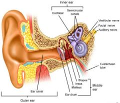
External and middle and Internal ear. Eustachian tube - where air enters the typanic membrane. Permitting the equalization of pressure on each side of the eardrum. |
|
|
From which nerve does the "Nerve to stapedius" branch from? What does this nerve do? |
Nerve to stapedius branches from the facial nerve. It attached to the stapedius mescle which works to "dampen" the vibrations of the stapeus bone. If you clap in front of a persons ear, and they complain its loud. This may be an indication that this muscle/nerve isn't working. |
|
|
When a child has "rhinitis", what other structures are effected? What does this cause? |
rhinitis is a mucosal irritation caused commonly by allergies in the nasal cavities. The Eustichian tube is effected in this state and causes inflammation. This can put pressure and cause pain. If a FRACTURE was to occur to the Petrous part of the temporal bone, this could cause somatic sensory and somatic motor loss for the fasical and vestibulocochlear nerves. |
|
What nerve is exiting the Petrotympanic fissure? |
The petrotemporal fissure is located behind the TMJ. it provides passage for the Chorda tympani which provides taste sensation (Seneory special) to the anterior 2/3 of the tongue. |
|

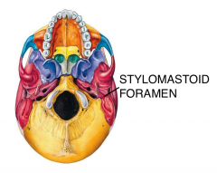
What nerve exits the Stylomastoid foramen? |
The fasical Nerve |
|
|
What is contained in the Tympanomastoid fissue? |
The Auricular Branch of the vagus nerve (Arnold's nerve). Stimulating this nerve (acupuncutr) can help stimulate the vagus nerve. |
|
|
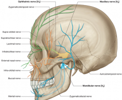
Where is the role of the infraorbital nerve? |
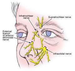
Off from the maxillary nerve and provides sensory innervation for the upper lip, lower eyelid and Anterior Nasal cavity. |
|
|
What is the branch of the Zygomatico-orbital foramina from? |
It is a branch from the Zygomaticofacial nerve (branch of maxillary N) |
|
|
Where is the Supraorbital foramen located? What exits it? |
Supraorbital A/V/N |
|
|
What bone contains the Cribiform plate? What nerve exits the cribform plate? |
Ethmoid bone. The olfactory nerve leaves the cribiform plate. |
|
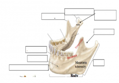
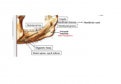
label the mandible |
|
|

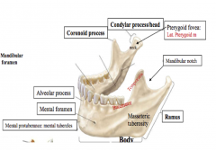
label the mandible |
|
|
|
What enters the Pterygopalantine fossa? |
Acts as a union station for the Nasal cavity Foramen rotundum (Trigamental nerve) Greater and Lesser Palatine fossa Pterygoid fossa |
|
|
Describe the path of the Tigeminal nerve (Opthalmic) |
The trigeminal nerve is composed of 4 nuclei (3 sensory, 1 motor) The trigeminal nerve comes from the midbrain, upper SC and the PONS (mAIN CORD). The cord branches to the Trigeminal ganglion and creates three branches; Opthalmic, Maxillary, Mandibular. Opthalmic branch enters the Superior orbital fissure and splits into branches: Frontal, nasocilary, lacrimal branch. The FRONTAL branch supplies the Forehead, Scaplp and Ethmoidal sinuses The Nasocilary branch supplies sensory innervation to the Cornea, upper eyelid, and the Dorsum of the Nose. The Licrimal Branch supplies the Lacrimal Gland |
|
|
Describe the path of the Tigeminal nerve (Maxillary) |
Maxillary branch enters the foramen rotundum. It divides into 14 branches. It supplies many parts of the face; Lower eyelid, cheek, maxillary sinus, nasal cavity, upper lip, upper teeth, superior palate. |
|
|
Describe the path of the Tigeminal nerve (Mandible branch) |
Exits the Foramen Ovale. Branches into 4 branches: Buccal, inferior alveolar, aurticulartemporal, linguinal. Suppleis the Mucos membrane of the Oral cavity, external ear, lower lip, chin, anterior 2/3rd of tongue., Mandibular branch also supplies motor innervation to the first brachial arch; Mastiffication muscles. These include: Temporalis, med/lat ptyergoid, Masseter, Tensor Tempani and Tensor palinti. The mandibular brnaches withthe Fascial nerve to form the "LINGUINAL NERVE" that enters the submandibular ganglia and the postganglionic fibers innervate the Submandibular and Sublinguinal Salivary Glands |
|
|
What is the significance of the Pterygopalatine fossa? |
It acts like a union station that connects the Interior Orbital Fissure. sPHENOPALATINE fORAMEN Greater/Lesser palatine foramen Pterygoid canal Foramen rotundun (Trigamental, Maxillary nerve exiting) |
|
|
What nerve is in the Pterygoid canal? |
Greater petrosal N of CNVII + Deep Petrosal N sympathetic nerve. |
|
|
Where doe the Facial Nerve originate (Neurons origins), what structure does it enter? What branches from it? What does it innervate? |
The fascial nerve arrises from the Pons and Medulla junction. The Fascial nerve enters the internal auditory meatus (Along with the Vestibulocochlear nerve). It travels through the Acustic Canal until it reaches the Genicular Ganglion. From there it branches into the Greater Petrosal Nerve. This nerve turns up into the internal skull and enter the Foramen Lacerum. The fibers meet with the Pterygopalatine Ganglion (Parasympathetic) and than innervates the Lacriminal Gland The Facial nerve (from the Acustic canal) will run a branch called the "Horizontal segment". From here, Nerve to Stapedius and Chorda Tympani branch. The nerve to stapedius innervates the Stapedius muscle, and the Chorda Tympani will exit through the Petrotempanic fissure. It becomes the Lingual Nerve joining with a branch from the Mandibular nerve. The Facial nerve picks up taste from the anterior 2/3rds via the Tensor Tympani. The Facial nerve from the horizontal segment it exits the Stylomastoid Foramen. Here it gives off three branches. Posterior Auricular nerve Nerve to posterior digastric Nerve to Stylomastoid. These muscles originate from the 2nd brachial arch. The nerve carries on and enters the Partoid gland where it branches into 5 branches: Tempral Zygomatic Buccal Mandibular Cervical. These branches supply the muscles of fasical expression. |
|
|
What nerves enter through the internal auditory meatus? |
CN VII (facial) CN IX (VESTIBULOCOCHLEAR) |
|
Label the following foramen |
|
|
What nerve exits the Peterotempanic fissure? |
Chorda Tympani which is a branch from the Fascial nerve. |
|
|
What is the Lingual Nerve made up of? WHAT DOES it do? |
The lingual nerve is composed of the Fascial and Mandibular nerve (CN VII and CNV3). The Lingual nerve innervates the tongue. The Mandibular branch recieves Sensory Innervation from the Anterior 2/3rds of the tongue. The Facial Nerve picks up TASTE sensation from the anterior 2/3rds. |
|
Label the Sphenoid bone |

|
|
|
What nerve does the Varicella virus (Chickenpox) lay latent in? |
The trigomental nerve (Mc in the Opthalmic nerve). Called Herpes zosters opthalmicus |
|
|
What are the attachments for the Buccinator muscle? |
Origin: from the alveolar processes of the maxillary bone and mandible, temporomandibular joint Insertion: in the fibers of the orbicularis oris |
|
|
What are 5 important features of the Muscles of Fascial Expression? |
1. Attached to the skin 2. In the Subcutaneous fasica 3. Surround openings (Nasal, orbit, oral) 4. Developed from the 2nd brachial arch 5. Innervated by the Facial nerve. |
|
|
What are the CN "ganglia"? |
The parasympathetic ganglia and met by preganglionic nerons within the spinal cord/Medulla/Pons that synapse at the Ganglia and emerge Post ganglionic nerves to Smooth muscle and glands. |
|
|
What pre/pot ganglionic nerurons are associated with the Cillary Ganglia? |
The Cillary Ganglia is located in the posterior Orbit cavity. The preganglionic nerves emerge from the Edinger-Westphal necleus in the midbrain which is associated with the Occulomotor Nerve (Inferior division) The Short Cillary nerve emerges as the postganglionic nerve and innervates the Cillary mm. Iris, Cornea. |
|
|
What post ganglionic neurons are associated with the Trigeminal Ganglia? |
Opthalmic which enters the Superior orbital fissure. Maxillry which enters the Foramen rotundum Mandibular which enters the Foramen Ovale |
|
|
Whee does the Fascial nerve enter and emerge from? |
Fascial nerve enters through the internal auditory meadius and emerges through the stylomastoid fossa (Between the Styloid and Mastoid processes of the temporal bone) |
|
|
Describe the Greater petrosal nerves role. |
The Greater Petrosal nerve carries Preganglionic nerve fibers from the Superior Salvitory Neuron in the PONS. The greater petrosal nerve travels through the Internal Auditory Meatus along with the facial nerve and exits through a Fossa in the petrous part of the temporal bone. The nerve carries to the Foramen Lacerum which is near the apex of the petrous part of the temporal bone. There it emerges with the DEEP Petrosal nerve through the Pterygoid canal (NOT THROUGH THE FORAMEN LACERUM, nothing emerges this foramen!) The DEEP petrosal nerves are Post Sympathetic nerve fibers that arise from the superior cervical sympathetic ganglia |
|
|
What post ganglionic neurons are associated with the Superior Cervical Sympathetic Ganglion? |
The deep petrosal nerve is a sympathetic postganglionic nerve that emerges from the Superior Cervical Sympathetic Ganglion. The deep petrosal nerve wraps around the Internal Carotid Artery and enters the petrous part of the temporal bone. There it branches off to the Foramen Lacerum where it joins with the fibers of the Superior Petrous nerve and form the Vidian/Pterygoid Canal nerve. This nerve passes through the Pterygoid canal. The Vidian/Pterygoid canal nerve emerges through the Pterygoid fossa and synapases with the Pterygopalatine Ganglia. This pterygopalatine Ganglia does not relay the Sympathetic cervical fibers. Only the parasympathetic fibers from the Superficial Petrosal nerve. |
|
|
What nerve merges with the Lacriminal Nerve (Opthalmic VI)? |
The Zygomatic branch from the Maxillary branch, off the Pterygoidpalatine Ganglia communicates with the Lacriminal Nerve. |
|
|
What nerves emerge from the Pterygopalatine ganglia? |
The Greater palantine N. which has nasal branches that enter via the Sphenopalantine fossa. The Lesser Palantine N. which sends Pharngeal branches. This nerve can stimulate Hay fever due to its conenctions with the Pterygopalatine fossa which has Lacrimal Branches, Basal Branches, and Pharynx Branches. The Zygomatic n. from the Maxillary branch which leads to the Lacrimal Gland. The Greater Petrosal and Deep Petrosal nerve are the preganglionic parsympathetic (sympathetic) neurons that synpase at the pterygopalatine ganglia. |
|
|
What nerves emerge from the Otic ganglia? |
The Otic ganglia is located next to the Mandibular Nerve near the tensor palantine muscle. It recieve Preganglionic Parasympathetic neuclus from the Inferior Salvivary nuclus located in the PONS. These preganglionic fibers leaves the PONS via the GLossopharngeal nerve. The Glossopharyngeal nerve passes through the Jugular FOramen, and gives rise to a Typanic Branch that returns back up through a small foremen near the Jugular foramen and forms the Tympanic Plexus. Emerging off the Tympanic Plexus is the Lesser Petrosal Nerve, this nerve enters the Foramen Ovale and synapses with the Otic Ganglia. The postganglionic parasympathetic fibers will innervate the Parotid Gland via the Auriculotemporal N. (Branch of the mandibular nerve) |
|
|
What is contained within the Tympanic Plexus?Where does it arise from? |
tYPMANIC PLEXUS is from a branch from the Glossopharyngeal near, that contains preganglionic parasympatheitic fibers from the Inferior Salvitory Nuceli within the PONS. |
|
|
Explain the Submandibular ganglia. |
It is attached to the Mandibular nerve, more specifically the Linguinal Nerve. Its Preganglionic parasympathetic nerve fibers are derived from the mid brain Superior Salivary neuron. The parasympathetic neurons travel within the Inferior Auditory Meiatus along the facial nerve. Just before the facial nerve exits the Stylomastoid foramen, it sends off a branch that enters the bone and passes over the tempanic membrane. This nerve is called the Chorda Tympani. It supplies taste sensation to the anterior 2/3rds of the tongue. |
|
|
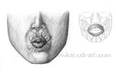
What muscles in the face cause wrinkling of the forehead and/or raising of the eye brows? |
Frontalis muscle - wrinkles and raises eye brows Corrugator - wrinkles forehead |
|
|
What muscles causes the lips to be puckered? |
Orbicularis Oris |
|
|
What is the role of the Buccinator muscle? What are its attachments? |
Buccinator sucks the cheeks inward close to the teeth. Atachments: alveolar processes from the mandible and the maxilla and inserts to the Obicularis oris muscle of the face. |
|
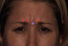
What muscle is involved in making a made face in the eyes and wrinkling the skin b/w the eyes? |
Procerus "Pro-se-rus" |
|
What muscle is responsible for flaring of the nostrils? |
Nasalis |
|
What muscle is responsible for doing this fascial action? What is the muscles innervations (2 branches) |
Orbicularis Oculi. The palpebral part of the muscle performs gentle closure of the eyelid, whereas the orbital portion closes more forcefully. Temporal and Zygomatic branches of the facial nerve. |
|
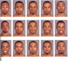
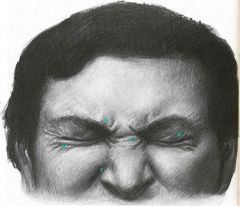
Label the muscles used in these images of facial expression |
|
|
|
Which branch communicates with the Lacrimal Nerve branch (Opthalmic nerve) to innervate that Lacrimal gland? |
Zygomatic branch from the Facial nerve carrying parasympathetic postganglionic fibers from the Pterygopalatine ganglia. |
|
|
What nerves branch off from the Maxillary nerve? |
Nasopalatine n Palatine n Zygomaticotempral n Zygomaticofacial n Infraorbital n. |
|
|
What branches off from the Mandibular nerve? What is the nerve course/anatomy when it passes through the mandibular foramen? |
Med + Lat Pterygoid. Temporal n. Buccal n. Massentric n. Linguinal n (merge with Chorda Tympani n) Mandibular foramen -> Inferior Alveolar nerve -> Mental foramen -> Mental nerve. |
|
|
What nerves innervate the posterior and anterior Digastric muscle? |
Posterior Digastric - Facial nerve. Anterior Digastric - Mandibular nerve (N. to Mylohyoid) |
|
|
Muscle nerves supply innervation to the tongue? |
Anteriro 2/3rds of tongue - Facial (chorda Tympani) nerve. + Mandibular n (Linguinal N) Posterior 1/3rd - Glossopharyngeal nerve. |
|
|
What is the parotid gland innervated by? |
Postganglionic neurons from the Otic Ganglia supplied by the Glossopharyngeal nerve |
|
|
What nerve innervates the levator palpebrae? |
Occulomotor nerve. |
|
|
What nerve is the Auriculotempral nerve a branch of? |
Mandibular nerve (trigeminal) |
|
|
What are the 3 meninges of the skull cavity? Between what two layers is the CSF? What are they innervated by? |
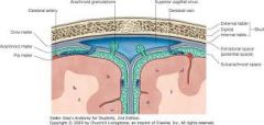
Dura matter - right under the skull. Made up of two layers. Periostium layer (Most superfical layer in the skull) and Menengeal layer. They are stuck together except at sinuses. Arachnoid matter - Next layer over the dura layer Pia matter - layer over the arachnid layer that covers the convolution of the brain. The CSF is between the Pia and Arachnid matter layers The opthalmic nerve innervates the tentrium dural sac. The VAGUS NERVE sends a recurrent menigeal branch from the Jugular ganglia to the posterior dural sac. |
|
|
where does the emissary vein run through? |
It runs through the nasal cavity (Cribiform plate) when it helps drain fluid from the superior sagital sinus. |
|
|
What are the three Dural folds in the skull? |
Falx Cerbri - separating the cerebrum into left asnd right hemispheres Falx Cerebelli - Separating the cerebellum into left and right hemispheres Tentorium cerebelli - separates the cerebellum from the cerebrum. |
|
|
Where does the temporalis muscle insert? |
Cornoid process (Anterior tubercle on mandible) So performs some closing of the jaw. |
|
|
What two arteries form the circle of willis? |
Vertebral artery and the Internal carotid artery |
|
|
what does the common carotid artery branch in too? What is the vertebral level this happens at? |
internal and external carotid arteries at level of c4 |
|
|
what are the main branches of the internal carotid artery? |
The internal carotid artery splits into the Middle Cerebral Artery, which runs through the lateral sulcus/fissure and terminates, and the Anterior Cerebral Artery which runs through the interhemspheric fissure. The aanterior cerebral arteries merge together via the Anterior communicating branch and they also merge with the posterior cerebral arteries via the posterior communicating branches The artery also supplies a branch for the eyes called the Opthalmic artery that runs with the optic nerve. As well as the Hypophseal branch to the pituitary gland Ophthalmic artery Superior hypophyseal Posterior communicating artery Anterior choroidal artery Anterior cerebral artery (a terminal branch) Middle cerebral artery (a terminal branch) |
|
|
what are the main branches of the external carotid artery? |
The internal carotid has no branches in the neck, but the external carotid artery has 8 branches 3 anterior branches 2 posterior branches 1 middles branch 2 terminal branches "Some Ancient Lovers Find Old Positions More Stimulating" Superior thyroid Ascending pharyngeal Lingual Facial - at the edge of the mandible (palpable) Occipital Maxillary Superficial Temporal |
|
|
what are the main branches of the vertebral artery? |
Vertebral artery join at the Pons and form the basilar artery that travels up the mid line of the Pons and than splits into the POSTERIOR Cerebral Arteries that merge with the anterior Cerebral arteries in the brain. |
|
|
What enters /exits the Mastoid foramen? |
Posterior meningeal artery Small branch for optic artery Mastoid emissary vein. |
|
|
What nerve is the Frontal nerve a continuation of? What does the Frontal nerve split into? |
It is a continuation of the Opthalmic nerve that runs throug hthe Superior orbital fissure, outside the COmmon tendinous ring, and runs over the Levator palpebrae Superiorus. It splits into the Supraorbital n. and the Supratrochlear n. |
|
|
The Levator Palpabrae Superioris is a striated muscle that also has a smooth muscle section called the Superior tarsal muscle. What are the innervation for the smooth and striated muscle? |
The Striated muscle is innervated by the Occulomotior nerve. While the Superior tarsal muscle is innervated by the sympathetic nervous system. |
|
|
What is the conjunctiva? It has two layers, what are they? What are they innervated by? |
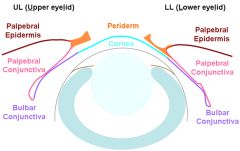
The conjuctiva is a muscus membrane that surrounds the eye. It has two layers, a Palpebral layer and a bulbar layer. The two layers are continuous with one another, with the Palpebral layer coming off the Palpebral Epidermis and the Bulbar layer surrounding the eye. The Conjuctiva is innervated by many nerves that is arranged in 4 sections: Superior: -Supraorbital n - Supratrochlear n -Infratrochlear n Inferior: Infraorbital n Lateral: -lacrimal n. Cornea: - Long Cillary Nerve (Off the Nasocillary) |
|
|
What are the components of the Eyeballs 1. Nucleous 2. Capsule. What is the innervation of the Iris (smooth muscle)? |
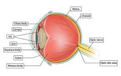
1. The nucleus is composed of the Lens and the Vitreous body, 2. The Capsule is composed of a Fibrous layer: Sclera, Cornea. A vascular Layer: Choroid, and smooth m Cillary bodies + Iris (Long Cillary N. Inn) And a internal layer: Retina. |
|
|
What causes Midriasis (Pupil Diolation) (What nerve) What causes Mitosis (Pupil constriction. |
Midriasis is caused by postganglionic sympathetic nerves that stimulate the dilator pupil muscle (Long cillary nerve) |
|
|
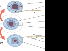
Explain the accommodation reflex. What muscle is being stretched/relaxed? What feedback is causing the change in DISTANT or NEAR vision? |
The accomidation reflex is a change in the lens shape, lens convergence, and pupil size to accommodate for near or distant focus. When focuses is on a distant object the Zonular ligament is under tension (It is relaxed) and causes the lens to be stretched. When focuses is on a near image, the Parasympathetic nervous system acts on the Zonular ligament, causing it to contract, and causes the lens to widen. |
|
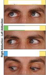
Label the following eye moments for the muscles being recruited. |
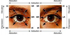
Top left - IO Top Right - SR Middle Left - LR Middle Right - MR Bottom left - SO Bottom right - IR |
|
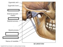
Label the following ligaments for the mandible. What are the roles of each of the ligaments? |
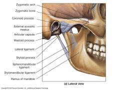
Lateral ligament - prevents posterior dislocation. Sphenomandibular + Stylomandibular ligament - prevent excessive opening of the jaw. |
|
|
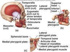
Where do the medial and lateral pterygoid muscles attach? What is their role? |
The medial pterygoid attaches to the Lateral Pterygoid plate. and the Palate The Lateral pterygoid plate is superior to the middle pterygoid and is attached to the Articular Disk of the jaw and the Synovial sheath and the Condyloid process |
|
|
What vessels run under the Sphenomandibular ligament? |
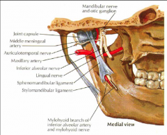
Auriculotemportal N. Maxillary artery Middle meningeal artery Inferior alveolar artery and mylohyoid nerve. |
|
|
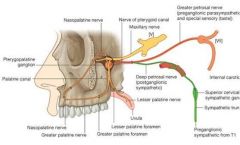
What preganglionic nerves innervate the Pterygopalatine ganglia? What nuceli from the pons/midbrain are carried. What Postganglianioc nerves come off the Pterygopalatine Ganglia? |
The greater petrosal, from CN7/Facial Nerve, carries Parasympathetic Pregangliatic nerves from the Inferior Salvitory Nuceli to the Pterygopalatine ganglia. These parasympathetic neurons supply the Lacrimal Gland The Ganglia is also innervated by sympathetic nerve (efferent) fibers from the Deep Petrosal nerve from the Superior Cervical ganglia. Postganglionic Parasympathetic+Sensory+Sympathetic nerves from the pterygopalatine ganglia follow the Maxillary nerve path branches. These include; Greater Palatine, Lesser Palatine, Nasopalatine, Sphenopalatine, N. to pharyngeal, Zygomatic (Zygomatictemporal/facial), Communicating branch from Pterygopalatine ganglia to Lingual nerve to Lingual gland |
|
|
What are the terminal branches off the maxillary artery? |
Maxillary artery is abranch off the EC aa. The maxillary artery has 3 regions of ateries it splits into. Mandibular - Deep Auricular, Anterior tympanic, Inferior alveolar, Middle meningeal, Accessory Minengeal. Pterygoid - Pt (med+lat), Deep temporal, Massenteric, Buccal, Pterygopalatine - Sphenopalatine, superior alveolar, Infraorbital, Pterygocanal. |
|
|
What nerve does the Pharyngeal N branch from? What are the innervations of the pharyngeal nerve? |
Pharyngeal nerve is branched from the Maxillary nerve (CNV2) and innervates the Tympanic cavity, Pharynx mucosa (Auditory tube) |
|
|
What is the terminal branch of the Nasopalatine nerve? |
Incisive nerve, passign throug hthe Incisive foramen. |
|
|
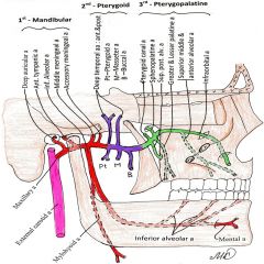
What are the branches of the Maxillary Nerve and the Pterygopalatine ganglia? |
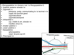
Blue is sensory from Maxilalry nerve Red is Deep petrosal Green is Greater petrosal. |
|
|
What are the 5 contents of the Pterygopalatine Fossa? (Nerves, Artery/vein, Ganglia) |
1.) Maxillary Nerve and its branches 2.) Pterygopalatine Ganglion 3.) Nerve to pterygoid canal 4.) Maxillary artery (3rd branch, Ptergopalatine) 5.) Maxillary Vein. |
|
|
What are the Parasympathetic nerve roots for the Pterygopalatine ganglia? |
Sensory Root - Pterygopalatine nerve (Maxillary) Parasympathetic Root - Greater petrosal nerve ( Facial nerve) |
|
|
Name all the nerves that innervates the Lacrimal Gland? |
Everything that synpases with the Ptyergopalatine ganglia. Parasympathetic secretomotor fibers from the Greater petrosal nerve from that Facial nerve. Sympathetic fibers from the Deep petrosal nerve. Maxillary nerve branch. |
|
|
What are the Preg. parasymp nerve for the Otic ganglia? Pterygopalatine G? Submandibular G? Ciliary G? |
Greater Petrosal < Pterygopalatine G Lesser Petrosal < Otic G. Chorda Tympani < Submandibular G. Inf. Ramus < Cililary G. |
|
|
Describe all the neurological components that synapse with the Cillary Ganglia? (Pre+Post) |
The Cillary Ganglia contains 3 components: Sympathetic pre-ganglionic neurons from the Cervical sympathetic plexus. Somatosensory preganlionic neurons from the Nasocillary N. Parasympathetic preganglionic neurons from the Edinger-Westphal neuron from the brain stem, that is carried via the Occulomotor nerve with branches into the Inf. Ramus nerve synapse with the Ganglia. Short Ciliary neurons branch from the Ciliary ganglia which carry the sympathetic and parasympathetic nerve fibers to the Sclera, Iris and Cornuea of the eye. |
|
|
Describe all the neurological components that synapse with the Otic Ganglia? (Pre+Post) |
The Otic ganglia contains the following: Sympathetic nerve fibers from the Middle Meningeal artery plexus from the Superior Cervical Ganglia. These post ganglionic fibers innervate the Parotid gland. Parasympathetic nerve fibers from the Inferior Salivary Nuclei in the Pons that are carried via the Glossopharyngeal nerve, through the tympanic cavity and carried further by the Lesser Petrosal nerve. These supply the Partoid gland and control secretomotor functions of it. Somatosensory nerves from the Mandibular nerve (Auriculotemporal division) that supply the Parotid gland |
|
|
Describe all the neurological components that synapse with the Submandibular/maxillary Ganglia? (Pre+Post) |
The Ganglia is synapsed with - Preganglionic parasympathetic nerve fibers from the Superior Salivtory Nuceus within the Pons. These are carried via the Facial nerve (CN7) and the Chorda Tympani. - Somatosensory nerve fibers from the Lingual Nerve. (Mandibular nerve) - Sympathetic nerves from the External carotid Cervical plexus. This Ganglia sends parasympathetic nerve fibers to the Submandibular gland and the Sublingual gland. |
|
|
Describe all the neurological components that synapse with the Pterygopalatine Ganglia? (Pre+Post) |
Pterygopalatine Ganglia is synapses with - Sympathetic nerves from the Deep Petrosal nerve from the Inferior Salivary ganglia - Parasympathetic nerves from the Facial nerve which is than carried further by the Greater Petrosal nerve to the ganglia. There is a connecting branch to the Lacrimal nerve that helps innervate the Lacrimal gland. The Greater Petrosal nerve also innervates the Nasal Glands. - Somatosensory nerves from the Maxillary nerve (Pterygopalatine nerve). |
|
|
What supplies somatosenosry innervation of the Dura matter? |
The sensory innervation of the meninges is primarily by meningeal branches of both the trigeminal and vagus nerves with a smaller contribution from the upper cervical spinal nerves 1,2. The supratentorialdura mater is mainly supplied by the ophthalmic division of the trigeminal nerve 3. |
|
|
Maxillary sinusitis and ethmoidal sinus inlammation will cause what symptoms? |
Headaches Becuase they share the maxillary somatosensory innervation to the Dura Matter. |
|
|
What is a potential cause of a nose bleed? |
High blood pressure. |
|
|
What is the source for the Ethmoidal arteries? Grerater palatine and Sphenopalatine? |
Opthalimic artey Maxillary artery branch from Extenal Carotid. |
|
|
What arteries anatomose at the Kisselback Septal Area? |
Anterior and posterior Ethmoidal aa. Sphenopalatine. Insivsive (Sphenopalatine continuation) Greatger Palatine Septal branch of the Superior labial branch of the facial aa. |
|
|
What are some influences of a deviated septum? |
Decreased air circulation on one side. And all sinuses (Ethmoidal, maxillary, etc on same side) are affected and increases chances of infection |
|
|
WHAT is Rhinitis? |
Inflammation of the nasal muscoa. Causing a loss of Smell. (Bloackage of the Olfactory mucosa cells) |
|
|
Since the Superior Alveolar nerves supply both the maxillary teeth and the mucosa of maxillary sinus, what will occur with a sinusitus issue? |
A tooth ache probably. |
|
|
What is the membrane at the bottom of the tongue? What is it a landmark for? |
Frenulum Submandiublar ducts |
|
|
Soft palate muscles? Supplied by which nerve? |
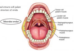
Palatoglossal muscle (continuous with tonuge muscle, attach to palatine apernorosus) Palatopharyngeus muscle Musculus Uvulae Levator Veli Palatini Tensor Veli Palatini Pharyngeal branch of Vagus nerve. |
|
|
If the Uvulae is deviated to the left side, where is the lesion? |
The right Vagus nerve lesion. |
|
|
What innervqates the tongue? What are the Extrisic muscles? |
Hypoglossal nerve CN 12. Styloglossus - Hyoglossus - Will flatten tongue Genoglossus - Vertical fibers of tongue, cause tongue to be pushed forward. Genohyoid muscle will retract tongue (NOT AN EXTRINSIC MUSCLE OF TONGUE THO) Palatoglossus is not innverated by the Hypoglossus, it is suppllied by the vagus, as it is considered a muscle of the Pharynx. |
|
|
Posterior 1/3rd of tongue is inervated by? (Special sensory and somatosensory) Anterior 2/3rd of tongue? |
CN 9 Glossopharyngeal Special sensory for taste from Tympani Nerve CN7 Somatosensory from Lingual nereve CN5(3) |
|
|
What muscle does the APR root of C1 innervate? What clinical problems can this cause? |
Genohyoid Stabilizes the Hyoid bone. May cause difficulty swolling if not functioning. |
|
|
If the tongue is protruded, and deviated to 1 side, what CN has lesion? |
Ipsilateral paralysis to the Hypoglossal N, Genuglossus muscle pushes the tongue to the weak side. |
|
|
After the greater petrosal nerve/vidian nerve (CN7/Facial nerve) synapses with the Pterygopaslatine ganglia, what postganglianic parasympathetic branches does it send? |
Pharyngeal nn Lacrimal gland branches Pos. Lat. Sup+Inf Nasal nn. Palatine branches. (greater/lesser) Alveolar branches (anterior/posterior/middle) |
|
|
if the maxillary nerve was cut off after it entered the IOF, what would occur? |
After the IOF the maxillary nerve branches the; Anterior/middle alveolar nerves, Infraorbital, zygomaticfacial and zygomtictemporal. There would be decreased sensation to the middle and anterior teeth but not the posterior. |
|
|
What are the connecting/emerging branches from the Nasopalatine nerve off the Pterygopalatine Ganglia. |
The Pterygopalatine ganglia gives off the Nasopalatine branch. This nerve branches into the - Posterior superior lateral + Posterior Inferior Lateral nasal branches. - Sphenopalatine banch that tavels alog the anterior nasal septum, diverges through the Incisive foramen and anastomoses with the greater palatine branch. |
|
|
this nerve branches into the Zygomaticotemporal/facial nerves? |
Zygomatic branch off maxillary nerve. |
|
|
What are the somatosensory actions of the Zygomatictemporal/facial nerves? (Where do they emerge) |
The Zygomaticofacial nerve emerges from the Orbicularis occuli muscle and supplies the skin over the cheek bone The Zygomaticotemporal nerve emerges 2cm above the zygomatic arch and supples the Anterior 1/3rd of the Temporal fossa. |
|
|
The Zygomatic nerve, in the pterygopalatine fossa,receives the Communicating postganglionicparasympathetic branch from the parasympathetic pterygopalatine ganglion! What does it innervate? |
Postganglionic parasympathetic nerve fibers from the Greater Petrosal nerve from the Pterygopalatine ganglia send communicating branch to the Zygomatic nerve that than branches off and sends post ganglionic parasympathetic nerurons to the Lacrimal gland |
|
|
what is the infraorbital nerve a direct continuation of? What branches does it give off? |
A direct continuation of the Maxillary nerve. If gives branches; superior middle and anterior alveolar branches (supplying mucosa of the maxillary sinus and superior middleand anterior maxillary teeth) |
|
|
What nerve rusn through the Greater Palatine foramen? What does it anastomose with? |
The greater palatine nerve enters the Greater palatine foreman, runs anterior along the hard palate and anastomoses with the Incisive nerve. |
|
|
Facial nerve --> Greater petrosal nerve --> Chorda tympani. |
xc |
|
|
Where do the deep and Greater petrosal nerve join? |
Foramen lacerum. |
|
|
What are the branches of the maxillary, mandibular region aa.? (MMA MMA) What are the branches of the maxillary, PTERYGOID region aa.? (MMA MMA) What are the branches of the maxillary, Peterygopalatine region aa.? (MMA MMA) |
-InferiorAlveolar Deep Auricular Middle Meningeal Accessory meningeal Mental Mylohyoid“MMA MMA” Deep temporal Buccal Pterygoid Masseter Pterygoid canal Shenopalatine Sup. Pos. Alveolar Greater and leser palatine Infraorbital |
|
|
Which Nasal Meatus/Concha is 1cm anterior to the Eustian tube? |
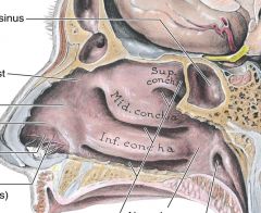
The inferior Conchae /Inferior meataus |
|
|
What sinuses are connected with the Superior Nasal Conchae/Meataus? What sinuses are connected with the MIDDLE Nasal Conchae/Meataus? What duct is connected with the Inferior Nasal Conchae/Meataus? |
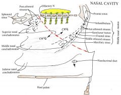
The Superior Nasal Conchae lets air into the SPHENOID SINUS into Sphenoethmoidal recess. It also leads into POSTERIOR ETHMOIDAL Sinuses. The Middle Nasal Meataus is longer and deeper. The frontal sinus is lead from the Infundibulum - smilunar hiatus - maxillary sinus The Inferior meataus contains the Lacrimal duct anterior to it |
|
|
Para-nasal Sinuses = Sphenoid, Frontal, Ethmoidal, Maxillary. What are the Parasympathetic innervation of these sinuses? |
All sinuses parasympathetic secromotor Innervated by the Pterygopalatine ganglia Frontal Sinus - Supraorbital n Ethmoidal sinuses - Ant/post Ethmoidal n Sphenoid Sinus - pos Ethmoidal nn Maxillary sinus - Ant/mid alveolar branches |
|
|
What nerve supplies the postero-inferior poriton of the nasal mucosa? What nerve supplies the anterr-superior porition? |
Postero-inferior = Maxillary nerve (Nasopalatine branch) Antero-superior = Ant/Post ethmoidal nerve (Nasocillary branch/Opthalmic nn) |
|
|
Describe the path and innervations of the Palatine nerves |
Palatine nerves:nasopalatine, greater palatine, lesser palatine. The palatine nerves arise from the maxillary division in thepterygopalatine fossa ; they run via the pterygopalatine canal ; within the canal the Nasopalatine nerves branch off such as the : Posterior superior lateral and Posterior inferior lateral nasal branches to mucosa of posterior-inferior aspect of lateral nasal wall Spenopalatine branch traverses the sphenopalatie foramen and runsanteriorly along the nasal septum (innervates postero -inferiorly nasalseptum), then it passes towards the incisive foramen as the Incisive nerve into the hard palate anastomising with the Greaterpalatine nerve. o Greaterpalatine nerve exits the pterygopalatine canal via the greater palatineforamen, then runs anteriorly along the hard palate innervating it andanastomosing with the incisive nerve o Lesserpalatine nerve exits the pterygopalatine canal via the lesser palatine foramenand innervates the soft palate |
|
|
What does the Pharyngeal branch from the Maxillary nerve supply? |
The maxillary nerve appears to transmit the principal sensorysupply from the tympanic=auditory= Eustachian tube and middle ear cavity,presumably through the pharyngeal branch. |
|
|
What does the Incisive nerve anastomose with? |
Greater petrosal nerve in the hard palate. |
|

The area in red is known as the Kiesselback anastomoses area where all the 5 Arterial branch come together. What are the 5 branches? |
1. Anterior ethmoidal artery (ophthalmic artery) 2. Posteriro ethmoidal artery (ophthalmic artery) 3. Sphenopalatine artery (maxillary artery, 3rd part) 4. Greater palatine artery – incisiveartery (maxillary artery, 3rd part) 5. Septal branches of the superiorlabial artery (facial artery) |
|
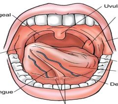
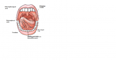
Label this Oral Cavity (Palatopharyngeal/glossal Arches) |
|
|
|
Other than the tongue, where may there be other Taste buds? |
On the Mucosal lining of the - Epiglottis - Posterior Oropharynx wall - Soft palate |
|
|
What are the innervation for the upper and lower lips? Cheeks? |
Upper Lip: Infraorbital nerve (Maxillary nerve) Lower lip: Mental nerve (Mandibular nerve) Buccal branches from the Facial nerve (Pharyngeal Arch 2. The Somatic Sensory component of Buccal is from the Mandibular nerve) |
|
|
What is the Somatic sensory and SOmatic motor innervations of the buccal nerve? What is the blood supply to the cheeks? |
Somatic sensory = Mandibular nerve Somatic Motor = Facial nerve Maxillary Artery, Buccal branch. |
|
|
What are the boarders of the Isthmus of the Fauces? What is the Isthmus of the Fauces? |
The palatopharyngeal and Palatoglossal arches (lateral) (Superior) Soft palate Inferior) Tongue The Isthmus of the Fauces is the short contricted space that connects the oral cavity with the oropharynx. |
|
|
What are the muscles that make up the Palatoglossal and palatopharyngeal arches and where do they attach? |
Palatoglossal Arch = Palatoglossal muscle. Attches to the side of the tongue. Palatopharyngeal Arch = Palatopharyngeal muscle. Attaches to the lateral side of the Oropharynx. |
|
|
The soft palate is movable fibromuscularposterior third of the palate and is suspended from the posterior border of the hard palate. It attachesby its aponeurotic part (palatineaponeurosis) to the posterior edge of the hard palate. Its sides blend withthe pharyngeal wall, and its inferior border is ..... what? |
uvula |
|
|
When a person swallows,the soft palate initially is tensed to allow the tongue to press against it,squeezing the bolus of food to the back of the mouth. The soft palate is thenelevated posteriorly and superiorly against the wall of the pharynx, therebypreventing passage of food into the nasal cavity. |
Fact |
|
|
Sphenopalatine nn (branches ofNasopalatine nn CNV2) afte they innervate the nasal septum, traverse theIncisive canal as ....which nerve |
incisive nerve to supply the hard palate |
|
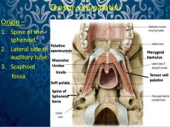
See where Levator Veli Palatine & Tensor Veli Palatine attach. What are all the Palatine muscles? What is the nerve that innervates the soft palate muscles? |
Levator palatine Veli attaches to the petrous part of temporal bone -> palatine apernousis (soft palate) Innervated b the Vagus nerve (CN X) Tensor Veli palatine attaches to the Medial pterygoid plate + Sphenoid spine + Cartilage of Euschian Tube. Innervated by Nerve to Medial Pterygoid (CNV3) Other muscles of the palatine are: Palatoglossal/pharyngeal and Uvulae. The innervations for the soft palate msucles is the Vagus Nerve pharyngeal branches |
|
|
What muscles/landmarks is the root of the tongue attached too? |
Hyoid bone, mandible, mylohyoid and Genuhyoid. |
|
|
WHAT nerve supplies taste to the posterior 1/3 of the tongue? Anterior 2/3rds? |
Posterior 1/3rd is supplied by the Glossopharyngeal Anterior 2/3rds by Chorda Tympani. |
|
|
General Visceral motor nerves to Salivary glands within the Oral Region: |
1. Chorda tympani CNVII preganglionic parasympathetic via lingualnerve innervates postganglionic parasympathetic Submandibular Ganglion and thenpostganglionic parasympathetic of Submandibular ganglion joining the Lingualnerve innervate Submandibular, Sublingual and Intralingual salivary glands andmucosal glands of the oral cavity. 2. Greater petrosal nerve CNVII pregagnlionicparasympathetic via the nerve of pterygoid canal innervates the postganglionicparasympathetic Pterygopalatine ganglion and then postganglionic parasympatheticaxons of this ganglion join the Greater and Lesser palatine nerves (CN VMaxillary nerve) to innervate the mucosal glands of the hard and soft palates. SSSSSSSSS5~WF |
|
|
What are the Extrinsic tongue muscles? Which muscle does the Hypoglossus nerve decend superfically to? |
Genioglossus Hypoglossus ** Styloglossus Palatoglossus (Vagus, Pharyngeal branches innervation) |
|
|
What are the Intrinsic muscles of the tongue? What is the innervation of all these muscles? What things pass deep to the Hypoglossus muscle? (3 things) |
Superior Longitudinal Inferior Longitudinal Transverse Vertical muscle. Hypoglossal 3 things that pass deep Hypoglossus muscle are the 1. Glossopharyngeal n 2. Stylohyoid ligament 3. Lingual A. |
|
|
What is the path of the hypoglossal nerve? |
Hypoglossalnerve CN XII leaves the cranium via the hypoglossal canal and passes deep tothe mandible to enter the tongue, where it supplies: all intrinsic andextrinsic lingual mm except the palatoglossus. CN XII is joined (distal tohypoglossal canal) by a branch conveying fibers from the C1 APR of cervicalplexus. These fibers hitch a ride with CN XII, leaving it as: a) superior rootof the ansa cervicalis/descending hypoglossi; b) nerve to thyrohyoid m; c)nerve to Geniohyoid muscle. The Hypoglossal nerve arises aspurely somatomotor nerve by several rootlets from the medulla and leaves thecranium through the hypoglossal canal. It emerges from its canal in a planemedial to the internal jugular vein , internal carotid artery, ninth, tenth andeleventh cranial nerves to the interval btw the internal jugular vein andinternal carotid artery. It has connection with the inferior vagal ganglion. Itbecomes superficial below the posterior belly of the digastric emerging btwinternal jugular vein and internal carotid artery. Then, it loops round theoccipital artery, crosses lateral to both internal and external arteries andthe loop of the lingual artery a little above the tip of the greater cornu ofthe hyoid bone. It inclines up and forwards on the hyoglossus by passing deep to the digastrics’ tendon,stylohyoid and the posterior border of the mylohyoid. At this point the nerve is inferior to thedeep part of the submandibular gland, submandibular duct and the lingual nerve.It then passes on the lateral aspect of the genioglossus, continuing forwards in its substance as far as thetip of the tongue and distributing fibers in this muscle. CN XII ends in manylingual branches that supply all intrinsic and the extrinsic muscles of thetongue (styloglossus, hyoglossus, and genioglossus, except the palatoglossus). v After exiting the cranial cavity, CN XII isjoined by the branches of the cervical plexus conveying general somatic motorfibers from C1 (small C2) spinal nerves and somatic general sensory fibers fromthe dorsal spinal ganglion C2. These spinal nerve fibers “hitch a ride” with CNXII to reach the hyoid muscles , with some of the sensory fibers passing retrograde along it to reach the dura materof the posterior cranial fossa. v A meningeal branch returns to the cranium through the hypoglossal canal and innervatesthe dura mater on the floor and posterior wall of the posterior cranial fossa.The nerve fibers conveyed are from the sensory spinal ganglion of spinal nerveC2 and are NOT hypoglossal nerve. v Branches continue:o Descending hypoglossi or superior root of the ansacervicalis (C1) leaves the hypoglossal nerve were it curves round the occipitalartery and then descends anterior to or in the carotid sheath. It contains only fibers from the first cervicalspinal nerve. After giving a branch to the superior belly of omohyoid, it isjoined by the inferior root of the ansa from the second and third cervicalspinal nerves. The two roots form the ansa cervicalis from which branchessupply the sternohyoid, sternothyroid, and inferior belly of the omohyoid. o The nerves to the thyrohyoid andgeniohyoid arisenear the posterior border of the hyoglossus and cross obliquely the greatercornu of the hyoid to supply the thyrohyoid and geniohyoid; they contain thefibers of the APR of C1 spinal nerve.5V.&F@~Z@ |
|
|
What does the Ans Cervicalis supply? |
Superior belly of Omohyoid, Sternohyoid, Sternothyroid, and inferior belly of omohyoid. Also Geniohyoid and Thyrohyoid. |
|
|
Which branches of the Lingual artery do not communicate with each other in the tongue? |
o Lingual artery brings the chief supply to the tongue and oral floorof the mouth, arises anteromedially from the external carotid artery oppositethe tip of the hyoid’ greater cornu. It passes medial/deep to the posteriorborder of hyoglossus muscle and then deep to it, and courses sinuouslyforwards on the tongue’s inferiorsurface as far as its tip. Branches:sublingual a, suprahyoid, and terminal: dorsal lingual and deep lingual aa. o Dorsal lingual aa supply the Root ofthe tongue; and Deep lingual aa supply the body of the tongue. o The deep lingual aa communicate witheach other near the apex of the tongue. o The dorsal lingual aa are prevented fromcommunicating by the lingual septum. |
|
|
When the Hypoglossal Nerve is lessioned, what side will the Apex of the tongue deviate too? |
The paralytic side, because of the actions of the Genioglossus muscle. |
|
|
Nerve supply to parts of Tongue: Sensory Nerves: 1. Lingual nerve : somatosensory toanterior 2/3 of tongue 2. Chorda tympani via Lingual nerve:special visceral sensory for taste from anterior 2/3 of tongue 3. Glossopharyngeal n CN IX (lingualbranches) : somatosensory and special visceral for taste to posterior 1/3 oftongue, including papillae vallatae 4. Vagus CN X (Internal laryngeal nerve): somatosensory and special visceral for taste to posterior 1/3 of tongue closeto epiglottis (epiglottis and oropharynx) ; Motor nerves: 1. Hypoglossal nerve CN XII, somatomotorcomponent : to all intrinsic and extrinsic tongue muscles except palatoglosssus muslce;2. Vagus CN X (pharyngeal branch viapharyngeal plexus), branchiamotor component, from branhial/pharyngealmesodermal arch ## 4-6: to Palatoglossus muscle.; |
FACT |
|
|
What are the three Pharyngeal constrictor muscles? What are they innervated by? What are their attachments? |
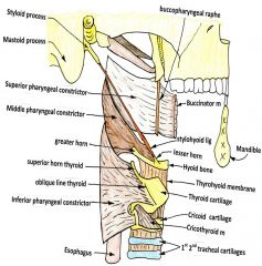
Superior Pharyngeal constrictor -> Attaches to the Pterygomanidular raphe (Line after Buccinator) and attaches to the Pharyngeal raphe Middle Pheryngeal constrictor -> Attaches to the Pharyngeal raphe and the Hyoid Bone. Inferior Pharyngea lraphe -> Attaches to the Pharyngeal raphe and the Cricoid and Thyroid cartilage. All three are innverated by the Pharyngeal branches of the Vagus nerve. |
|
|
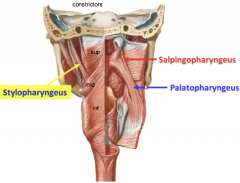
What are the three Pharyngeal Diolatorss? What are their origins, insertions and innervations? |
Palatopharyngeus - Hard palate & Palatine aponeurosis to the side of pharynx. CN X brachial motor component Salpingopharyngeus - Attaches to cartilaginous part of Eustichian tube to the side of pharynx. CN X brachial motor component Stylopharyngeus - attaches the styoloid process to in between inferior and superior pharyngeal constrictors. Innervated by the CN IX, brachialmotor component. |
|
|
Nasopharynx is home to which tube? |
Eustachian's tube. Muscle that attaches to the carillagenous portion of this tube is the Salpingopharyngeus. The muscle opens the tube while sqallowing to help equilize pressure in the ear. |
|
|
What is Waldeyer’s lymphatic ring: compossed of? How many tonsils of the following; Nasopharynx tonsil, Tubular tonsils, Lingual tonsils, Palatine tonsils? |
Waldeyer’s lymphatic ring:1.Lingual tonsil (one)2.Palatine tonsils (two)3.Tubal tonsils (two)4.Pharyngeal tonsil=adenoids (at junction of roof &posterior wall of nasopharynx) (one). |
|
|
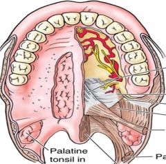
What is the blood supply to the tonsils? |
Tonsil’sblood supply: 1.Ascending pharyngeal a (Ext. carotid a) 2.Ascending palatine a (Facial a) 3.Tonsillar aa ( Facial a) |
|
|
How will you identify the Internal Laryngeal nerve (Branch off the Superior Laryngeal Nerve) How will you identify the External Laryngeal nerve (Branch off the Superior Laryngeal Nerve) How will you identify the recurrent Laryngeal nerve (Branch off the Superior Laryngeal Nerve) |
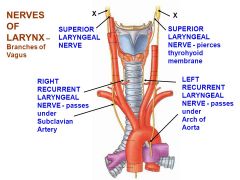
It pierces b/w the superior and Middle Laryngeal Constrictor muscles, providing somato-sensory branches laryngeal mucosa down to the vocal folds The external laryngeal nerve also branches off the Superior Pharyngeal nerve and perforates the inferior pharyngeal constrictor. It supplies the Cricothyroid Muscle The recurrent laryngeal nerve is a branch off the Vagus nerve that supplies the intrinsic muscles, including the vocal cords of the Larnyx except the Cricothyroid muscle |
|
|
Where is the palatine fossa situated b/w? |
The palatine fossa is situated between the Paloglossal arch, and the palatopharyngeal arch. |
|
|
At what level does the Pharynx bec0me the Epiglotis? |
C6 |
|
|
what brain nuceli contains the cell bodies of nerves that deliver Brachial Motor innervation to the muscles of the soft palate,pharynx, and larynx which are strongly associated with speech and swallowing. |
the nucleus ambiguous which arries from the medulla and roots from the accessory nerve |
|
|
What 3 muscles are supplied by the Medial Pterygoid nerve, from the Mandibular branch? |
Tensot Veli Palatini, Tensor Tymapni, Medial pterygoid. |
|
|
What are the Nucela associated with the Vagus nerve? (4 Nuceli in Medulla, 3 ganglia) |
Afferent General Somatic Sensory from the Auricle (Meningeal and Auricular branches) -> Superior Vagal ganglia -> Spinal trigemental Nuceli Afferent Sepecial visceral sensory + General Visceral sensory from the Trachea mucosa, Soft palate, Oropharynx, Root of tongue, Larynx, esophagus and thoracic viscera -> Inferior Vagal ganglia -> Solitarius nuceli Special visceral motor from Nuceli Ambiguous --> Muscle of soft palate (except Tensor Veli palatine), muscle of pharynx, muscles of larynx. General visceral motor preganglionic parasympathetic fibers from the Dorsalis nucelus --> glands and smooth muscle of the trachea/larynx/pharynx. |
|
|
What artery does the vertebral artery branch from? What artery does the Inferiorthyroid artery branch from? |
THe veretebral artery branches off the subclavian artery. The Inferior thyroid artery is a branch off the Thyrocervical trunk that branches from the Subclavian aa off of the Brachiocephalic artery. |
|
|
At what level does the Common carotid artery split? What branches come off the External carotid artery? The internal carotid artery? |
The coomon cartoid splits at the level of C4, where chemo (CAROTID body) and baroreceptors (Carotid sinus) The BODY is innervated by the Vagus nerve. The SINUS is innervated by the Glossopharyngeal nerve, which relays information fro the NTS (Nucelous tractus soltarius) to modulated blood flow and reads 02 content in blood. Off the external carotid Artery branches the Superior thyroid artry to supply the anterior aspect of the Thyroid cartilage and the Superior Laryngeal branch. Superior to these branches, branch above the hyoid bone is the Superior Hyoid Branch, the Deep Lingual artery and the Sublingual Arteries. The next main branch off the External Carotid Artery is the Facial Artery, Posterior Auricular, Occipital branch, and Maxillary Branch Off the Internal Carotid artery is the Opthalmic artery to supply the eyes. |
|
|
What major artery is the Facial artery branched from? |
External Carotid Artery |