![]()
![]()
![]()
Use LEFT and RIGHT arrow keys to navigate between flashcards;
Use UP and DOWN arrow keys to flip the card;
H to show hint;
A reads text to speech;
147 Cards in this Set
- Front
- Back
|
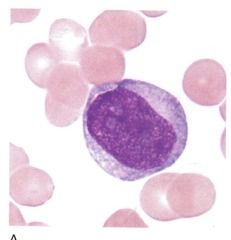
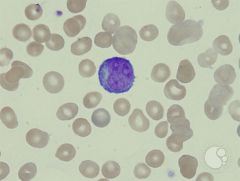
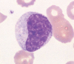
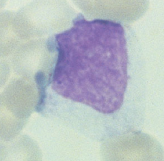
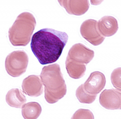
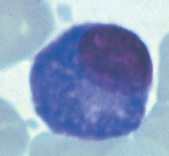
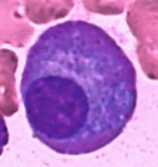
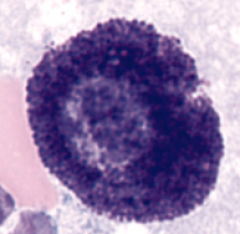
Myeloblast Cellsize: 12-20 um N/Cratio 4:1•Round-ovalnucleus, “round refrigerator corners”*•Centralor eccentric nucleus•Lightred-purple fine meshwork chromatin with no aggregation of material •1-5nucleoli*•Scantybasophilic cytoplasm•Nogranules•Maycontain Auer rods |
|
|
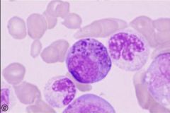
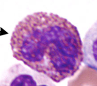
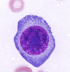
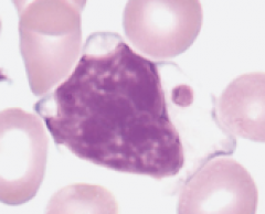
Promyelocyte (pretty much a blast with granules) •Coarse azurophilic nonspecific (primary) granules* •Cellsize: 15-23 um•N/Cratio 3:1•Round-ovalnucleus •Lightred-purple fine meshwork chromatin, possibly slight aggregation at nuclearmembrane•1-3nucleoli•Basophiliccytoplasm (may be slightly increased over blast) |
|
|
Myelocyte•Cellsize: 10-18 um•N/Cratio 2:1 or 1:1•Moreround-oval nucleus*•Centralor eccentric nucleus•Red-purplefine chromatin with slightly aggregated pattern*•Mayor may not have nucleoli Moderatebluish - pink cytoplasm nonspecific(primary=purple) and specific (secondary) granules* |
|
|
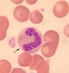
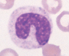
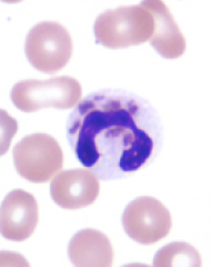
Metamyelocyte •Cellsize: 10-18 um•N/Cratio 1:1•Indented(kidney shaped) nucleus* •Centralor eccentric nucleus•Lightblue-purple with chromatin clumps easily distinguishable•Nonucleoli•Moderatebluish - pink cytoplasm (occasionally clear pink)•Specific(secondary) granules (few non-specific)• |
|
|
Band•Cellsize: 10-16 um•N/Cratio 1:1•Elongatednarrow band shape of uniform thickness, singular lobe*•Centralor eccentric nucleus•Deepblue-purple coarsely granular nuclear chromatin (distinctparachromatin)•Nonucleoli•Abundantpink cytoplasm•Fineviolet-pink specific granules• |
|
|
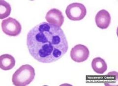
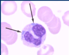
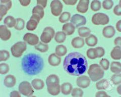
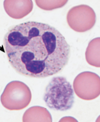
PMN•Cellsize: 10-16 um•N/Cratio 1:1•2-5distinct nuclear lobes (filament connecting lobes)*•Centralor eccentric nucleus•Deepblue-purple coarsely granular nuclear chromatin (distinct parachromatin)•Nonucleoli•Abundantpink cytoplasm•Fineviolet-pink specific granules |
|
|
Basophilic Myelocyte |
|
|
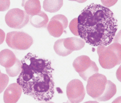
Eosinophilic Myelocyte |
|
|
Monoblast•Cellsize: 12-20 um•N/Cratio 4:1•Round-oval,or slightly folded nucleus•Centralor eccentric nucleus•Palered-purple, minimal, fine threadychromatin •Usually1-2 nucleoli•Moderatebasophilic cytoplasm, regular border•Nogranules Maycontain Auer rods |
|
|
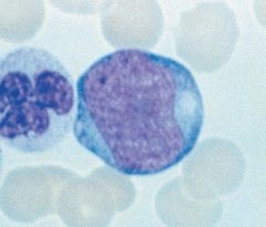
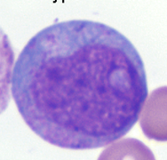
Promonocyte•Cellsize: 10-16 um•N/Cratio 3:1 or 2:1•Roundnucleus with chromatin creases or cerebriformfolding more distinct•Centralor eccentric nucleus•Palered-purple, very fine pattern, (aerated network of threads)•0-2nucleoli•Paleropaque gray-blue cytoplasm, more abundant, may see pseudopodia and vacuoles |
|
|
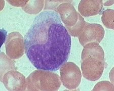
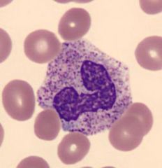
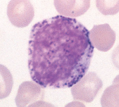
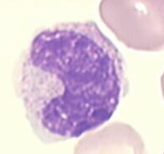
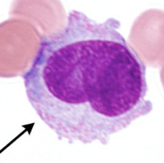
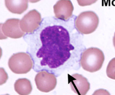
Monocyte•Cellsize: 12-20 um•N/Cratio 2:1 or 1:1•Cerebriformor horseshoe shaped nucleus•Centralnucleus•Blue-purple,fine reticular nuclear chromatin (less distinct parachromatin)•0-2Nucleoli •Abundantpale gray-blue cytoplasm, may seepseudopodia & vacuoles•Morenumerous very fine (dust like) red granules evenly dispersed• |
|
|
Megakaryoblast |
|
|
Promegakaryocyte |
|
|
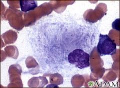
Megakaryocyte |
|
|
Thrombocytes |
|
|
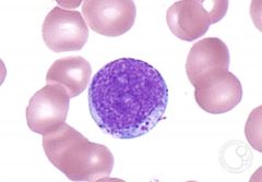
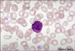
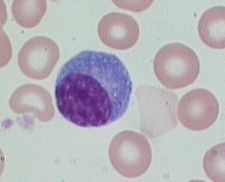
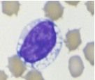
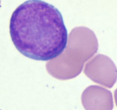
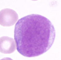
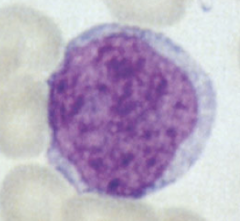
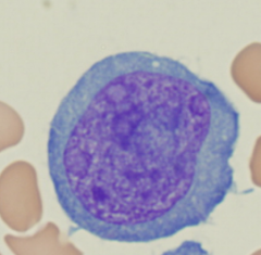
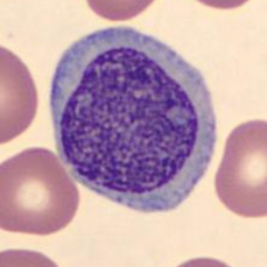
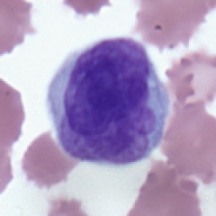
Lymphoblast•Cellsize: 10-20 um•N/Cratio 4:1•Roundor oval nucleus•Centralor eccentric nucleus•Undifferentiatedsparse red purple chromatin •1-2indistinct nucleoli•Scanty,often nearly absent, clear basophilic cytoplasm* •Nogranules |
|
|
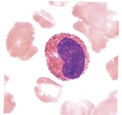
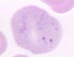
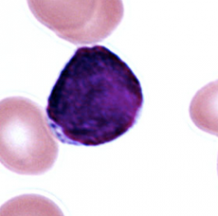
Prolymphocyte•Cellsize: 9 - 18 um•N/Cratio 4:1 (occasionally 3:1)•Roundor oval and flat nucleus•Centralor eccentric nucleus•Combinationof condensed clumped blue-purple chromatin with red-purple parachromatin*•0-1more distinct nucleoli*•Oftenscanty, but visableclear basophilic cytoplasm •Nogranules• |
|
|
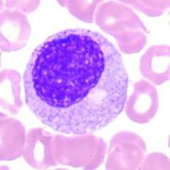
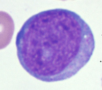
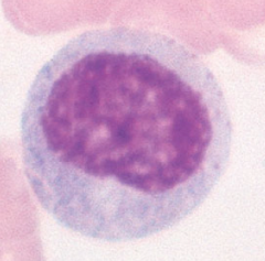
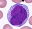
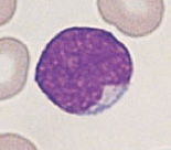
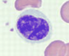
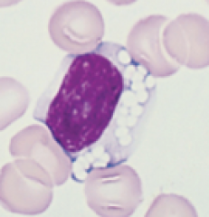
Lymphocyte•Cellsize: 7-18 um (usually 9-12)•N/Cratio 4:1 (occasionally 3:1)•Roundor indented nucleus•Eccentricnucleus often with scanty cytoplasm •Homogeneous,coarse blue-purple nuclear chromatin (smudged)•Nucleoliusually absent , rarely one seen in mature forms•Lightto dark blue cytoplasm, scanty to moderate•Occasionallya few azurophilicgranules seen• |
|
|
Reactive Lymphocyte |
|
|
Plasmacytoid Cell |
|
|
Plasma Cell•Cellsize: 8 - 20 um•N/Cratio 2:1 or 1:1•Roundor oval nucleus•Usuallyeccentric nucleus•Blue- purple, dense chromatin w/ largeclumps near nuclear margin•Nonucleoli•Moderate,basophilic cytoplasm, with perinuclearclear zone adjacent to nucleus, may contain vacuoles•Nogranules (may see Russell Bodies)• |
|
|
Flame Cell Brilliantred staining cytoplasm (Flame Cell, produces IgA) |
|
|
Mott Cell (IG's stuck in there) |
|
|
Auer Rods (maligancy/malformed granules) |
|

|
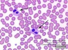
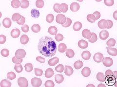
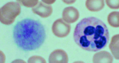
Hypersegmented neutrophils (megaloblastic change) will also see macrocytic anemia |
|
|
Pelger Huet (congenital) hyposegmentation |
|
|
Toxic granulation (too few cell divisions) shift to the left from infection |
|
|
Toxic vacuoles (response to infection) |
|
|
Dohle bodies (leftover RNA) associated with a left shift |
|
|
May-Hegglin- Dole like bodies+giant platelets |
|
|
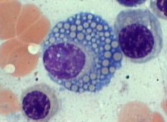
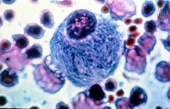
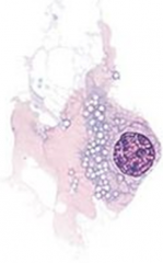
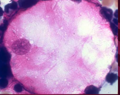
Foam cell (Nieman-Pick) macrophages accumulate fats |
|
|
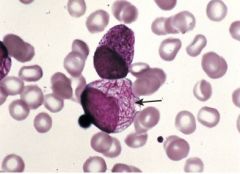
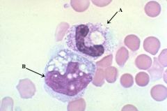
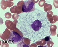
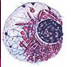
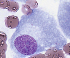
Gaucher cell (lipid storage disorder) •Inabilityto degrade glucocerebroside= accumulation in monocytesand macrophages Deficient enzyme: beta-glucocerebrosidase |
|
|
Gaucher cell (lipid storage disorder) •Inability to degrade glucocerebroside = accumulation in monocytes and macrophages Deficient enzyme: beta-glucocerebrosidase |
|
|
May-Hegglin- Dole like bodies+giant platelets |
|
|
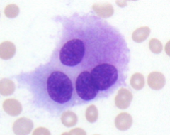
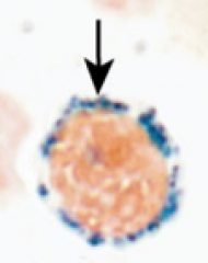
Tay-Sachs (vacuolated lymphs) congenital nerve disease |
|
|
Tay-Sachs (vacuolated lymphs) congenital nerve disease |
|
|
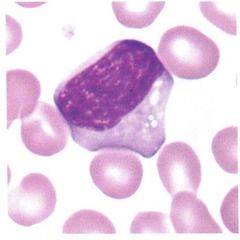
Promyelocyte |
|
|
Myelocyte •Cell size: 10-18 um •N/C ratio 2:1 or 1:1 •More round-oval nucleus* •Central or eccentric nucleus •Red-purple fine chromatin with slightly aggregated pattern* •May or may not have nucleoli Moderate bluish - pink cytoplasm nonspecific (primary=purple) and specific (secondary) granules* Chromatin=white spaces |
|
|
Which blast cell(s) can contain Auer Rods? What are the rods? Example of a pathology: |
Monoblast Myleoblast Abnormally formed lysosomal granules Found in pathologies: acute myeloid leukemia (AML) |
|
|
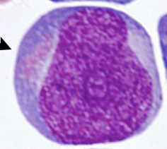
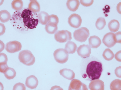
MyeloblastCell size: 12-20 um N/C ratio 4:1 •Round-oval nucleus, “round refrigerator corners” * •Central or eccentric nucleus •Light red-purple fine meshwork chromatin with no aggregation of material •1-5 nucleoli* •Scanty basophilic cytoplasm •No granules •May contain Auer rods |
|

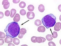
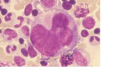
|
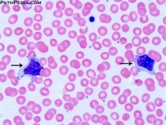
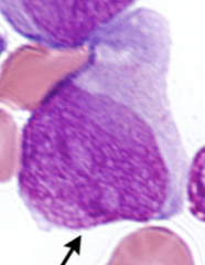
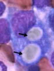
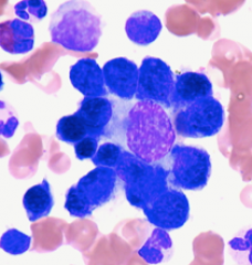
Myeloblast with Auer Rods (top left) MyeloblastCell size: 12-20 um N/C ratio 4:1 •Round-oval nucleus, “round refrigerator corners” * •Central or eccentric nucleus •Light red-purple fine meshwork chromatin with no aggregation of material •1-5 nucleoli* •Scanty basophilic cytoplasm •No granules |
|
|
What is the lineage of the granulocytic series? Also, what's the other name for it? |
Granulocytic (or Myelocytic series) Myleoblast Promyelocyte Myelocyte Metamyelocyte Band Neutrophil |
|
|
Promyelocyte (pretty much a blast with granules) •Coarse azurophilic nonspecific (primary) granules* |
|
|
Type I blast (myeloblast) is devoid of granules and referred to as an agranularblast. |
|
|
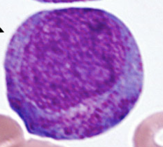
Type II blast (myeloblast) has <20 granules and is referred to as granular blast |
|
|
Type III blast (myeloblast) has >20 granules but is still an immature cell thatdoes not fit the definition of a promyelocyte. Also referred to as granular blast. |
|
|
Pappenheimer bodies |
|
|
Myelobast Type II |
|
|
Myelobast |
|
|
PROMYELOCYTE |
|
|
Myelocyte •Cell size: 10-18 um •N/C ratio 2:1 or 1:1 •More round-oval nucleus* •Central or eccentric nucleus •Red-purple fine chromatin with slightly aggregated pattern* •May or may not have nucleoli Moderate bluish - pink cytoplasm nonspecific (primary=purple) and specific (secondary) granules* |
|
|
Metamyelocyte •Cell size: 10-18 um •N/C ratio 1:1 •Indented (kidney shaped) nucleus* •Central or eccentric nucleus •Light blue-purple with chromatin clumps easily distinguishable •No nucleoli •Moderate bluish - pink cytoplasm (occasionally clear pink) •Specific (secondary) granules (few non-specific) • White spaces=chromatin |
|
|
Metamyelocyte •Cell size: 10-18 um •N/C ratio 1:1 •Indented (kidney shaped) nucleus* •Central or eccentric nucleus •Light blue-purple with chromatin clumps easily distinguishable •No nucleoli •Moderate bluish - pink cytoplasm (occasionally clear pink) •Specific (secondary) granules (few non-specific) White spaces=chromatin |
|
|
Band •Cell size: 10-16 um •N/C ratio 1:1 •Elongated narrow band shape of uniform thickness, singular lobe* •Central or eccentric nucleus •Deep blue-purple coarsely granular nuclear chromatin (distinct parachromatin) •No nucleoli •Abundant pink cytoplasm •Fine violet-pink specific granules |
|
|
Eosinophilic band |
|
|
What is a Leukomoid Reaction |
Alteration in Granulocyte # & Maturity Transient,reactive condition characterized by moderate to severe increased WBC and a Shift to the Left (increasedimmature granulocytes in blood) –Infection –Physiologicalleukocytosis |
|
|
Neutrophils in the peripheral blood exist ineither the ___________ or the __________ pool. |
Neutrophils in the peripheral blood exist ineither the circulating or the marginating pool. Themarginated neutrophils roll along a vessel wall, (surface carbs interact with selectins on the endothelial cell). Afteractivation, they change shape and affinity of their integrin molecules for endothelial cellintercellular adhesion molecules. The neutrophils then crawl and undergo diapedesis by interacting withplatelet-endothelial cell adhesion molecules on the endothelial surface and byliberating hydrolases that permit passage of the neutrophils through the capillary basementmembrane. |
|
|
Which white blood cells of the peripheral blood is actually considered immature? |
Monocytes Afteronly a few hours in the blood it migrates into the tissues and matures furtherinto the macrophage or histiocyte. |
|
|
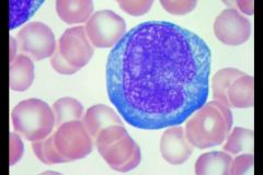
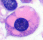
Monoblast Although almost identical to a myeloblast, mono blasts can be identified by the presence of a single large nucleolus, an irregularly shaped nucleus and a delicate chromatin May have Auer rods |
|
|
Monoblast Although almost identical to a myeloblast, mono blasts can be identified by the presence of a single large nucleolus, an irregularly shaped nucleus and a delicate chromatin May have Auer rods |
|
|
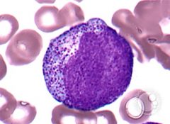
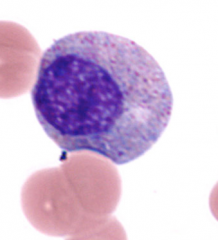
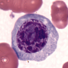
Promonocyte •Cell size: 10-16 um •N/C ratio 3:1 or 2:1 •Round nucleus with chromatin creases or cerebriform folding more distinct •Central or eccentric nucleus •Pale red-purple, very fine pattern, (aerated network of threads) •0-2 nucleoli •Paler opaque gray-blue cytoplasm, more abundant, may see pseudopodia and vacuoles |
|
|
What are the two categories of macrophages? |
1. Free–foundin sites of inflammation and repair –Bodyfluids 2. Fixed– tissue macrophages –Foundin specific sites –Ex: CNS – microglialcells Liver – Kupffercells |
|
|
Lymphoblast |
|
|
Lymphoblast |
|
|
"small lymphoblast" |
|
|
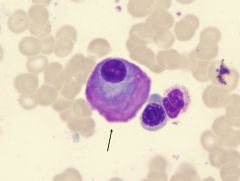
Prolymphocyte •Cell size: 9 - 18 um •N/C ratio 4:1 (occasionally 3:1) •Round or oval and flat nucleus •Central or eccentric nucleus •Combination of condensed clumped blue-purple chromatin with red-purple parachromatin* •0-1 more distinct nucleoli* •Often scanty, but visable clear basophilic cytoplasm •No granules • |
|
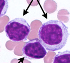
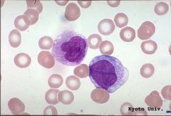
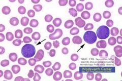
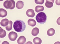
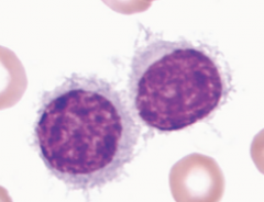
What are the top two cells (Same)? What is the bottom? |
Top two = prolymphocytes bottom = lymphocytes |
|
|
If a B lymphoblast produces antibody it is called a ________________. |
Plasmablast |
|
|
If a B-prolymphocyte produces antibody it is called a _______________. |
Proplasmacyte |
|
|
What is the normal development series of plasma cells? |
1.B-lymphoblast ---> 2.B-Prolymph---> 3. B-lymph---> 4. Plasmacytoid lymph ---> (Ab production)---> 5. Plasma cell |
|
|
What is the abnormal development series of plasma cells? |
Plasma blast---> (Ab production)---> Proplasmacyte---> (Ab production)---> Plasma cell |
|
|
Plasmablast •Cellsize: 16-18 um•N/Cratio 4:1•Roundnucleus•Centralor eccentric nucleus•Palered- purple, fine stippled chromatin •Usually1-3 nucleoli•Scantyto moderate, paler bluethan more mature forms, occasionally perinuclearclear zone •Nogranules• |
|
|
Proplasmacyte •Cellsize: 15 -25 um•N/Cratio 3:1•Roundor oval nucleus•Centralor eccentric nucleus•Red-purple, increased granularity of chromatin •Usually1-3 nucleoli•Moderate,basophilic cytoplasm, may or may not have perinuclearclear zone adjacent to nucleus•Nogranules• |
|
|
Plasma Cell •Cell size: 8 - 20 um •N/C ratio 2:1 or 1:1 •Round or oval nucleus •Usually eccentric nucleus •Blue - purple, dense chromatin w/ large clumps near nuclear margin •No nucleoli •Moderate, basophilic cytoplasm, with perinuclear clear zone adjacent to nucleus, may contain vacuoles •No granules (may see Russell Bodies) • |
|
|
Incertain pathological states and when manufacturing immunoglobulins, the plasmacells may undergo striking morphological changes. These include: |
•Flame Cell •Russell Bodies •Giant cell size (> 20um) •Multinucleated forms •Rouleaux formation of RBCs |
|
|
Russell Bodies Redor white globular inclusions of Ig in the cytoplasm. Maybe so voluminous as to obliterate the other cell structures (mott cell, morulacell or grape cell) May also be in crystals or rods |
|
|
Russell Bodies Red or white globular inclusions of Ig in the cytoplasm. May be so voluminous as to obliterate the other cell structures (mott cell, morula cell or grape cell) May also be in crystals or rods |
|
|
Russell Bodies Red or white globular inclusions of Ig in the cytoplasm. May be so voluminous as to obliterate the other cell structures (mott cell, morula cell or grape cell) May also be in crystals or rods |
|
|
Russell Bodies Red or white globular inclusions of Ig in the cytoplasm. May be so voluminous as to obliterate the other cell structures (mott cell, morula cell or grape cell) May also be in crystals or rods |
|
|
Russell Bodies Red or white globular inclusions of Ig in the cytoplasm. May be so voluminous as to obliterate the other cell structures (mott cell, morula cell or grape cell) May also be in crystals or rods |
|
|
Mott cell |
|
|
Flame Cell Brilliant red staining cytoplasm (Flame Cell, produces IgA) |
|
|
Dohle bodies are made of what? What pathologies are associated? Which cells can have them? |
Remnant RNA Severe infection and tissue destruction. Shift to left granulocytes and monocytes |
|
|
What does dysplasia mean? |
1. abnormality of development. 2. in pathology, alteration in size, shape, and organization of adult cells=dysplastic |
|
|
What does megaloblastic mean? |
AbnormalDNA synthesis, usually due to vitamin B12 or folatedeficiencies Results in delayed nuclear development, causing larger cells |
|
|
What are the three cellular pools? |
Stem cell pool Bone marrow pool Peripheral blood pool |
|
|
Name this: Transient reactive condition with increased WBCs and a shift to the left. Response to infection. |
Leukomoid reaction |
|
|
List the stages of lymphopoiesis in the order of maturation. |
Lymphoblast Prolymphocyte Lymphocyte |
|
|
An activated B lymph is called a... |
plasmacytoid lymph |
|
|
HUMORAL: An activated B lymph (plasmacytoid) goes on to be either a _______ cell to create Ab OR a _________ cell (memory). |
An activated B lymph (plasmacytoid) goes on to be either a PLASMA cell to create Ab OR a Sensitized B-lymph cell (memory). |
|
|
What are mott cells |
The inclusions of Mott cells represent immunoglobulins within vesicular structures. These inclusions are Russell bodies which are dilated endoplasmic reticulum cisternae containing condensed immunoglobulins (Ig). |
|
|
State the function of the following cell types: Segmented neutrophil |
They are the body's primary defense against bacterial infection and physiologic stress. |
|
|
State the function of the following cell types:Eosinophil |
Associated with allergies and fight multicellular parasites. |
|
|
State the function of the following cell type: Basophil |
Generate histamine, heparin like substances and other mediators of inflammatory response. |
|
|
State the function of the following cell type: Monocyte |
Phagocytic cell that destroys extracellular organisms, some viruses, and old, mutated or damaged cells. |
|
|
State the function of the following cell type: Macrophage |
Wound repair.Process foreign antigens and present them to lymphs. |
|
|
State the function of the following cell type: Free Macrophage |
Circulating macrophage in body fluids. |
|
|
State the function of the following cell type: Fixed Macrophage |
In connective tissues, lymph nodes, spleen, and bone marrow. |
|
|
State the function of the following cell type: B-lymphocyte |
B-Lymphocyte responsible for synthesis and excretion of antibodies |
|
|
State the function of the following cell type: T-lymphocyte |
responsible for producing many substances which regulate the activities of other cells in the immune response |
|
|
State the function of the following cell type: Plasma cell |
Plasma Cell A fully differentiated B cell that produces a single type of antibody. |
|
|
List causes of absolute increases and decreases in number of the following cells: Increase in neutrophils: Decrease in neutrophils: |
Increase in neutrophils: infections Decrease in neutrophils: prolonged infections |
|
|
List causes of absolute increases and decreases in number of the following cells: Increase in eosinophils: Decrease in eosinophils: |
Increase in eosinophils: helminth infections Decrease in eosinophils: not that common |
|
|
List causes of absolute increases and decreases in number of the following cells: Increase in basophils: Decrease in basophils: |
Increase in basophils: allergies Decrease in basophils: not common |
|
|
List causes of absolute increases and decreases in number of the following cells: Increase in monos: Decrease in monos: |
Increase in monos: mononucleosis, chronic inflammation Decrease in monos: acute inflammation, stress, aplastic anemia |
|
|
List causes of absolute increases and decreases in number of the following cells: Increase in lymphs: Decrease in lymphs: |
Increase in lymphs: viral infections Decrease in lymphs: ALL or prolonged viral illnesses |
|
|
List the reference range for plasma cells in peripheral blood and bone marrow. |
Not usually seen in peripheral blood. Make up ~5% of bone marrow. . |
|
|
Define: Petechia purpura endomitosis |
Bleeding lesions under the skin. Petechia small, purpura and endomitosis large. Endomitosis - form of mitosis that lacks telophase and cytokinesis (separation into daughter cells); Unique to Megakaryocytes |
|
|
Alder- Reilly Inclusions Hematologicalmanifestationof agroup of inherited recessivedisorders–Deficiencyin enzymes to break down mucopolysaccharidesaccumulate in WBCs–Hurlerssyndrome, Hunters syndrome and other varientsof “gargoylism”–•Prominentred to purple granules mayappear in allwbctypes (occsurrounded by a halo) –Maybe difficult to distinguish from toxic granulation (Manypatients diebefore age 10 years) |
|
|
Alder- Reilly Inclusions seen in Hurler's anomaly –Hurlers syndrome, Hunters syndrome and other varients of “gargoylism” Hematological manifestation of a group of inherited recessive disorders –Deficiency in enzymes to break down mucopolysaccharides accumulate in WBCs |
|
|
Chediak- Higashi Syndrome Rareautosomalrec disorder: giant lysosomesin most cells of the body. HematologicalManifestations–Abnormallyformed lysosomalgranules in wbc. –Decreasedplts w/ abnormal large granulesanddefective function–•Increasedsusceptibility to infections and bleeding problems=shortened lifespan. |
|
|
Chediak - Higashi Syndrome Rare autosomal rec disorder: giant lysosomes in most cells of the body.Hematological Manifestations –Abnormally formed lysosomal granules in wbc. –Decreased plts w/ abnormal large granules and defective function – •Increased susceptibility to infections and bleeding problems=shortened lifespan. |
|
|
Chediak - Higashi Syndrome Rare autosomal rec disorder: giant lysosomes in most cells of the body.Hematological Manifestations –Abnormally formed lysosomal granules in wbc. –Decreased plts w/ abnormal large granules and defective function – •Increased susceptibility to infections and bleeding problems=shortened lifespan. |
|
The presence of this inclusion and the bizarre plt = which disorder? |
May Hegglin Anomaly •RareAutosomal Dominant condition Hematologicalmanifestations–Dohlelike bodies. Platelets: Decreased, Giantand Bizzarreforms •Atrisk for infections and bleeding, but doesn’t usually lead to early death. |
|
The presence of this inclusion and the bizarre plt = which disorder? |
May Hegglin Anomaly RareAutosomal Dominant condition Hematologicalmanifestations–Dohlelike bodies–Platelets•Decreased•Giantand Bizzarreforms Atrisk for infections and bleeding, but doesn’t usually lead to early death. |
|
|
How do they diagnose: Chronic Granulomatous Disease |
Diagnosis:–Nitro blue tetrazolium test •Normalcells can reduce the water soluble dye and form a precipitate Treatment:prophylactic antibiotics, bone marrow transplant |
|
|
What is: Chronic Granulomatous Disease? |
PMNscan phagocytize bacteria - but defectin enzyme (NADPH oxidase) responsible for respiratory burst and generation ofsuperoxide to kill them. Patientsusuallydie frombacterialinfection from5- 7 years old. |
|
|
What is: Myeloperoxidase (MPO) Deficiency |
Neutrophilshave decreased enzyme resulting in longer killing time of bacteria Genemutation (Relativelymild in most patients) |
|
|
What are two Diabetesmellitus -Associated Dysfunctions? |
•Poor neutrophil function •High glucose levels results in abnormal oxidative burst. |
|
|
What is "LeukocyteAdhesion Disorder"? |
•Inability of neutrophils and monocytes to adhere to endothelial cells and migrate fromblood into the tissues. •Gene mutation •Increased and potentially lethal bacterial infections. |
|
|
What is LazyLeukocyte Syndrome? |
•Cells have poor directional and random movement •Patientshave a history of recurrent infections |
|
What's Hairy Cell Leukemia? What are the lab findings? |
•Few too many “hairy cells” - hairy cytoplasmic projections... Confused cell– B-lymph origin but has features of monocytes Lab Findings: splenomegally, moderate to severe pancytopenia, WBCoften less that 4,000/ mm3, often “dry tap" and TRAPpositive |
|
What is Sezary Syndrome? |
Sezary cells–malignant cells that appear as mature lymphs with convoluted, cerebriform nuclear folds. |
|
|
Sezary cells – malignant cells that appear as mature lymphs with convoluted, cerebriform nuclear folds. |
|
|
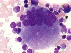
What disease is characterized by: Cellularappearance of Macrophages: crumpledsilk, striated, chickenscratch |
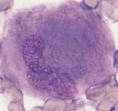
Gaucher's disease, duh. Lipid storage disorder, Deficientenzyme: beta-glucocerebrosidase |
|
|
What disease is characterized by: Inabilityto degrade sphingomyelin= accumulates in spleen, liver, lungs, brain, bone marrow |
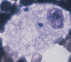
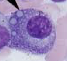
Niemann- Pick Disease (Lipid storage disorder) Foam cell Deficientenzyme: sphingomyelinase |
|
|
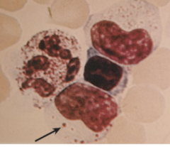
What disease is characterized by: Accumulationof unmetabolizedgangliosidein almost all tissues |
Tay- Sachs Disease (Lipid storage disorder) VacuolatedLymphs Deficientenzyme: HexosaminidaseA |
|

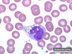
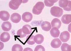
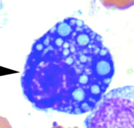
This type of cell abnormality is seen in which lipid storage disorder? |
Tay-Sachs Disease Vacuolated Lymphs Deficient enzyme: Hexosaminidase A |
|
|
List reference ranges for WBC, RBC, and Plt counts |
WBC: 4.4-11.0 x 10³/mm3 Platelets: 150-400 x 103/mm3 |
|
|
State the volume in mm3 of any primary, secondary or tertiary squares. |
Primary 0.1 mm3 (For WBCs) Secondary 0.004 mm3 (For RBCs) Tertiary 0.00025 mm3 |
|
|
State the typical dilutions used for WBC, RBC, Plt. |
WBC 1:100 Plt 1:100 RBC 1:200 |
|
|
Calculate the WBC, RBC, and Plt count. |
Av. # of cells x dilution factor x depth (10) _______________________________________________
(Number of primary squares counted) |
|
|
Express the WBC, RBC, and Plt counts in scientific notation with the correct number of significant digits and correct units. |
WBC - one decimal place x 103/ mm3 RBC - two decimal places x 106 / mm3 Eos - whole number / mm3 Plt - no decimals x 103 / mm3 |
|
|
Osteoclast •responsiblefor bone reabsorption•mostresembles a megakaryocyte–itis multinucleated and nuclei are very round and clearly separate from oneanother |
|
|
Osteoblast •boneforming cell•mostresembles a plasma cell–slightlylarger–nucleusso eccentric as to appear almost extruded–haloseparate from nucleus |
|
|
FatCell |
|
|
MastCell (Tissue Basophil) •Morphologysimilar to blood basophils •Somethink it may arise from a separate stem cell line •Increasedin –chronicinfections–autoimmunediseases–systemicmastocytosis |
|
|
nonnucleatedrbcwith Fe granules: |
•siderocyte- nonnucleatedrbcwith Fe granules |
|
|
nucleated rbcwith Fe granules: |
•sideroblast- nucleated rbc with Fe granules |
|
|
•Stainfor Iron stores |
•Stain for Iron stores–Prussian Blue |
|
|
Ringed sideroblast shown with Prussian Blue stain |
|
|
A NurseCell is a centrally located macrophagesurrounded by a ring of maturing erythroid cells. Itis thought to provide substances important for the growth of the erythroid cells. Together these cells form a structure known as an erythroblastic island. |
|
|
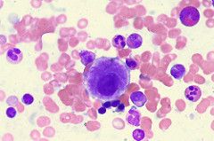
MarrowInterpretation (1000X) Determine M:E ratio... |
Ex:300 wbcdifferential and 75 nrbcswere noted M:Eratio = 300:75 or 4:1 Reference range: 3:1 to 4:1 |