Reading...
![]()
Play button
![]()
Play button
![]()
Use LEFT and RIGHT arrow keys to navigate between flashcards;
Use UP and DOWN arrow keys to flip the card;
H to show hint;
A reads text to speech;
49 Cards in this Set
- Front
- Back
- 3rd side (hint)
|

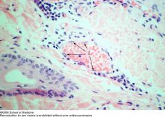
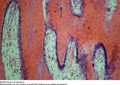
1. Simple squamous cell nuclei
|
Simple Squamous epithelium
|
|

|
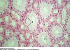
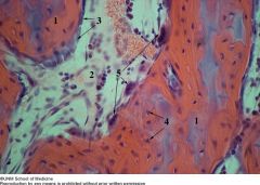
1. Nuclei of cuboidal epithelium
2. Cell borders of cuboidal epithelium 3. Basal lamina |
Simple squamous epithelium (Kidney)
|
|

|
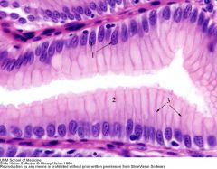
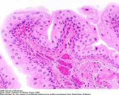
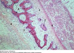
1.Nuclei of columnar epithelial cells
2.Cytoplasm containing mucous vesicles 3.Lateral cell borders |
Simple columnar epithelium (stomach)
|
|

|
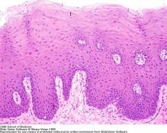
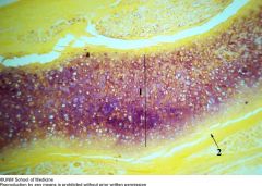
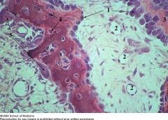
1.Squamous epithelium of the epidermis
2.Cuboidal epithelium of the epidermis |
Stratified squamous epithelium (epidermis)
|
|

|
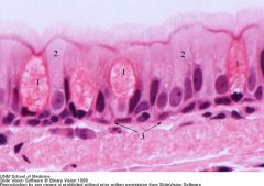
1.Lumen
2.Basal epithelial nuclei 3.Columnar epithelial nuclei |
Transitional epithelium (Bladder)
|
|

|
PCCE (Trachea)
1.Goblet cells 2.Non-secretory epithelium 3.Location of basal lamina |
PCCE (Trachea)
|
|
|

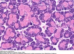
1.Apical cytoplasm with zymogen granules
2.Basal cytoplasm 3.Ducts |
Pancreas
|
|

|
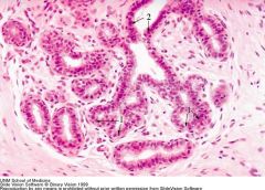
Mammary gland
1.Glandular epithelium 2.Ducts |
Mammary gland
|
|
|

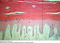
Epidermis
1.Stratum corneum 2.Stratum lucidum 3.Stratum granulosum 4.Stratum spinosum 5.Stratum basale 6.Dermal ridges 7.Dermis 8.Epidermis |
Epidermis
|
|

|
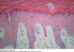
Epidermis/dermis
1.Stratum corneum 2.Stratum lucidum 3.Stratum granulosum 4.Stratum spinosum 5.Stratum basale 6.Dermal ridges 7.Epidermal ridges |
Epidermis/dermis
|
|

|
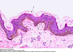
Epidermis
1.Keratin layer 2.Epidermis 3.Pigmented layer of epidermis 4.Dermis |
Epidermis
|
|

|
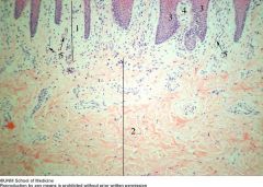
Dermis
1.Papillary layer of dermis 2.Reticular layer of dermis 3.Epidermal ridges 4.Dermal ridges 5.Dermal capillaries |
Dermis
|
|
|

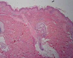
Hair follicle
1.Epidermis 2.Dermis 3.Hair follicles 4.Adipose tissues 5.Sebaceous gland 6.Sweat Gland 7.Arrector pili muscle |
Hair follicle
|
|

|
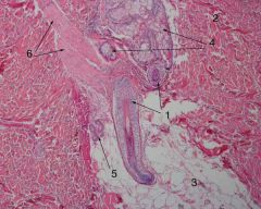
Hair follicle
1.Hair follicles 2.Dermis 3.Adipose tissue 4.Sebaceous glands 5.Sweat gland 6.Arrector pili muscle |
Hair follicle
|
|
|

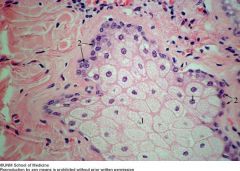
Sebaceous gland
1.Secretory cells 2.Basal cells |
Sebaceous gland
|
|
|

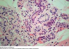
Sweat gland
1.Secretory cells 2.Duct 3.Myoepithelial cells 4.Basal lamina |
Sweat gland
|
|
|

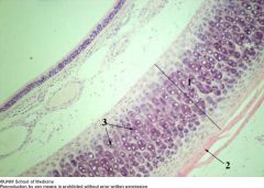
Hyaline cartilage (trachea)
1.Hyaline cartilage 2.Perichondrium 3.Chondrocytes in lacunae |
Hyaline cartilage (trachea)
|
|
|

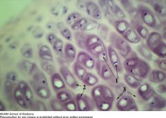
Hyaline cartilage (trachea)
1.Chondrocytes in lacunae 2.Territorial matrix 3.Interterritorial matrix |
Hyaline cartilage (trachea)
|
|

|
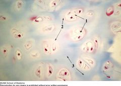
Hyaline cartilage (trachea)
1.Chondrocytes in lacunae 2.Territorial matrix 3.Interterritorial matrix |
Hyaline cartilage (trachea)
|
|

|
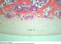
Hyaline cartilage (articular)
1.Chondrocytes 2.Subchondral bone 3.Bone marrow |
Hyaline cartilage (articular)
|
|
|

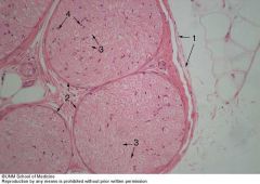
Elastic cartilage (epiglottis)
1.Elastic cartilage 2.Pericondrium |
Elastic cartilage (epiglottis)
|
|

|
Elastic cartilage (epiglottis)
1.Chondrocytes in lacunae 2.Elastic fibers |
Elastic cartilage (epiglottis)
|
|
|

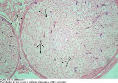
Fibrous cartilage (meniscus)
1.Chondrocytes 2.Fibrous collagen matrix |
Fibrous cartilage (meniscus)
|
|

|
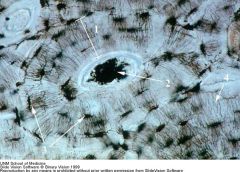
Compact bone
1.Osteons 2.Haversian canal 3.Osteocytes 4.Canaliculi |
Compact bone
|
|
|

Decalcified bone
1.Skeletal muscle fibers 2.Periosteum 3.Circumferential lamellae 4.Osteons 5.Haversian canals 6.Osteocytes |
Decalcified bone
|
|

|
Decalcified bone
1.Osteons 2.Haversian canal 3.Volkmann's canal 4.Osteocytes |
Decalcified bone
|
|
|

Decalcified trabecular bone
1.Trabeculae 2.Osteocytes 3.Marrow tissue |
Decalcified trabecular bone
|
|

|
Decalcified trabecular bone
1. Trabeculae 2.Osteocytes 3.Osteoblasts 4.Marrow tissue |
Decalcified trabecular bone
|
|
|

Decalcified trabecular bone
1.Trabeculae 2.Bone marrow 3.Osteoblasts and osteoprogenitor 4.Osteocytes |
Decalcified trabecular bone
|
|

|
Decalcified bone
1.Trabeculae 2.Bone marrow 3.Osteoblasts and osteoprogenitor 4.Osteocytes 5.Osteoclasts |
Decalcified bone
|
|
|

Trabecular bone (Developing skull)
1.Embryonic connective with mesenhymal cells 2.Newly formed trabeculae with osteoblasts and osteoid 3.Older anastomosing trabeculae covered by osteoblasts 4.Forming periosteum |
Trabecular bone (Developing skull)
|
|

|
Developing skull
1.Osteoblasts 2.Connective tissue capillaries 3.Osteoid 4.Osteocytes 5.Osteoclasts |
Developing skull
|
|

|
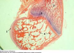
Developing long bone
1.Articular cartilage 2.Epiphysis 3.Diaphysis 4.Trabecular bone 5.Epiphyseal / Growth Plate |
Developing long bone
|
|
|

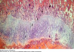
Developing long bone
1.Zone of proliferation 2.Zone of maturation and hypertrophy 3.Zone of ossification 4.Region of vascular invasion |
Developing long bone
|
|
|

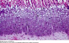
1.Chondrocytes
2.Zone of proliferation 3.Zone of maturation and hypertrophy 4.Zone of ossification 5.Region of vascular invasion |
Epiphyseal plate
|
|

|

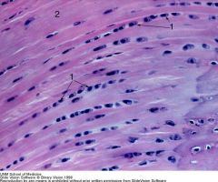
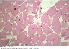
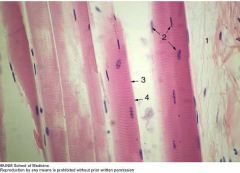
Skeletal muscle
1.Perimysium 2.Muscle fiber nuclei 3.A band 4.I band |
Skeletal muscle
|
|
|

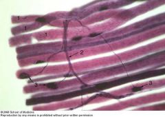
Skeletal muscle
1.Perimysium 2.Muscle fibers 3.Endomysium |
Skeletal muscle
|
|
|

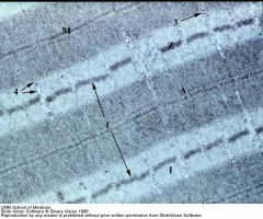
Skeletal muscle
1.Perimysium 2.Muscle fiber nuclei 3.A band 4.I band |
Skeletal muscle
|
|
|

Skeletal muscle1.Sarcolemma
2.Endomysium and basal lamina 3.Nucleus and Nucleoli 4.Mitochondria |
Skeletal muscle
|
|

|
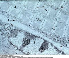
Skeletal muscle
1.Sacromere 2.Transverse tubules 3.Sacroplasmic reticulum |
Skeletal muscle
|
|

|
Skeletal muscle fiber
1.Muscle fibers 2.Motor neuron 3.Motor end plates |
Skeletal muscle fiber
|
|

|
Neuromuscular junction
1.Motor nerve axon 2.Motor endplate / nerve terminal 3.Myelin sheath 4.Junctional folds |
Neuromuscular junction
|
|
|

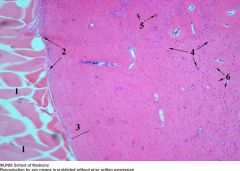
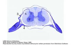
Spinal cord
1.Dorsal Horn 2.Ventral Horn 3.Dorsal Root 4.Ventral Root 5.Dorsal Root Ganglion 6.White Matter |
Spinal cord
|
|
|

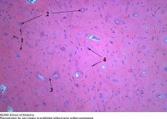
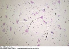
Ventral horn
1.Neuron cell body 2.Neuroglial cell nuclei 3.Neuropil |
Ventral horn
|
|

|
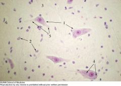
Ventral horn
1.Neuron Cell Body 2.Neuroglial Cell Nuclei 3.Neuropil 4.Nissl Bodies 5.Neuronal Processes (Dendrites / Axon) |
Ventral horn
|
|
|

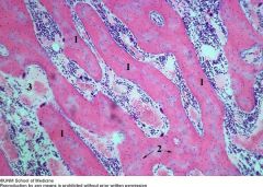
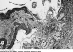
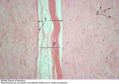
Peripheral nerve
1.Epineurium 2.Perineurium 3.Axons 4.Schwann cell nuclei |
Peripheral nerve
|
|

|
Peripheral nerve
1.Epineurium 2.Perineurium 3.Axons 4.Schwann cell nuclei |
Peripheral nerve
|
|

|
Peripheral nerve
1.Perineurium 2.Schwann cell nuclei 3.Epineurium |
Peripheral nerve
|
|

|
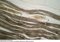
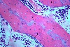
Peripheral nerve
1.Axons 2.Node of Ranvier |
Peripheral nerve
|