![]()
![]()
![]()
Use LEFT and RIGHT arrow keys to navigate between flashcards;
Use UP and DOWN arrow keys to flip the card;
H to show hint;
A reads text to speech;
33 Cards in this Set
- Front
- Back
|
What is the nervous system? |
-includes all neural tissue in the body including neurons (communication, control) and neuroglia (supporting cells) -two divisions: Central Nervous System (CNS) and Peripheral Nervous System (PNS) |
|
|
What is the CNS? |
•Consists of the spinal cord and brain •Contain neural tissue, connective tissues, and blood vessels Processes & coordinates: •sensory data (incoming info) •motor commands (controls activities if peripheral body) •higher functions (intelligence, memory) |
|
|
What is the PNS? |
•All neural tissue outside CNS
•Cranial nerves (connect to brain) & spinal nerves (connect to spine) Functions of peripheral nerves: 1.Sensory neurons deliver sensory information to the CNS 2.Motor neurons carry motor commands to peripheral tissues and systems |
|
|
What are the divisions of the PNS? |
>the CNS signals and coordinates a response >the PNS sends the info to the CNS, then collects motor commands (afferent or efferent) Afferent division: •carries sensory information (incoming info) •from PNS to CNS Efferent division: •carries motor commands (outgoing info) •from CNS to PNS muscles and glands •Divided into the somatic nervous system (controls skeletal muscle contractions an reflexes) & the autonomic nervous system (regulates smooth muscle contractions, glandular secretions, things you are not aware of) |
|
|
What are receptors and effectors? |
Receptors: •detect changes or respond to stimuli •neurons and specialized cells •complex sensory organs (e.g.,eyes, ears) Effectors: •respond to efferent signals •cells and organs |
|
|
How does information transfer work? |
Input versus output affect/efferent sensory/motor dendrites or soma/axon (at the cellular level) |
|
|
Structure or neuron: cell body, dendrites, axons, axon hillock, initial segment of axon, nucleus, axon, synaptic terminals Cell body Dendrites Axons |
Cell Body: main part of cell •Perikaryon: cytoplasm in the cell body •Cytoskeleton extends into dendrites (tree like, sense info outside neuron) and axons •Lack centrioles Dendrites: •Highly branched with dendritic spines (branches of dendrites) •Receive information from other neurons Axons: long extension •Carry electrical signal (action potential) to target Axon hillock: connects dendrite to axon Initial segment of axon: name Synaptic terminals: where info crosses from one neuron to another |
|
|
The Synapse: the parts |
Presynaptic cell: neuron that sends message Postsynaptic cell: cell that receives message -Neuromuscular junctions -Neuroglandular junctions -Other neurons Synaptic Cleft: gap that separates the presynaptic membrane and postsynaptic membrane |
|
|
The Synapse: the types. |
1. Electrical synapses: •direct physical contact between cells -presynaptic neuron to postsynaptic cell connected through integral membrane proteins and share a member/pore, producing a continous current -action potential freely propagated -rare 2. Chemical synapses: •signal transmitted across a gap by chemical neurotransmitters -more common, no direct contact though, rely on neurotransmitter *Action potentials are transmitted from presynaptic neuron to postsynapticcell across a synapse. |
|
|
What is a synpatic delay? |
Synaptic delay: occurs between the arrival of action potential at synaptic knob and the effect on postsynaptic membrane. |
|
|
What is a neurotransmitter? |
•Chemical messengers released at presynaptic membrane •Affect receptors of postsynaptic membrane •Broken down by enzymes & reassembled at synaptic knob |
|
|
What are the structural classifications of neurons? |
1. Anaxonic neurons: found in brain and sense organs, small, lots of dendrites, no axon 2. Bipolar neurons: found in special sensory organs (sight, smell, hearing), small, 1 dendrite, 1 axon 3. Unipolar neurons: found in sensory neurons of PNS, very long axons, fused dendrites and axon, cell body off to one side, end at synapses in CNS 4. Multipolar neurons: common in CNS, include all skeletal muscle motor neurons, have very long axons, one axon and multiple dendrites |
|
|
Function classifications of neurons |
Sensory neurons: •Info from sensory receptors to CNS •Monitor internal and external environment• Motor neurons: •Info from CNS to peripheral effectors Interneurons: •Coordinate between sensory and motor neuron •Most are located in brain, spinal cord•Involvedin higher functions |
|
|
What are the types of neuroglia? |
1. Ependymal cells (CNS) 2. Astrocytes (CNS) 3. Oligodendrocytes(CNS) 4. Microglia (CNS) 5. Satellite cells (PNS) 6. Schwann cells (PNS) |
|
|
1. Ependymal cells (CNS): |
1. Ependymal cells (CNS)
Line central canal of spinal cord and ventricles of brain: -secrete cerebrospinal fluid (CSF) -have cilia or microvilli that circulate CSF -monitor CSF |
|
|
2. Astrocytes (CNS): |
2. Astrocytes (CNS): -Maintain blood–brain barrier (isolates CNS) -Create 3-dimensional framework for CNS -Repair damaged neural tissue -Guide neuron development -Control interstitial environment *largest and most numerous neuroglia, star shape |
|
|
3. Oligodendrocytes (CNS): |
3. Oligodendrocytes(CNS): -Processes contact other neuron cell bodies •Wrap around axons to form myelin sheaths •Insulates axons from extracellular fluid •Myelin sheaths are internodes •Nodes are gaps between internodes *increases speed of action potential •Myelinated axons makes nerves appear white > •White matter: learning, speed of communication •High concentration of cell bodies + unmyelinated axons makes tissue appear grey >•Grey matter: cognition, understanding, awareness *male brain has more white while female grey |
|
|
4. Microglia (CNS) |
•Smallest & least numerous in CNS •Migrate through neural tissue •Clean up cellular debris, waste products, and pathogens *smallest neuroglia |
|
|
5. Satellite cells (PNS) |
Surround cell bodies and regulate environment around neuron |
|
|
6. Schwann cells (PNS) |
-form myelin sheath around peripheral axons -participate in repair processes after injury -surround all neurons in PNS |
|
|
Passive process: What are chemical, electrical and electrochemical gradients? |
Chemical gradients: concentration gradients of ions Electrical gradients: charges of positive & negative ions2 *Difference in ions gives membrane ability to conduct action potential Electrochemical gradients: sum of chemical and electrical forces acting on the ion across a cell membrane. |
|
|
Active forces: ion exchange pump |
•Ion exchange pumps actively move substances across the membrane •Maintains homeostasis inside the cell Example: outside cell has more sodium (+), inside cell (-), electrical gradient wants to move it inside. In a resting cell sodium permeability is low and there is an active mechanism to get it out (sodium potassium ATPase pumps sodium out to maintain homeostasis). *resting cell potential is -70mV |
|
|
Graded/local potentials |
•Chemically gated sodium ion channels are closed =voltage gated channel (need voltage to open) •Open when chemical binds to receptors •Sodium enters cell causing depolarization *inside cell negative, outside positive, cell at rest *when chemical shows up and binds, opens channel, + ions flow in, membrane changes from resting potential *shift in membrane potential is depolarization -movement of sodium through channel, produces local current, depolarizes nearby cell membrane (graded/local potential), change in potential is proportional to stimulus |
|
|
What is repolarization and hyperpolarization? |
Repolarization: Stimulus is removed -Resting membrane potential is restored Hyperpolarization: Increasing the negativity of the resting potential -Result of opening a potassium channel -Opposite effect of opening a sodium channel -Positive ions move out, not into cell |
|
|
Action potentials and |
•Propagated changes in membrane potential •Affect an entire excitable membrane •Stronger than graded potential •Can result from repetitive graded potentials *as sodium binds and depolarizes it is self propagating, voltage opens channel and spreads |
|
|
All or none principle |
•Threshold: the membrane potential at which an action potential begins •A stimulus below the threshold creates a graded depolarization. •A stimulus at or above the threshold results in an action potential. •The action potential is either generated by a stimulus or it is not (no inbetween, grey area) |
|
|
Refractory period |
Refractory period: from beginning of action potential to return to resting state (channel closes), membrane will not respond normally to additional stimuli •From beginning of action potential to return to resting state •Membrane will not respond normally to additional stimuli |
|
|
Steps in an action potential & frequency of APs |
1.Depolarization to threshold 2.Activation of Na+ channels: •rapid depolarization; Na+ions rush into cytoplasm •inner membrane changes from negative to positive 3.Inactivation of Na+ channels: •Na+gates close •K+channels open & repolarization begins 4.Return to normal permeability: •K+channels begin to close when membrane reaches normal resting potential •K+channels finish closing: membrane is hyperpolarized •Membrane potential returns to resting level: action potential is over Frequency: Degree of sensory stimulation or strength of motor response is related to frequency ofaction potentials |
|
|
Propagation: continous or saltatory |
Continuous propagation: action potential travels along unmyelinated axons one segment at a time Saltatory propagation: action potential travels along myelinated axons, jumping from node to node |
|
|
Types of neurotransmitters: excitatory and inhibitory |
1.Excitatory neurotransmitters: •cause depolarization of postsynaptic membranes •promote action potentials •Cause facilitation 2.Inhibitory neurotransmitters: •cause hyperpolarization of postsynaptic membranes •suppress action potentials |
|
|
Temporal versus Spatial summation |
Temporal: multiple times, rapid, repeated stimuli at a single synapse Spatial: multiple locations, many stimuli, arrive at multiple synapses |
|
|
Information processing: What is net effect? |
•Many dendrites receive neurotransmitter messages simultaneously: some excitatory, some inhibitory •Net effect on axon hillock determines if action potential is produced |
|
|
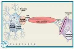
What are the main processes in neural activities? |
1.Resting potential: the membrane potential of resting cell 2.Graded potential: temporary, localized change in resting potential caused by stimulus 3.Action potential: electrical impulse produced by graded potential; propagates along surface of axon to synapse 4. Synaptic activity: releases neurotransmitters at presynaptic membrane; produces graded potentials in postsynaptic membrane 5. Information processing: response of postsynaptic cell |