Reading...
![]()
Play button
![]()
Play button
![]()
Use LEFT and RIGHT arrow keys to navigate between flashcards;
Use UP and DOWN arrow keys to flip the card;
H to show hint;
A reads text to speech;
13 Cards in this Set
- Front
- Back
- 3rd side (hint)
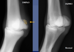
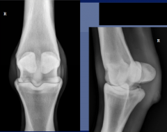
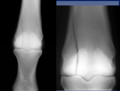
Fetlock joint
standard views dorsopalmar/dorsoplantar |
Lateral medial
and flexed lateral |
DLPMO/DMPLO
|
|

|
soft tissue swelling
|
location:
soft tissue joint tendon sheath repeart radiographs in 6 - 8 wks may show early DJD and/or periarticular dystrophic calcification palmar/plantar soft tissue swelling - digital flexor tendon sheath effusion (windpuffs) soft tissue swelling and annular ligament constriction - annular ligament desmitits - tenosynovitis/tendonitis |
|
|
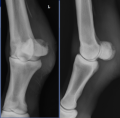
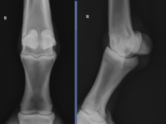
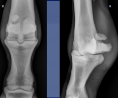
degenerative joint disease
|
- periarticular osteophyte formation
-subchondral bone sclerosis - ill defined small lucent zones in the subchondral bone - narrowing of the joint space - joint capsule distension - periarticular soft tissue swelling - supracondylar lysis - fibrous proliferation of synovial membrane |
|
|

|
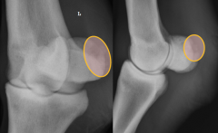
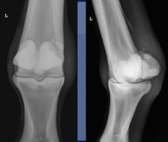
degenerative joint disease
osteophytes soft tissue swelling - joint capsule distention narrowed joint -sclerosis supracondylar lysis |
|
|
|
degenerative joint disease
soft tissue swelling osteophytes joint space narrowing -sclerosis -subchondral bone lysis |
osteophytes
joint space narrowing sclerosis soft tissue swelling |
|

|
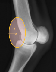
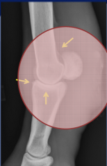
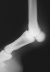
villonodular synovitis
-secondary to repetitive trauma -thickening of the bilobed synovial pad .with extension of the fetlock, P1 impinges on the pad leading to inflammation (capsulitis/synovitis) -radiographic findings .asymmetric soft tissue swelling usually more pronounced on the dorsal aspect .crescent shaped radiolucent "cut out" on the dorsal surface of the distal cannon bone .dystrophic mineralization within the joint |
asymmetrical soft tissue swelling
focal osseous resorption/lysis dystrophic mineralization |
|
|
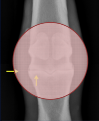
supracondylar lysis
-osteophytes -enthesophyte capsular attachment -dorsal soft tissue swelling -palmar/plantar resportion of MC III |
|
|

|
Sesamoiditis
- proximal sesamoids - ligament attachments . suspensory branches .distal sesamoidean ligaments - radiographic findings . enthesophytes .widened vascular changes .focal osteolysis .periosteal proliferation |
|
|
|
septic sesamoiditis
secondary to puncture wound |
injury to suspensory ligament
|
|
|
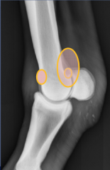
Osteochondrosis
|
failure of endochondral ossificiation/subchondral bone defect
etiology unknown, may be traumatic main forms in fetlock: -fragmentation of the sagittal ridge - fragments on the dorsoproximal aspect of the proximal phalanx - palmar/plantar osteochondral fragments - flattening palmar/plantar condyle of MC/MT III |
|
|
|
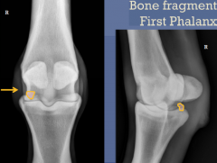
osteochondrosis
bone fragment proximal phalanx |
|
|
|
condylar fractures
- difficult to visualize -radiographic findings uneven joint surface interruption of the metaphyseal cortex presence of a radiolucent line extending from the joint surface to the cortex complete displaced, complete nondisplaced, incomplete possible concurrent sesamoid fractures |
|
|
|
proximal sesamoid fractures
Apical -common in racehorses midbody - medial sesamoid forelimb TB -lateral sesamoid hindlimb STB Basilar - origin of the distal sesamoideal ligaments Abaxial - insertion injuries of suspensory ligament braches |
|