Reading...
![]()
Play button
![]()
Play button
![]()
Use LEFT and RIGHT arrow keys to navigate between flashcards;
Use UP and DOWN arrow keys to flip the card;
H to show hint;
A reads text to speech;
13 Cards in this Set
- Front
- Back
|
What is binocular vision?
|
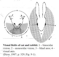
-occurs where visual fields of both eyes overlap
-allows for focus on near objects and for depth perception -greater in carnivores than in their prey (ie. herbivores) |
|
|
What does the eyeball consist of?
|
-consists of:
3 thin tunics: *outer fibrous tunic *middle vascular tunic *internal nervous tunic -lens for focusing light on receptors -partly liquid, partly gelatinous centers |
|
|
Describe the fibrous tunic
|

-dense collagneous tissues
-resists internal pressure -consists of sclera (posteriorly) and cornea (anteriorly) which meet at limbus ✩SCLERA -opaque -penetrated by fibers of optic nerve -continuous with dura mater of optic nerve -provides attachment for tendons of extrinsic muscles of eye ✩CORNEA -specialized transparent, dense connective tissue -covered by anterior and posterior epithelial layers -transparency dependent on: →highly organized layers of collagen fibers →continuous pumping out of interstitial fluid by posterior epithelium -avascular → nutrition by diffusion from lacrimal fluid (tears) and aqueous fluid of anterior chamber -very sensitive due to nerve endings (branches of ophthalmic division of CN V) |
|
|
Describe the vascular tunic
|
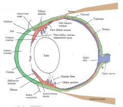
-also known as uvea
-consists of choroid, ciliary body, and iris (from posterior to anterior) ✩CHOROID -lines sclera from optic nerve almost to limbus -contains blood vessels in pigmented connective tissue -provides nutrients for outer layers of retina -contains avascular tapetum lucidum: →area of dorsal choroid →reflective iridescent →not present in pigs (or humans) -probably assists in nocturnal vision ✩CILIARY BODY -thickening of choroid anteriorly in radial ridges (ciliary processes) -provides anchoring point for zonular fibers, which suspend lens -ciliary muscles (smooth) at base of ciliary body: →contraction causes relaxation of zonular fibers and rounding of lens → focus on near objects (accomadation) -produces aqueous humour ✩IRIS -ring of tissue suspended between cornea and lens -opening in center is pupil -consists of pigmented connective tissue with epithelium (pigmented posteriorly) →connective tissue layer contains smooth muscle sphincter (circular - parasympathetic) and dilator (sympathetic) which regulate pupillary size -separates anterior from posterior chamber -colour dependent on amount of pigment -species variation in shape of pupil when constricted → DOG = round, cat = vertical slit, horse, ox = oval |
|
|
Describe the internal tunic
|
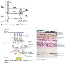
-contains light-sensitive receptor cells (posterior 2/3rds of retina)
-lines vascular tunic from pupillary margin posteriorly to optic nerve -thin outer layer (pigmented except over tapetum) and thick inner layer (neuroepithelium) ♦photoreceptor layer = rods & cones ♦horizontal cells ♦bipolar cells ♦amacrine cells ♦ganglion cells - non-myelinated axons extend across inner surface of retina to optic disc, where they meet to form optic nerve ie. ganglion cells are cell bodies of CN II -photoreceptors synapse with bipolar cells which synapse with ganglion cells -horizontal cells modify transmission between photoreceptors and bipolar cells -amacrine cells modify transmission between bipolar cells and ganglion cells -blood vessels enter retina at optic disc with optic nerve and spread across retina →supply inner layer of retina ✩PHOTORECEPTOR CELLS -outer segment contains an array of stacked membranous discs with photopigments in membranes -when light hits photoreceptor, photopigment is transformed, leading to hyperpolarization and inhibition of transmission of action potential to bipolar cells -rods highly sensitive → adapted for night vision -cones less sensitive to light, but responsible for colour perception →dogs & horses have fewer rods and limited colour perception compared to humans |
|
|
Describe the lens
|
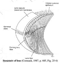
-soft, transparent, biconvex structure
-composed of 'lens fibers' - epithelial cells running in layers from anterior to posterior poles of lens -avascular - nourished by aqueous and vitreous humour -focuses images on retina -surrounded by an elastic capsule (basement membrane) |
|
|
Describe the aqueous humour and vitreous body
|
✩AQUEOUS HUMOUR
-fills space between cornea and lens (anterior and posterior chambers) -produced by cells of ciliary body -passes from posterior chamber to anterior chamber -drains into venous sinuses in sclera at iridocorneal angle ✩VITREOUS BODY -occupies space between lens and retina -gel-like mass consisting of stroma of fine transparent fibers filled with glycosaminoglycans and water |
|
|
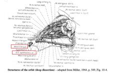
Describe the structure of the periorbita
|
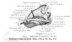
-fibrous fascial sheath, which blends with the periosteum medially and dorsally
-inserts in eyelids and surrounds eyeball and extrinsic muscles of eye -attached to skull near optic foramen -contains smooth muscle which keeps eyeball slightly protruded with normal tone (under sympathetic control) -contains and surrounded by periorbital fat (cushions contents of orbit) |
|
|
Describe the extrinsic muscles of the eye
|
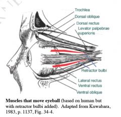
-seven extrinsic muscles act together to move eye within orbit
-all except ventral oblique originate on orbital wall near optic foramen -insert on fibrous tunic ✩RECTUS MUSCLES -dorsal, ventral, lateral, and medial rectus muscles ✩OBLIQUE MUSCLES -rotate eye about visual axis -dorsal oblique: →travels anteriorly along dorsomedial wall of orbit; tendon passes through trochlea (cartilaginous thickening in dorsomedial periorbita - acts as pully), then inserts on dorsolateral aspect of fibrous tunic →contraction pulls dorsolateral aspect of eye medially (ie. medial rotation) -ventral oblique: →arises from ventromedial wall of bony orbit and passes laterally to insert on ventrolateral aspect of eyevall →contraction pulls ventrolateral part of eyeball medially ✩Retractor bulbi -several slips of muscle which insert on posterior aspect of eye -surrounds optic nerve -retracts globe in socket -absent in humans ✩LEVATOR PALPEBRAE SUPERIORIS -originates on posterior wall of orbit and inserts in upper eyelid, with periorbita -raises eyelid |
|
|
Describe the eyelids
|
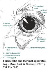
✩UPPER AND LOWER EYELIDS (palpebrae)
-meet at medial and lateral angles (canthi) -surrounded by palpebral fissures -formed by the following layers ♦skin ♦musculofibrous layer: →contains orbicularis oculi, fibrous periorbita and smooth muscle →tarsal glands - open at free edge of lid; secrete fatty material (contribution to lacrimal fluid) ♦conjunctiva →thin mucous membrane lining posterior surface of eyelids (palpebral conjunctiva) and reflected onto sclera (bulbar conjunctiva) -lacrimal caruncle → mucosal elevation at medial canthus -puncta lacrimalia: →minute slits on upper and lower lids adjacent to lacrimal caruncle →openings to canaliculi leading to nasolacrimal duct (in lateral wall of nasal cavity) ✩THIRD EYELID -between lower lid and eyeball -covered with conjunctiva on both surfaces -supported by T shaped piece of cartilage -gland of third eyelid surrounds stem of T: →secretes towards eyeball from posterior surface -while eyes open, normally held retracted by smooth muscle (sympathetic control) -provides additional protection and moisture for eyeball |
|
|
Describe lacrimation
|

-lacrimal fluid distributed over anterior aspect of eye by blinking
-required to moisten and nourish cornea and flush away foreign objects -secretion stimulated by conjunctival, corneal or nasal irritation -lacrimal secretion consists of 3 layers: ♦outermost lipid layer (from tarsal glands) spreads tear film evenly and prevents evaporation ♦aqueous layer from: →lacrimal gland (located on dorsolateral aspect of eye within periorbita; secretes through several fine openings into palpebral sac ie. space between palpebral and bulbar conjunctivae) →gland from third eyelid ♦inner mucoid layer (from goblet cells in conjuctiva) - binds tears to cornea -lacrimal fluid drains via lacrimal puncta and canaliculi to nasolacrimal duct |
|
|
Describe the vascular supply to the eye
|
♦external ophthalmic artery - principle supply to eye
→branches from maxillary artery then penetrates the apex of periorbita →gives off 3 groups of branches which penetrate the sclera at different levels to rach the vascular tunic and retina *short posterior ciliary arteries *long posterior ciliary arteries *anterior ciliary arteries ♦internal opthalimic artery - supplies CN II and spreads over retina from optic disc -venous drainage via several vorticose veins that emerge through sclera |
|
|
Describe the nerve supply to the eye and related structures
|
CNII - perception of light
CNIII - somatic efferent: - dorsal, medial and ventral recti - ventral oblique - levator palpebrae superioris – visceral efferent: -parasympathetic innervation to smooth muscle of iris and ciliary body CNIV - dorsal oblique CNV (ophthalmic and maxillary divisions) – sensory to eyeball (especially cornea), eyelids and conjunctivae CNVI - lateral rectus, retractor bulbi CNVII – motor to orbicularis oculi Sympathetic innervation • exits cranial thoracic spinal cord and ascends in vagosympathetic trunk to cranial cervical ganglion near tympanic bulla • postsynaptic neurons travel with CNIII to supply smooth muscle of eye: - periorbita (protrudes eyeball) - pupillary dilator - eyelids (keeps palpebral fissure open) - third eyelid (retracts) |