![]()
![]()
![]()
Use LEFT and RIGHT arrow keys to navigate between flashcards;
Use UP and DOWN arrow keys to flip the card;
H to show hint;
A reads text to speech;
61 Cards in this Set
- Front
- Back
|
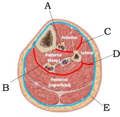
A) Interosseous Membrane
B) Transverse intermuscular septum C) Anterior intermuscular septum D) Lateral intermuscular septum E) Crural fascia |
|
|
A) Anterior tibial artery and deep peroneal nerve |
|
|
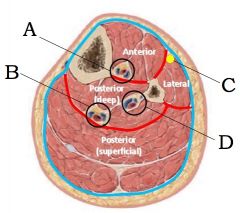
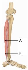
A) Tibialis anterior
B) Extensor digitorum longus C) Extensor hallucis longus |
|
|
What is the action, innervation, and blood supply of the muscles of the anterior compartment of the leg? |
Action: dorsiflexion of foot and extension of toes
Innervation: deep peroneal nerve
Blood supply: anterior tibial artery |
|
|
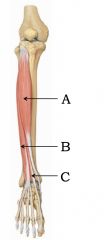
A) Superior extensor retinaculum
B) Inferior extensor retinaculum C) Peroneus tertius |
|
|
What are the actions of the tibialis anterior? |
dorsiflexes and inverts the foot |
|
|
What is dorsiflexion of the foot? |
moving your foot so that your toes go up |
|
|
What is the action of the peroneus tertius? |
dorsiflexes and everts the foot |
|
|
What does everting the foot mean? |
rotating foot laterally so that smallest toes are higher than big toe |
|
|
What are shin splints? |
Small tears in the periosteum (mild form of compartment syndrome) due to over extension or trauma |
|
|
What nerve if damaged leads to drop foot syndrome? |
Deep peroneal nerve |
|
|
A) Peroneus Longus
B) Peroneus Brevis |
|
|
What are the innervation and blood supply of the muscles of the lateral compartment of the leg? |
Innervation: Superficial peroneal nerve
Blood supply: peroneal artery |
|
|
Where do the tendons of the muscles of the lateral compartment of the leg run distally? |
They run posterior to the lateral malleolus (like a tibial epicondyle) and deep to the superior and inferior retinacula |
|
|
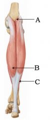
A) Plantaris
B) Gastrocnemius C) Soleus (deep to gastrocnemius) |
|
|
What two muscles' tendons become the achilles tendon?
What is the scientific name for this tendon? |
- Gastrocnemius and Soleus
- Tendo calcaneus |
|
|
What are the actions of the gastrocnemius? |
Plantar flexes foot and flexes leg |
|
|
What are the actions of the soleus? |
plantarflexes the foot |
|
|
What are the actions of the plantaris? |
very weak plantarflexion and flexion of the leg |
|
|
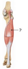
Soleus
|
|
|
How does plantarflexion help walking? |
It helps push the back foot off the ground. |
|
|
What kind of patients are most likely to rupture their calcaneal tendon? |
Achilles tendon rupture is most common with patients who have a history of calcaneal tendinitis. |
|
|
What 4 muscles make up the boundaries of the popliteal fossa? |
Superior medial: semimembranosus Superior lateral: biceps femoris
Inferior medial: gastrocnemius Inferior lateral: plantaris |
|
|
What nerves, artery, and vein run through the popliteal fossa? |
Tibial nerve, common peroneal nerve, popliteal artery and vein |
|
|
Of what artery is the popliteal artery an extension? |
femoral artery |
|
|
What does the femoral artery pass through when entering the popliteal fossa? |
adductor hiatus |
|
|
What does the popliteal artery divide into at the lower boundary of the popliteal fossa? |
Anterior and posterior tibial arteries |
|
|
What 5 branches does the popliteal artery give off in the popliteal fossa to form anastomoses? |
Two Superior, one middle, and two inferior genicular arteries |
|
|
What vein drains into the popliteal vein in the popliteal fossa? |
Small saphenous vein |
|
|
What two nerves that make up the sciatic nerve split at the popliteal fossa? |
Common peroneal nerve and tibial nerve |
|
|
What does the latereal sural cutaneous nerve innervate?
From what larger nerve does it branch? |
- Skin of the calf
- Common peroneal nerve |
|
|
What function do the muscles of the posterior compartment of the leg have besides plantarflexion and leg flexion? |
To steady the foot while standing |
|
|
What is the function of the popliteus? |
To basically unlock the knee by flexing it and rotating the tibia medially |
|
|
What muscle of the thigh rotates the leg externally while walking? |
Biceps femoris |
|
|
Which direction does the tibia rotate to lock the knee? |
Laterally |
|
|
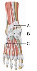
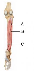
A) Tibialis posterior
B) Flexor hallucis longus C) Flexor digitorum longus |
|
|
What is the action of the tibialis posterior? |
plantarflex and invert the foot |
|
|
What is the action of the flexor digitorum longus? |
flexes lateral four toes and plantarflexes foot |
|
|
What is the action of the flexor hallucis longus? |
flexes big toe and plantar flexes foot |
|
|
From deep to superficial, what is the order of the tendons of the muscles in the deep posterior compartment including blood vessels and nerves? |
Tibiliasis posterior, flexor digitorum longus, vein, nerve, flexor hallucis longus |
|
|
What is the distal attachment of the tibiliasis posterior? |
median cunieform tarsal proximal to 1st metatarsal as well as 2nd, and 3rd metatarsal |
|
|
What direction does the femur rotate to lock the knee when it is full extended? |
medially |
|
|
Why are the condyles of the femur so much larger than the articular surface of the tibia? |
Because the femur rolls during leg flexion |
|
|
What ligament holds the lateral condyle of the femur to the head of the fibula? |
lateral collateral ligament |
|
|
Between the lateral and medial collateral ligaments, which one is stronger? |
Lateral collateral ligament (fibular) |
|
|
Between the lateral and medial collateral ligaments, which one is directly attached to its associated meniscus and knee capsule? |
Medial collateral ligament (tibial) |
|
|
Between the LCL and MCL, which one is wider? |
MCL, medial collateral ligament |
|
|
Does the lateral collateral ligament connect the femur to the fibula or tibia? |
fibula |
|
|
When the medial collateral ligament is damaged, what other two components are often damaged as well?
What is the grouping called? |
ACL and medial meniscus
Terrible triad |
|
|
What ligament connects medial and lateral menisci? |
transverse genicular ligament |
|
|
Where do the anterior and posterior cruciate ligaments reside? |
Inside the joint capsule of the knee |
|
|
What are the attachments of the ACL and PCL? |
ACL: Femur to the anterior tibia
PCL: Femur to the posterior tibia |
|
|
What is the function of the ACL? |
To prevent anterior displacement of the leg from the knee |
|
|
What is the function of the PCL? |
To prevent posterior displacement of the leg from the knee |
|
|
During what action are the ACL and PCL taut? |
ACL: taut during leg extension (standing)
PCL: taut during leg flexion (bent) |
|
|
What can damage the ACL and PCL? |
ACL: damage by excessive medial rotation or hyperextension
PCL: damage by excessive hyperextension |
|
|
What nerve innervates the posterior compartment of the leg? |
tibial nerve |
|
|
At what point does the tibial nerve divide into medial and lateral plantar nerves? |
when it reaches the medial malleolus |
|
|
What branch off the common peroneal nerve innervates the lateral compartment of the leg? |
superficial peroneal nerve |
|
|
What branch off the common peroneal nerve innervates the anterior compartment of the leg? |
deep peroneal nerve |
|
|
Besides the anterior compartment of the leg, what other muscles does the deep peroneal nerve innervate? |
extensor digitorum brevis |