Reading...
![]()
Play button
![]()
Play button
![]()
Use LEFT and RIGHT arrow keys to navigate between flashcards;
Use UP and DOWN arrow keys to flip the card;
H to show hint;
A reads text to speech;
42 Cards in this Set
- Front
- Back
- 3rd side (hint)
|
Anatomy of Respiratory system
Upper RS |
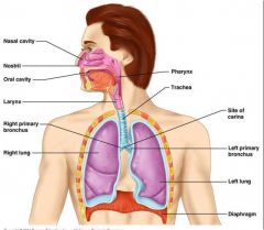
nasal cavities
nasopharynx |
|
|
|
Anatomy of Respiratory system
Lower RS 4ct |
larynx
trachea bronchi lungs |
|
|
|
pharynx
3ct |
Nasopharynx
Oropharynx Laryngopharynx |
|
|
|
the larynx is found at what vertebral level?
|
C3–C6
infants, C2–C3 |
|
|
|
The laryngeal skeleton consists of nine cartilages:
|
: three single
thyroid, cricoid, and epiglottic three paired arytenoid, corniculate, and cuneiform |
|
|
|
epiglottis is a flap of
|
elastic cartilage
covered with a mucus membrane, attached to the root of the = |
tongue
|
|
|
The epiglottis guards the entrance of the glottis, the opening between
|
vocal folds
|
|
|
|
Trachea
There are about ___/___ incomplete C-shaped cartilaginous rings which reinforce the anterior and lateral sides of the trachea to protect and maintain the airway. |
15-20
|
|
|
|
Info x2
The trachealis muscle connects the ends of the incomplete rings, and contracts during coughing, reducing the size of the lumen of the trachea to increase the air flow rate. |
The esophagus lies posteriorly to the trachea. The cartilaginous rings are incomplete to allow the trachea to collapse slightly so that food can pass down the esophagus.
|
|
|
|
first sign of development
respiratory diverticulum of the primitive foregut is during week __? |
wk 4
|
|
|
|
The distal end of the respiratory diverticulum enlarges to form the =
|
lung bud.
The lung bud divides into two bronchial buds that branch into the |
main right and left primary bronchus.
|
|
|
The respiratory diverticulum
become separated by indentations of *_________, becoming the = |
mesoderm
tracheoesophageal folds. |
|
|
|
the tracheoesophageal folds fuse in the midline to form the
|
tracheoesophageal septum,
|
|
|
|
The endoderm lining the groove (tracheoesophageal folds) gives rise to the epithelium and glands of the = 4ct
|
larynx
trachea bronchi pulmonary epithelium Connective tissue, cartilage and smooth muscle of these structures develop from the = |
the splanchnic mesenchym
mesoderm |
|
|
BMP-4
Hox-complex, FGF-10 N-myc |
bone morphogenetic protein
fibroblast growth factor proto-oncogene |
|
|
|
bone morphogenetic protein
fibroblast growth factor proto-oncogene |
BMP-4
Hox-complex, FGF-10 N-myc |
|
|
|
laryngeal epithelium and glands are derived from
|
endoderm
|
|
|
|
laryngeal muscles are derived from
|
mesoderm
|
|
|
|
laryngeal cartilages
thyroid cricoid arytenoid corniculate cuneiform are derived from = |
mesoderm
What Arches = |
pharyngeal arches 4 and 6.
|
|
|
The laryngeal epithelium proliferates rapidly, resulting in =
|
temporary occlusion of the laryngeal lumen.
Recanalization of the larynx normally occurs by the = |
tenth week
|
|
|
Recanalization of the larynx of the laryngeal lumen.,normally occurs by the =
|
tenth week
|
|
|
|
Vocal cords develop from folds of mucous membrane during the process of
|
recanalization
|
|
|
|
(CHAOS)
|
congenital high airway obstruction syndrome
|
|
|
|
This rare anomaly results from failure of recanalization of the larynx, and causes obstruction of the upper fetal airway
|
congenital high airway obstruction syndrome (CHAOS)
|
|
|
|
incomplete recanalization of the larynx is called =
|
Laryngeal web
|
|
|
|
Laryngeal web
Symptoms 4ct |
1. weak cry
2. Aphonia (inability to speak ) 3. Stridor (high pitched sound resulting from turbulent air flow in the upper airway ) 4. Hoarseness |
|
|
|
abnormal communication between the trachea and esophagus that results from improper division of foregut by the tracheoesophageal septum.
|
Tracheoesophageal fistula (TEF)
|
|
|
|
Tracheoesophageal fistula (TEF) :
generally associated with = 2ct |
esophageal atresia
polyhydramnios |
|
|
|
Development of Bronchi and Lungs
2 *________ bronchial buds Early in week __ each bronchial bud enlarges into the primordium of a ________ ____________ |
endodermal
wk 5 primary bronchus secondary bronchi are also called = |
lobar bronchi
|
|
|
The secondary bronchi
(lobar bronchi) further subdivide into segmental (tertiary) bronchi How many = |
10 on the right side
9 on the left side By week __, they divide another __times and the respiratory bronchioles have developed They will divide an additional __ more times before birth |
Wk 24
14x 7x |
|
|
As the bronchi develop, they expand laterally and caudally into a space known as
|
primitive pleural cavity
|
|
|
|
Maturation of the lungs is divided into four periods:
|
Pseudoglandular
Canalicular Terminal saccular Alveolar |
|
|
|
By week __ all major elements of the lungs have formed
|
Wk 16
except for those involved with gas exchange |
|
|
|
By week __all major elements of the lungs have formed except for those involved with gas exchange
|
16
|
|
|
|
Canalicular period (__ to __ weeks)
|
16-26 wks
|
|
|
|
By week __, respiratory bronchioles have developed and respiration becomes possible, although the chances of survival are less.
|
24
|
|
|
|
Terminal sac period
(__ weeks to birth) The intimate contact between epithelial and endothelial cells establishes the blood-air barrier, which permits adequate gas exchange for survival of the fetus if it is born prematurely |
26 wks
|
|
|
|
Respiratory distress syndrome (RDS) : Hyaline membrane disease
is caused by a = |
deficiency or absence of surfactant.
|
|
|
|
Surfactant is composed of =
|
cholesterol (50%),
di-palmitoyl-phosphatidyl-choline (DPPC; 40%), surfactant proteins A, B, and C (10%). |
|
|
|
Respiratory distress syndrome (RDS
Treatments include |
- betamethasone (a corticosteroid) to the mother for several days before delivery
-artificial surfactant solu -artificial ventilation |
|
|
|
Congenital lobar emphysema (CLE)
is characterized by progressive = |
overdistention of one lobe or the upper lobes or the right middle lobe with air.
due to failure of bronchial cartilage formation. *** |
|
|
|
In this situation, air can be inspired through collapsed bronchi, but cannot be expired.
what condition |
Congenital lobar emphysema (CLE)
*** During the first few days of life, fluid may be trapped in the involved lobe, producing an opaque, enlarged hemithorax. Later, the fluid is resorbed, and the classic radiologic appearance of an emphysematous lobe with generalized radiolucency (darkened images on x-ray films) is apparent. |
|