Reading...
![]()
Play button
![]()
Play button
![]()
Use LEFT and RIGHT arrow keys to navigate between flashcards;
Use UP and DOWN arrow keys to flip the card;
H to show hint;
A reads text to speech;
30 Cards in this Set
- Front
- Back
|
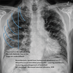
What three findings on a chest x-ray are characteristic of hyperinflation?
|
"1) Flattened diaphragms (more dramatic on the lateral view)
2) Air in the substernal space (causing hyperresonance) 3) Enlarged AP diameter" |
|
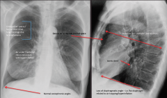
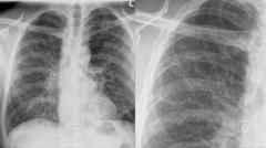
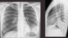
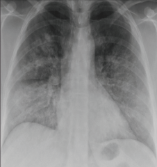
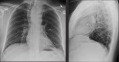
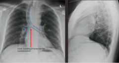
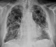
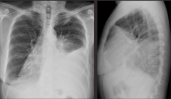
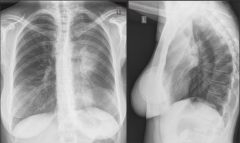
Take a look at the x-ray. What is your diagnosis? What are the abnormalities?
|
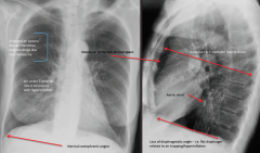
"Hyperinflation
7 ribs visible anteriorly, loss of interstitial lung markings, increased AP diameter, flattening of the diaphragms, air in retro sternal area. Bonus: Aortic stent" |
|
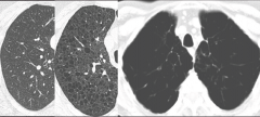
A CT progression of what condition is pictured?
|
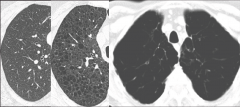
L to R: Normal lung, mild/moderate emphysema, severe emphysema.
|
|
Take a look at the x-ray. What is your diagnosis? What are the abnormalities?
|
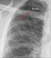
"Severe bollus emphysema.
Some scoliosis, upper lobes are missing lung tissue, a bunch of bollae are visible." |
|
Take a look at the x-ray. What is your diagnosis? What are the abnormalities?
|
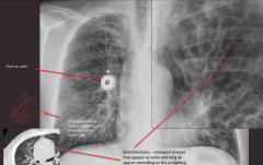
"Mixed airway disease and fibrosis from chronic infection - bronchiectasis
Long-ballooned bronchioles - tram-tracking Bonus: Port-o-cath going into the right atria" |
|
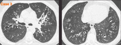
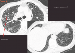
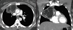
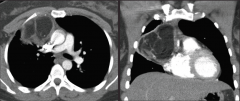
Take a look at the CTs. What is your diagnosis?
|
Bronchiectasis - visible dilated bronchioles
|
|
|
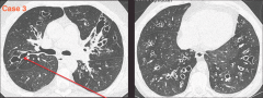
What is the Signet ring sign (seen on CT) and what does it indicate?
|
"Signet ring sign describes bronchioles that are bigger than arteries within the lung.
It is seen in bronchiectasis." |
|
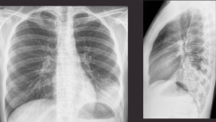
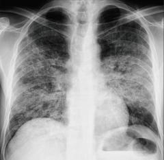
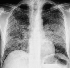
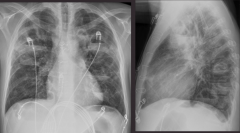
A 35 y.o. man is presenting with a fever. Take a look at the x-ray. What is your diagnosis? What are the abnormalities?
|
"Left lower lobe pneumonia
White mass on left lower lobe, same density as diaphragm (silouette sign), can see bronchi through the opacity (air bronchogram)" |
|
|
What is an air bronchogram and what does it indicate?
|
"Air bronchogram - visible bronchi through the opacity.
Indicative of consolidation What 4 things can cause consolidation?" |
|

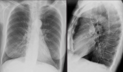
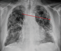
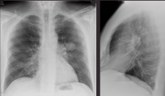
Take a look at the x-rays. What is your diagnosis? What are the abnormalities?
not exact matches on x-ray |
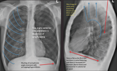
"Emphysema (hyperinflation)
Too many anterior ribs, increased air in substernal space, flattened diaphragms, increased AP diameter." |
|
Take a look at the x-ray. What is your diagnosis?
|
"Pneumocystis pneumonia.
(She didn't cover this one with our group, so I don't have any more info)" |
|
Take a look at the x-ray. What is your diagnosis?
|

Pulmonary hemorrhage.
|
|
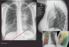
An IV drug user presents with a fever. Take a look at the x-ray. What is your diagnosis? What are the abnormalities.
|
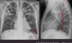
"Tuberculosis
Circular abcess in the left lower lobe that is filled halfway with fluid (likely exudate), there are all kinds on the AP to the left if you look closely." |
|
A 40 y.o. presents with fatigue and notes red painful lumps on their shins. Take a look at the x-ray. What is your diagnosis. What are the abnormalities?
|
"Sarcoidosis
Red lumps are erythema nodosum Hilar lymphadenopathy and calcifications," |
|

The x-rays are of a 35 y.o. woman with sarcoid. The film on the left is at diagnosis. The film on the right is four years later. What has changed on the x-ray?
|
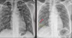
"At diagnosis - patchy, bilateral, symmetric densities.
4 years later - webs of lines, more linear, trachea deviated, diaphragms pulled up. All signs point to interstital disease and fibrosis, pulling on the structures." |
|
|
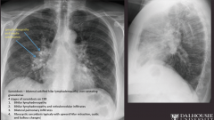
What is the definition of sarcoidosis? What are the four stages of sarcoidosis as seen on x-ray?
|
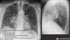
"Sarcoidosis -bilateral calcified hilar lymphadenopathy, non-caseating granulomas
Four stages of sarcoidosis on CXR 1. Bihilar lymphadenopathy 2. Bihilar lymphadenopathy and reticulonodular infiltrates 3. Bilateral pulmonary infiltrates 4. Fibrocystic sarcoidosis typicaly with upward hilar retraction, cystic and bullous changes" |
|
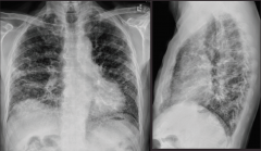
Take a look at the x-ray. What is your diagnosis? What are the abnormalities?
|
"Idiopathic pulmonary fibrosis (IPF) - aka Usual interstital pneumonitis (UIP)
Linear densities, hypoinflation (4.5 anterior ribs), shaggy heart and diaphragms" |
|
|
What kind of pattern would Idiopathic pulmonary fibrosis (IPF) have on CT?
|
Honeycombing and bronchiectasis.
|
|
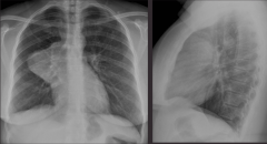
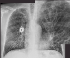
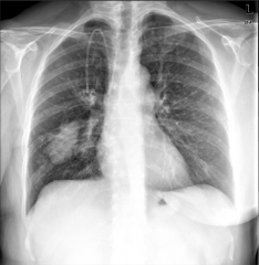
Take a look at the x-ray. What is your diagnosis? What are the abnormalities?
|
"Mediastinal mass.
Silouette sign with the mediastinum, lateral view shows an anterior mass" |
|
Take a look at the CT. What is the differential diagnosis for something like this?
|
"Anterior mediastinal mass.
4 T's 1)thymoma (tumor of thymus) 2) thyroid (would be more superior) 3) teratoma (germ cell tumor with calcium, hair, fat) 4) Terrible lymphoma" |
|
Take a look at the x-ray. What is the diagnosis? What are the abnormalities?
|
"Thyroid goiter or thyroid adenoma.
Trachea deviated to the right, getting pushed on by the enlarged thyroid gland." |
|
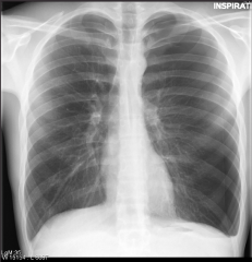
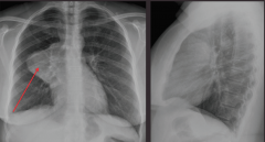
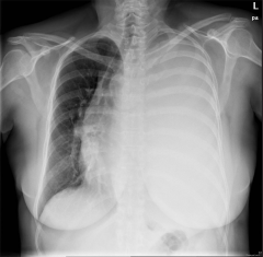
A 20 y.o. male presents with chest pain. Take a look at the x-ray. What is your diagnosis? What are the abnormalities?
|
"Left-sided pneumothorax.
Faint lung outline on the left side, blunting of the left costophrenic angle." |
|
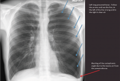
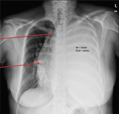
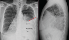
Take a look at the x-ray. What is your diagnosis? What are the abnormalities?
|
"Left tension pneumothorax.
Collapsed lung, structures pushed to the right (trachea, heart)" |
|
|
How and where anatomically do you treat a tension pneumothorax?
|
A needle aspiration with a large bore needle in the second intercostal space along the mid-clavicular line.
|
|
Take a look at the x-ray. What is your diagnosis? What are the abnormalities?
|
"Pleural plaques.
Densities in the lung periphery, on the surface of the diaphragm. Often caused by asbestos exposure." |
|
Take a look at the x-ray. What is your diagnosis? What are the abnormalities?
|
"Pleural effusion.
L side whited out, trachea deviated, more dense on the bottom due to gravity." |
|
Take a look at the x-ray. What is your diagnosis? What are the abnormalities?
|
"Malignant pleural effusion, secondary to breast cancer.
L lower lobe density, fluid forming a meniscule line," |
|
A 62 y.o. smoker presented to his family doctor due to confusion and found to have hypercalcemia. Take a look at the x-ray. What is your diagnosis? What are the abnormalities?
|
Left lung mass (cancer)
|
|
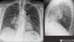
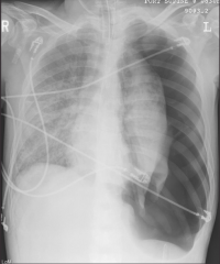
Take a look at the x-ray. What is your diagnosis? What are the abnormalities?
|
"Left upper lobe collapse.
Air on the lateral where the upper lobe should be, collapsed lung shows up as a white density." |
|
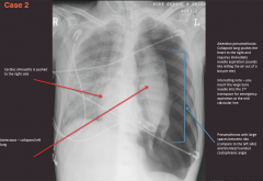
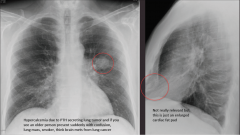
Take a look at the x-ray. What is your diagnosis? What are the abnormalities?
|
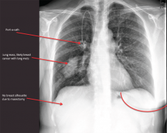
"Right lung mass, likely from breast cancer mets.
Density in R lung, port-o-cath, right breast missing" |