![]()
![]()
![]()
Use LEFT and RIGHT arrow keys to navigate between flashcards;
Use UP and DOWN arrow keys to flip the card;
H to show hint;
A reads text to speech;
117 Cards in this Set
- Front
- Back
|
Things on a microscope I should know:
|
Arm
Base Ocular lens Objective lens Stage Condenser Body tube Iris diaphragm Coarse and fine adjustment |
|
|
Function of coarse adjusement.
|
Raises and lowers stage only on scanning.
|
|
|
Iris diaphragm -
|
Controls the amount of light to the slide and through the condenser.
|
|
|
Condenser -
|
concentrates light on specimen.
|
|
|
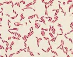
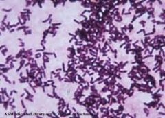
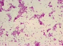
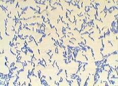
Three morphologies -
|
cocci
bacillus spirilum |
|
|
Total magnification of
Scanning - Low - High - Oil - |
40x 100x 400x 1000x |
|
|
Parfocal -
|
microscopes under focus in scanning will focus again with little adjustment.
|
|
|
True motility -
|
purposful and directional movement.
|
|
|
Brownian motion -
|
static vibration movement.
|
|
|
Streaming -
|
moving with the flow of water
|
|
|
Simple stains - 2 ex.
|
direct stain
negative stain |
|
|
direct stain -
|
stains the bacteria itself
|
|
|
negative stain -
|
stains the background.
|
|
|
what does a heat-fix do?
|
stops the bacteria from washing away during staining.
|
|
|
Differential stain ex. (1)
|
Gram stain
|
|
|
Steps of making a Gram stain -
|
1. Bacteria on slide
2. Air dry 3. Heat-fix 4. Crystal violet 5. Water 6. Gram's Iodine 7. Water 8. 95% ethanol alcohol 9. Water 10. Safranin 11. Water 12. bibulous paper. |
|
|
Mordant for Gram stain -
|
Gram's Iodine
|
|
|
Primary stain for Gram stain -
|
Crystal violet
|
|
|
Decolorizing agent for Gram stain -
|
Ethanol.
|
|
|
Gram positive chracterisics of cell wall - 2.
|
1. thick peptidoglycan
2. teichoic acid. |
|
|
Gram negative chacteristics- 2
|
1. thin peptidoglycan
2. lipopolysaccharides |
|
|
Color of:
Gram negative - Gram positive - |
Pink
purple |
|
|
Following the ethanol alchohol, before safranin, how would :
Gram positive look? Gram negative - |
- purple
- Colorless. |
|
|
Most important step of Gram stain?
|
Decolorizing, if crystal violet left on too long, it will cause both Gram negative and Gram positive to lose their color.
|
|
|
Acid fast stain:
Primary stain - Decolorzing agent - Counterstain - |
carbolfuchsin dye
acid alcohol Methylane blue |
|
|
Acid fast cells color -
non acid fast cells color - |
- red
- blue |
|
|
acid fast cell wall contain -
|
mycolic acid
|
|
|
Capsules - what these bacteria have to cause them
|
outside layer of polysaccharide
|
|
|
Endospore stain procedure:
|
1. Smear bacteria and one drop of water on slide.
2. airdry 3. Heat-fix. 4. bibulous paper on slide 5. malachite green 6. water 7. safranin 8. water |
|
|
Why do endospores require harsh treatment to be killed?
|
They live for survival. They don't allow particals to flow into it so harsh conditions need to open pours to allow stain in it.
|
|
|
Endospore appears what color?
The rest will appear ? |
Green
pink or red |
|
|
Disease caused by Acid Fast bacteria -
|
Tuberculosis
|
|
|
2reasons why capsules are helpful
|
1. increase virulence
2. attach to surfaces easily. |
|
|
Why are capsule forming bacteria harmful for humans?
|
Immune system doesn't respond to it quickly and immune against antibiotics.
|
|
|
Mordant in acid fast stain -
|
heat
|
|
|
Inoculate -
|
add bacteria to sterile medium
|
|
|
Incubate
|
put in favorable temps to allow growth
|
|
|
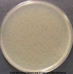
streak plate method -
|
allows million of bacteria to be so spread out that it forms distinguishable colonies
|
|
|
spread plate -
|
dilutes bacteria with 10 mL ofwater to count colonies
|
|
|
Pour plate -
|
diluted cells are mixed with agar then into petridish.
|
|
|
aseptic technique -
|
1. flame loop, cool
2. take off cap with pinky 3. flame top 4. get bacteria 5. flame top 6. put cap on 7. spread bacteria on slide 8. flame loop |
|
|
How many bacteria per ml were there in the original culture tube?
30 colonies in the 10^-7 |
30/10^-7 x (10^7/10^7) = 3.0x10^8 bacteria/mL
|
|
|
Nutreint broth tuves are sterile when...
|
the media is clear
|
|
|
Nutrient agar plates are sterile when...
|
the plate is clear even after incubation.
|
|
|
colony -
|
one cell grows and divides to make million of bacteria.
|
|
|
Plaque formation -
|
Fromed by bacteriophages because bacteriophage is a virus, so the virus kills part of the lawn to make the clear spots.
|
|
|
Metabolism -
|
all chemical reactions that occur within living organims.
|
|
|
oxygen requirements - 5
|
1. Obligate aerobes
2. Obligate anaerobes 3. Faculative anaerobes 4. Microaerophiles 5. aerotolerant |
|
|
Obligate aerobes -
|
require oxygen to grow
|
|
|
Obligate anaerobes -
|
Can only survive with no oxygen.
|
|
|
Faculative anaerobes -
|
grow with or without oxygen, but bette with oxygen
|
|
|
Microaerophiles -
|
small amount of oxygen
|
|
|
Aerotolerant -
|
grow with or without oxygen.
|
|
|
Catalase:
Media used? Reagent added? Positive test - if positive, what present? |
slide
hydrogen peroxide Bubbles catalase |
|
|
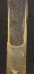
Gelatin Hydrolysis:
Media used: Reagent added? Positive test? If positve, what present? Negative test - |
test tube
ice Liquid Produces proteases Solid |
|
|
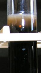
Hydrogen sulfide production:
Media used: Reagent added- Positive test - What present if positive? Negative - |
SIM tube
iron sulfate 75% black iron sulfide small amount or no black. |
|
|
Oxidase:
Media used- Positive test - What is present? Negative test - |
oxidase dryslide
color change to blue Cytochrome C No color change. |
|
|
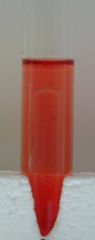
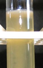
Glucose/Dextrose Fermentation/ Lactose/ Sucrose:
Media used Reagent added Positive? Whats in it? Negative? |
- Durham tubes
- Phenol Red - Yellow/Orange and bubbles - Acid - red/pink, so it's base and no bubbles |
|
|
Starch hydrolysis:
Media used: Reagent added Positive - what's in it? Negative? |
- Starch plate
- Iodine - clear zone/ yellow halo Amylase present - no clear zone. |
|
|
Urea production:
- Media used Reagent added - positive - what's in it? Negative - |
- Small tube
- phenol red - pink - Urease present - orange. |
|
|
Oxygen requirement:
- media used? Positive: What requirement is it? Negative? What requirement is it? |
- Thioglycollate tube (FTM)
- Green present - Faculative anaerobe - No green present - Obligate aerobe. |
|
|
Why use biochemical test to determine unknown?
|
Because Gram stain only determines characterstics in cell wall, while the biochemical tests test multiple things to distinguish between them.
|
|
|
Why can't we test oblifate anerobe?
|
We live in an oxygen rich environement.
|
|
|
What two compounds are produced by action of urease on urea?
|
Ammonia and carbon dioxide
|
|
|
Breaking down the previous to results in what to ph?
|
Increases it.
|
|
|
Disinfectants -
|
chemicals used on inanimate objects
|
|
|
Antiseptics -
|
chemicals that are used on the skin
|
|
|
Bactericidal agents -
|
killing bacteria.
|
|
|
Bacteriostatic agents -
|
temporary inhibiting growth/divide
|
|
|
Phenol coefficeint -
|
compare an antibiotic to phenol to determine if it's strong or weak.
|
|
|
Chemotherapeutic agents/ antibiotics -
|
antimicrobial compounds that can be taken internally.
|
|
|
MIC -
|
minimum inhibitory concentration
|
|
|
MIC - definition -
|
the smallest value of antibiotics that still do the job.
|
|
|
Kirby-Bauer agar method:
|
1. Microorganisms spread over plate to form lawn
2. Paper discs impregnated with various antibiotics are spread on surface. 3. incubation |
|
|
Zone of inhibition -
|
the area of no growth
|
|
|
Big zone -
|
bacteria is inhibited
|
|
|
Narrow spectrum -
|
antibiotics that can work against one type
|
|
|
Broad spectrum -
|
antibiotics that can work against multiple.
|
|
|
Why would it be important to know the pathogen before treating with antibiotics?
|
Certain antibiotics can work faster and better than others. Also some bacteria can resist certain antibiotics.
|
|
|
Sterile -
|
totally free from organisms.
|
|
|
Physical methods -
|
Radiation and Heat and UV
|
|
|
Chemical methods -
|
disinfectents
|
|
|
How does heat work?
|
denatures the enzymes to inhibit growth.
|
|
|
Thermal death time -
|
length of time required to kill all bacteria at a given temp
|
|
|
The higher the temp....
|
shorter the TDT
|
|
|
Autoclave requirements -
|
15 minutes
121 celcius 15 pounds of pressure |
|
|
UV kills by...
|
creating thymine dimers in the DNA to cause inhibition of growth.
|
|
|
Epidemiology -
|
study of how, when where, what and who are involved with the spread and distribution of diseases in a population
|
|
|
infectious disease -
|
a disease caused by microorganisms that enter the body and multiply in tissues.
|
|
|
Communcable -
|
able to be transferred.
|
|
|
Epidemic -
|
If the number of newly reported cases in a given period of time in a specific area is excessive.
|
|
|
Ex of epidemics -
|
Ebola, black plague, infuenza.
|
|
|
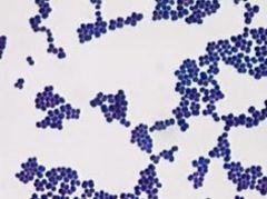
Staphylococci -
Streptococci |
clumps of circles
links of circles. |
|
|
Streptobacilli - |
Links of rods
|
|
|
- Gram positve
- Cocci - staphylococci |
|
|
- Gram negative
- bacillus - diplobacilli/streptobacilli |
|
|
- Gram positive
- bacillus - staphylobacilli |
|
|
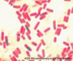
Acid Fast cells
bacillus |
|
|
non acid fast cells
bacillus |
|
|
Capsules
bacilli |
|
|
Endospores
bacilli vegetative cells - ones just pink. |
|
|
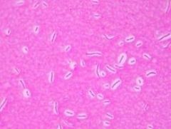
Negative sucrose/lactose/glucose test.
Basic/ no bubbles. |
|
|
Postive sucrose/lactose/glucose test
Acidic/ bubbles. |
|
|
Positive for sucrose/lactose/glucose
acidic , but no bubbles. |
|
|
Positive starch hydrolysis.
|
|
|
Negative starch hydrolysis.
|
|
|
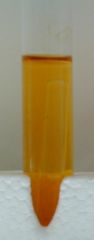
negative gelatin hydrolysis.
|
|

|
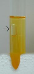
Positve geletin hydrolysis.
|
|
|
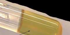
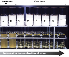
Minimum inhibitory concentration for antibiotics. The first clear one is the minumum anount needed.
|
|
|
positive urea production
|
|
|
Negative urea production
|
|

|
Positve catalase test.
|
|
|
Negative catalse test.
|
|
|
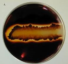
Positve hydrogen sulfide production
|
|
|
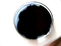
Negative hydrogen sulfide production
|
|
|
Growth in a plate?
|
|
|
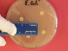
Measuring zone of inhibitionDiameter
|