Reading...
![]()
Play button
![]()
Play button
![]()
Use LEFT and RIGHT arrow keys to navigate between flashcards;
Use UP and DOWN arrow keys to flip the card;
H to show hint;
A reads text to speech;
52 Cards in this Set
- Front
- Back
|
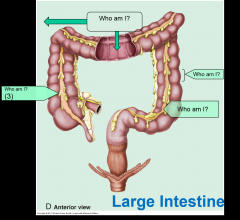
What structures are located in the mid gut?
|
1. Distal half of duodenum
2. Jejunum & ileum 3. Cecum & vermiform appendix 4. Ascending & transverse colon |
|
|
What structures are located in the hind gut?
|
1. descending colon
2. sigmoid colon 3. rectum |
|
|
What vertebral level does the jejunum begin at?
|
L2
|
|
|
Where does the ileum end?
|
Ends at the ileocecal junction/ right sacroiliac joint
|
|
|
What is the difference b/e the jejunum & ileum?
|
Jejunum Ileum
1. fewer arcades 1. more arcades 2. larger diameter 2. fat near bowel 3. fat near the root 3. smaller diameter 4. Thicker wall 4. thinner wall 5. more plica circularis 5.less plica circularis |
|
|
What holds the jejunum/ileum to the posterior abdominal wall?
|
The mesentary
|
|
|
What provides the pathway for vessels and nerves that serve the jejunum and ileum?
|
The mesentary proper
|
|
|
Where is the root of the mesentary proper located?
|
Extends from left of L2 diagonally to the right sacroiliac joint
|
|
|
What does the mesentary proper cross over?
|
1. The duodenum
2. Aorta & IVC 3. Right psoas major, ureter, gonadal vessels |
|
|
What does the mesentary proper contain?
|
Superior mesentaric artery/branches and vein/tributaries.
Superior mesentaric nerve |
|
|
|
|
|
Where is the right colic flexure/hepatic flexure?
|
The flexure b/e ascending and transverse colon.
|
|
|
What is the left colic flexure?
|
The "splenic flexure" b/e the transverse and descending colon.
|
|
|
What part of the large intestine is intraperitoneal?
|
1. Cecum
2. Vermiform appendix 3. Transverse colon 4. sigmoid colon |
|
|
What part of the large intestine is retroperitoneal?
|
1. ascending colon
2. descending colon |
|
|
What fossa contains the appendix?
|
The right iliac fossa
|
|
|
Where is the opening for the appendix?
|
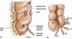
~3cm below the ileocecal orifice
|
|
|
What artery runs through the superior ileocecal fold/recess?
|
A branch of the ileocolic artery
|
|
|
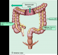
Where is the origin of the taenia coli?
|
At the base of the appendix.
|
|
|
Where is pain referred to from the appendix?
|
Afferent (pain) fibers refer pain to the T10 dermatome.
|
|
|
What Sy is caused by retrocecal appendicitis?
|
Psoas sign:
passive: pain on passive extension of right thigh active: flexion of the right hip against resistance |
|
|
what is the Sy that is caused by a pelvic appendix?
|
Obturator sign (pain upon internal rotation of leg w/ hip and knee flexed)
*caused by pressure of the inflamed appendix lying on the obturator muscle |
|
|
What is Rovsing's pain?
|
Pressure of lower left quadrant elicits pain in the right lower quadrant
|
|
|
Where is a meckel's diverticulum located?
|
Located 2 feet proximal to the ileocecal valve in the mesentaric border of ileum.
|
|
|
What artery supplies the transverse mesocolon?
|
middle colic artery
|
|
|
What do the branches of the superior mesentaric artery supply?
|
distal duodenum, jejunum/ileum, large intestine to left colic flexure.
|
|
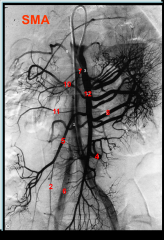
Who are we?
|
1: Aorta
2: Appendicular 3: catheter in SMA 4: ileal brs of SMA 5: ileocolic artery 6: common iliac artery 7: inferior pancreaticoduodenal 8: jejunal brs of SMA 9: lumbar artery 10: middle colic artery 11: SMA |
|
|
What do the branches of the inferior mesentaric artery supply?
|
From left colic flexure to the upper half of anal canal.
|
|
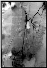
Who are 3, 5, 8 and 10?
|
Branches of IMA:
3: inferior mesentaric artery 5: left colic artery 8: sigmoidal br. 10: superior rectal artery |
|
|
What is the anastomosis b/e the celiac trunk and SMA?
|
Via pancreaticoduodenal artery
|
|
|
What is the anastomosis b/e the superior and inferior mesentaric arteries?
|
1. middle and left colic arteries
2. Riolan and marginal anastomosis |
|
|
What is the anastomosis b/e the IMA and internal iliac artery?
|
Superior rectal artery and middle or inferior rectal artery
|
|
|
What vertebral level does the rectum begin at?
|
S3
|
|
|
What does the ileocolic artery supply?
|
Cecum, appendix, proximal part of ascending colon and distal ileum
|
|
|
What does the middle colic artery supply?
|
The proximal half of the transverse colon.
|
|
|
What does the left colic artery supply?
|
Distal half of the transverse colon and descending colon.
|
|
|
What do the sigmoid arteries supply?
|
Sigmoid colon
|
|
|
What does the superior rectal artery supply?
|
The rectum and proximal half of the anal canal.
|
|
|
What is the marginal artery of Drummond?
|
A composite artery that courses along the inner border of the colon from the ileocecal to the rectosigmoid junction.
|
|
|
What is the clinical importance of the marginal artery of Drummond?
|
Provides collateral circulation b/e the left branch of the middle colic artery and the branches of the inferior mesentaric artery to the left side of the colon.
|
|
|
Where does the IMA run?
|
Descends retroperitoneally along the left side of the abdominal aorta
|
|
|
What does SMA cross anterior to along its course?
|
The left renal vein and the horizontal part of the duodenum
|
|
|
What is the lymphatic drainage above the upper half of rectum?
|
(celiac, superior mesentaric and inferior mesentaric nodes) => intestinal lymphatic trunk => cisterna chyle => thoracic duct
|
|
|
What vertebral level does the anal canal begin at?
|
Tip of the coccyx
|
|
|
What muscle forms a sling to form the anorectal angle?
|
The puborectalis muscle
|
|
|
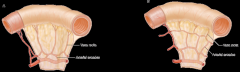
What are Houston's valves?
|
Two left transverse folds and one right transverse fold help prevent leaks.
|
|
|
What is the epithelium above and below the pectinate line?
|
Hindgut (above): stratified columnar
Proctodeum (below): stratified squamous |
|
|
What is the arterial supply above and below the pectinate line?
|
Hindgut (above): Superior rectal from IMA
Proctodeum (below): middle rectal (form internal iliac) and inferior rectal (from internal pudendal br of internal iliac) |
|
|
What is the venous drainage above and below the pectinate line?
|
Hindgut (above): Superior rectal to (IMV) to hepatic protal vein (liver)
Proctodeum (below): middle rectal (to internal iliac v) and inferior rectal veins to internal pudendal vein (IVC) |
|
|
What is the lymphatic drainage of above and below the pectinate line?
|
hindgut (above): internal iliac nodes
proctodeum (below): superficial inguinal nodes |
|
|
What is the innervation above and below the pectinate line?
|
Hindgut (above): autonomics (S2-4) pelvic splanchnic
Proctodeum (below): somatic via pudendal n. |
|
|
What is the afferent (sensation) above and below the pectinate line?
|
Hindgut (above): only to pressure and distention
Proctodeum (below): exquisite sensitive to touch and pain. |