Reading...
![]()
Play button
![]()
Play button
![]()
Use LEFT and RIGHT arrow keys to navigate between flashcards;
Use UP and DOWN arrow keys to flip the card;
H to show hint;
A reads text to speech;
124 Cards in this Set
- Front
- Back
|
How did scientists discover what part of the brain does what?
|
1.Electrically stimulating different cortical areas
2. Studying deficits in people with brain lesions |
|
|
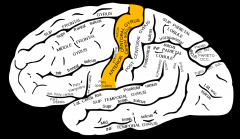
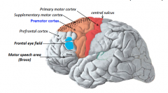
Where is the primary motor cortex located?
|
precentral gyrus
|
|
|
Show the precentral gyrus
|
|
|
|
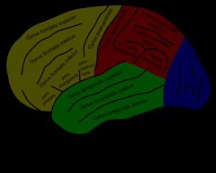
What are the three horizontal gyri in front?
|
Superior, Middle, and Inferior Frontal Gyrus
|
|
|
Show the 3 frontal gyri. (yellow)
|
|
|
|
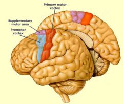
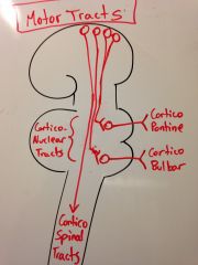
What are the three separate areas in the cortex that control motor function?
|
1. Primary motor cortex
2. Premotor area 3. Supplementary motor area |
|
|
Can you show what they look like in the brain?
|
|
|
|
How do you remember that the premotor cortex is the one that is anterior and the supplemental motor cortex is superior and medial?
|
Pre seems to mean anterior in the brain so premotor will go in front.
|
|
|
What do the pre and supplemental motor cortexes do?
|
They are involved in programming movements before they are sent to the primary cortex to be executed.
|
|
|
How are the pre and supplemental cortexes different in function?
|
premotor- advanced for limbs and talking
supplementary motor- primitive movement like the trunk and hip |
|
|
What does this mean to trying to actively resist an urge?
|
Your brain is already halfway down the road of giving in.
|
|
|
What kind of sensory input do you need before executing a planned conscious motor action?
|
You need proprioception information about where your body is so that you know where your starting point is.
|
|
|
What else do you need to have before executing an action?
|
An idea of what you would like to achieve, which can come from many different places in the brain.
|
|
|
Are you only moving the muscles that you plan to move?
|
No, on their way down, motor neurons send off collateral branches to adjust postural muscles to hold the rest of your body stable. This is unconscious.
|
|
|
So does this mean that there is one or multiple motor tracts going down?
|
Multiple. One straight from consciousness (cortical) and others that are subcortical.
|
|
|
What kinds of decision making takes place in the primary motor cortex?
|
This area houses the final motor neurons that are just told what to do by the pre and supplemental motor cortexes.
|
|
|
What happens when you stimulate one area of the primary motor cortex?
|
you get one meaningless movement like a knee jerk
|
|
|
What happens when you stimulate one area of the pre or supplemental motor cortex?
|
You get a meaningful program of actions like a word.
|
|
|
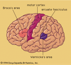
Imagine the area that allows you to execute a constellation of actions to speak. What is it called?
|
|
|
|
What muscles do you need in order to speak?
|
Larynx, tounge, facial muscles
|
|
|
So what types of neurons are on the primary motor cortex next to Broca's area?
|
Those very motor neurons!
|
|
|
Mnemonic to differentiate Broca's and Wernicke's areas?
|
Tom Broca is a reporter who really likes to talk!
|
|
|
What do pt's experience when they have damage to Broca's area?
|
They know what they want to say (planning is intact), but they can't say it (programming portion is missing)
|
|
|
What area is above Broca's area on the premotor cortex?
|
the frontal eye field
|
|
|
What does the frontal eye do?
|
It is the push button for the eyes and will cause eyes to shift to the contralateral side.
|
|
|
Show the frontal eye field.
|
|
|
|
How are the frontal eye fields operating normally?
|
Both sides are equally active.
|
|
|
What is you have a lesion on the right frontal eye field?
|
Your eyes will look to the left.
|
|
|
What happens to activity of neurons when they are irritated?
|
They will become over active.
|
|
|
What happens in the short and long term when you have a tumor in the right frontal eye field? Why?
|
Short term- look to left because tumor substances are iriitating the neurons
Long term- look to right side because tumor has destroyed it |
|
|
Mnemonic for what irritation vs destruction does to your eye position.
|
If someone is irritating you, you look away. But if they start to destroy your stuff, you have to look towards.
|
|
|
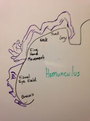
What is right above the frontal eye field? Mnemonic?
|
The area controlling neck movement because this usually accompanies eye movement.
|
|
|
What is above the area the neck control area?
|
The area for controlling fine hand movement.
|
|
|
Premotor cortex controls movement contralaterally. How does the supplemental motor cortex control movement?
|
bilaterally!
|
|
|
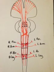
Draw the homunculus.
|
|
|
|
Why is the homunculus useful for diagnosing?
What do you notice about where the homunculus bends? |
Depending on the dysfunction you have, you can be having damage to that area of the brain.
The homunculus bends at the hips. |
|
|
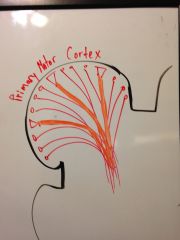
Where do descending motor fibers come from? (give percentages and layer)
|
5th layer of cerebral cortex
primary motor cortex- 30% supplemental and premotor cortexes- 30% Sensory motor cortex- 40% |
|
|
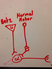
What is special about some of the motor neuron cell bodies? Which area do they come from and what are they called?
|
There are large cell bodies found only in the primary motor cortex called cells of Betz.
|
|
|
How large are cells of Betz? (compare to RBC)
|
60 um (about 8x a RBC)
|
|
|
Are most of the cells in the primary motor cortex cells of Betz?
|
No, only some.
|
|
|
What is special about the axons from cells of Betz?
|
they are large and heavily myelinated.
|
|
|
How much larger are the axons of the cells of Betz?
|
4 times larger than others at 16um.
|
|
|
What is special about the connection the Betz cells have with alpha motor neurons?
|
They directly stimulate alpha motor neurons whereas regular ones stimulate an interneuron to do the job.
|
|
|
Show the comparison of the Betz cell connection.
|
|
|
|
What are these interneurons called?
|
Internunchal neurons
|
|
|
How many descending motor neurons are there?
|
1 million come down from each cerebral hemisphere.
|
|
|
How many of each million are coming from the Betz cells?
|
30k
|
|
|
Where do all the descending cortical motor fibers converge on? What is the fanned part called?
|
The posterior limb of the internal capsule. The fanned part is also called the corona radiata.
|
|
|
What is the pathological implication of the convergence of all these fibers?
|
If you have damage at that level, you will have massive hemiparesis (and loss of sensation too), but if you get it further up, you will only lose some function.
|
|
|
What tends to go wrong pathologically at this convergence area for the motor and sensory fibers?
|
ischemia of the lenticulostriate arteries
|
|
|
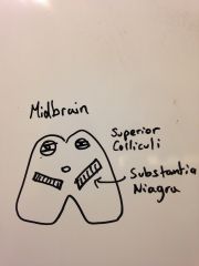
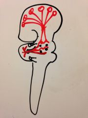
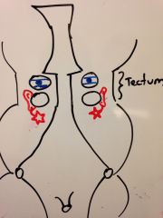
Draw the transection of the top of the midbrain. Include the substantia niagra.
|
|
|
|
Why are there eyes on top?
|
That is the superior colliculus, which is part of the tecta which are EYES because they regulate the visual reflex!
|
|
|
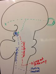
Give an example of the spinovisual reflex?
|
You step in mud and you immediately turn your eyes to look at it.
|
|
|
Give an example of the visual-spinal reflex?
|
you see a ball coming at you and you
|
|
|
Why is the substantia niagra named what it is?
|
Because it is a black substance.
|
|
|
What disease does damage to the sbstantia niagra result in?
|
Parkinson's.
|
|
|
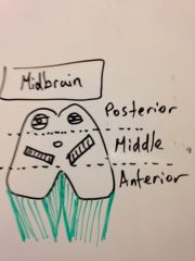
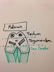
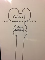
Draw the 3 different sections of the transection of the midbrain.
|
|
|
|
Now label each part.
|
|
|
|
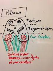
Show which part the cortical spinal tract goes through the midbrain. Does it go through this whole part?
|
|
|
|
What happens to the motor neurons in the pons?
|
Some get dispersed by the pontine nuclei and then converge again at the medulla.
Others terminate on the pontine nuclei and go to update the cerebellum. |
|
|
What are these motor fibers terminating in the pons called? Why?
|
cortical pontine fibers. (for where they originate and end)
|
|
|
Draw what the motor pathway from the cortex to the cerebellum look like.
|
The pontine nuclei send all the descending motor info posterior to the cerebellum.
|
|
|
What happens to the cortico-spinal motor neurons in the pon?
|
They get dispersed by all the pontine nuclei and then reconverge again.
|
|
|
If you get a lesion in the pons, how much motor loss will you get? Why?
|
Only some because they are spread out.
|
|
|
Show where the motor corticospinal tracts pass through the medulla? (transection) What are these tracts called?
|
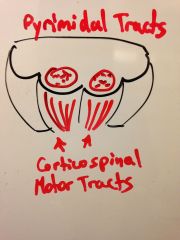
|
|
|
Show where the pyrimidal motor tracts cross. What is this called?
|
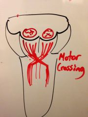
|
|
|
How many of the fibers decussate here at the Junction of medulla and spinal cord?
|
90%
|
|
|
What are the 10% that don't cross?
|
The medial 10% which will control axial muscles like the trunk.
|
|
|
What are the tracts that haven't crossed at the motor crossing called? Why?
|
Ventral or anterior descending motor tracts because they go through the anterior columns.
|
|
|
What are the tracts that have crossed at the motor crossing called? Why?
|
lateral descending motor tracts because they go through the anterior columns.
|
|
|
Will all of these motor neurons terminate at an alpha motor neuron?
|
Only the 3% that are from Betz cells. The rest will go through a internunchal neuron (interneuron).
|
|
|
Show where the cell bodies for the axial vs appendicular alpha motor neurons are.
|
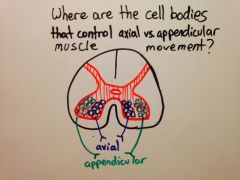
|
|
|
Where are the neurons that terminate in the medial anterior horn from? Why?
|
The supplemental motor cortex because they are more primitive motions!
|
|
|
When do the axial motor neurons decussate? Mnemonic?
|
The the level of their alpha motor neuron. They are those AX-holes that swerve off the interstate at the last minute to their exit.
|
|
|
Show the path of the motor neurons from the cortex to their exit points.
|
|
|
|
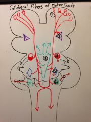
So overall, what is the point of making a lot of motor collateral connections in the brain?
|
It informs other parts of the brain about what it plans to do so they can support the movement!
|
|
|
Show the collateral neurons given by the descending motor neurons and where they send the signals to. (6)
|
|
|
|
Can you list all 6 of these structures?
|
1. Motor cortex 2. Lenticulate nucleus
3. Reticular formation 4. Red Nuclei 5. Vestibular nuclei 6. Olivary nuclei |
|
|
Why send collaterals of the motor fibers to the motor cortex? (Mnemonic?)
|
So it knows what you have decided to do. (Like telling your wife you are leaving the house)
|
|
|
Why send collaterals of the motor fibers to the lenticulate nucleus?
|
It's part of the basal ganglia and will help with execute the movement by providing background tone.
|
|
|
Why send collaterals of the motor fibers to the reticular formation?
|
It will wake up the cerebral cortex so you can pay more attention to what you are doing?
|
|
|
What implication does this have for exercise's effect on your alertness?
|
Going for a jog will increase your brain capacity on a physical neurological level.
|
|
|
Why send collaterals of the motor fibers to the red nucleus? What functions does it help with?
|
this is where you will control wrist tone which will help execute functions like writing.
|
|
|
Why send collaterals of the motor fibers to the vestibular nuclei?
|
They are antigravity muscles that make sure you don't fall while executing the function.
|
|
|
Why send collaterals of the motor fibers to the olivary nuclei?
|
They will inform the cerebellum of your plan so it can integrate that with it's information about where your body is and compare that with what happens.
|
|
|
What is a lower motor neuron?
|
Neurons that come out of the CNS and terminate on a neuromuscular junction.
|
|
|
So are preganglionic, S, and PS neurons considered lower motor neurons?
|
NO! Because they end on a ganglion.
|
|
|
Can lower motor neurons only come out of the CNS from the spinal cord?
|
No, they can also come out of the brainstem.
|
|
|
What is the definition of an upper motor neuron?
|
All the neurons that come from the upper level and connect with lower motor neurons and altering their activity (directly or indirectly)
|
|
|
What are the upper motor neurons that control the cranial nerves called? Why? (Hint, the ones we already learned about are corticospinal neurons)
|
Corticonuclear neurons because they terminate in nuclei of the brainstem.
|
|
|
What are the special corticonuclei in the medulla called?
|
corticobulbar
|
|
|
Draw out the two kinds like corticonuclear fibers.
|
|
|
|
Which nerves are lower motor neurons that come from the brainstem and thus the corticonuclear tracts?
|
Cranial nerves
|
|
|
What do the lower colliculi regulate?
|
auditory reflexes
|
|
|
Draw all the neurons that go to the superior coliculi and label them.
|
|
|
|
Draw the posterior view of the whole tectum and what functions they serve. (Image mnemonics)
|
|
|
|
So if a tract is ascending to the tecta from the spine, where is it going? Why?
|
To the superior colliculi because it is the spinotectal tract for the visual reflex. (hearing does come from spine)
|
|
|
What other sensory fiber goes to the superior colliculi? What other part of the brain are they on their way to?
|
sensory info from the eyes which are on their way o the occipital lobe.
|
|
|
What is the tract that coordinates reflexive muscle movement to avoid a car that you see speeding by called?
|
the tectospinal tract.
|
|
|
Do the tectospinal fibers cross? Where? Mnemonic?
|
Yes, they cross at the level they exit (think primitive reflex)
|
|
|
What are the tracts comiing from the red nucleus called? Where do they cross? Mnemonic?
|
rubospinal tracts. In the brain stem (think about it, they support modern hand movements like writing)
|
|
|
What are the tracts comiing from the vestibular nuclei called?
|
vestibulospinal tract
|
|
|
What are the tracts comiing from the medullary olives called?
|
olivaryspinal tract
|
|
|
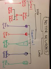
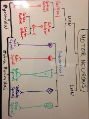
Show the locational difference between a upper motor neuron of cortical origin and subsortical origin?
|
|
|
|
Draw out a flow chart of the type of motor neurons and which tracts are involved in each. (8 tracts)
|
|
|
|
Show which tracts in this flow chart are pyrimidal vs extrapyrimidal.
|
|
|
|
What is another name for the corticospinal tracts? Why?
|
Pyrimidal tracts because they go through the pyrimids of the medulla.
|
|
|
Why are the corticonuclear tracts considered pyrimidal tracts?
|
they are functionally very similar to the corticospinal tracts (originate from cortex) so they are grouped together even though they don't anatomically pass through the pyrimids.
|
|
|
Why did scientists find it useful classify tracts as pyrimidal or extrapyrimidal in the past?
|
They though pyrimidal tracts were responsible for more advanced motor function and extrapyrimidal for more primitive ones like posture and reflexes.
|
|
|
Why are we moving away from these terms?
|
Because we are discovering that these rules that distinguish the two tracts don't hold true.
|
|
|
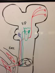
What do you do when a ViP person walks into the room?
|
You STAND UP!
|
|
|
What are the two major groups of muscles in our bodies?
|
Flexor and extensor muscles
|
|
|
What general motions are each group good for?
|
extensor- standing up
flexor- sitting down |
|
|
What worldly force do the extensor muscles counter? Why?
|
gravity because they help you stand up against it
|
|
|
What does ViP stand for?
|
Vestibule nuclei and Pontine-reticular nuclei
|
|
|
Draw out how the ViP's help you stand up. (include muscle involved, the column they travel down, and whether or not they cross sides)
|
|
|
|
What is the working mnemonic of ViP?
|
The vestibulonuclei and pontine-reticular nuclei send fibers down to enhance the tone of extensor muscles to help you stand up.
|
|
|
How do you remember what column the ViP tracts go down?
|
They go down the ipsilateral anterior column because ViPs always go in front and they don't move for anyone!
|
|
|
Which out of those two standing supportive nuclei help more?
|
The vestibulonuclear spinal tract.
|
|
|
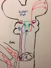
What is the mnemonic for what enhances flexor muscle action?
|
Sit on a RUBBER MAT!
|
|
|
What is the working explanation of sitting on the rubber mat?
|
The rubellonuclei and medullary reticular nuclei will send signals to enhance the tone of flexor neurons.
|
|
|
Draw out how how the rubber mat help the flexor system. (include the column they travel down and whether or not they cross sides)
|
|
|
|
Did I get the orientation of the flexors and extensors right in the knee?
|
No, I reversed them. Extensors are quadripceps and flexors are hamstrings.
|
|
|
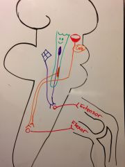
Draw and lateral view of how these assistant tracts connect. (make sure to get their positions correct and note which cross.
|
|