Reading...
![]()
Play button
![]()
Play button
![]()
Use LEFT and RIGHT arrow keys to navigate between flashcards;
Use UP and DOWN arrow keys to flip the card;
H to show hint;
A reads text to speech;
19 Cards in this Set
- Front
- Back
|
Describe the culture and microscopy of microsporum canis.
|
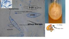
long spindle macroconidia with terminal knob often seen
5-15 cells with most oftenn more than 6 some microconidia seen wart like protusions from thick walls in macroconidia flat mycelium groth in 2-3 days matures in 6-10 days culutre is often white flat mycelial growth with yellow reverse scalp lesion/lesion swab |
|
|
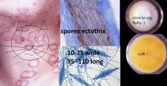
What does Microsporum canis cause? who is it acquired from?
|
- infections of the scalp and skin, sometimes nails
- dogs and cars - clinical presentation - scaling erythematous borders, can be crusting, or cause of suppurative folliculitis - kerion - ecto thix infection |
|
|
What is the clinical picture of the Microsporum canis?
|

Kerion
suppurative folliculitis scalp lesion swab raised erythematous advancing borders xudate |
|
|
Describe the culutre growth of Microsporum canis
|
non pigmented strains can occur
|
|
|
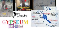
Describe Microsporum gypseum
|
|
|
|
Describe the microscopic picture of M. gypseum
|
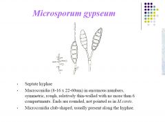
- septate hyphae
- +++ macroconidia - symmetirc rought - thin walls with =/>6 compartments - rounded ends - microconidia club shaped ususally along hyphae |
|
|
Differentiate M. gypseum from M. canis microscopically
|

|
|
|
What is the pathogenicity of M gypseum?
|
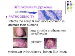
|
|
|
Describe the colonial morphology of M. gypseum
|
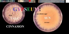
- surface flat and spreading
- powdery/granular - irregular fring border - buff --> tan ---> cinnamon brown |
|
|
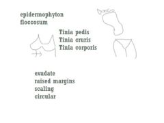
Describ the morphologyical appearance of epidermophyton floccosum
|
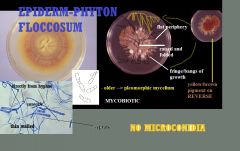
khaki
suede finge raised folded centre flat periphery yellowish brown reverse present growth in 1 week |
|
|
Describe microscopic morphology of Epidermophyton floccosum
|
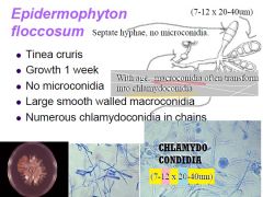
septate hyphae
chlamydospores forming chains from hyphae in older cultrues thin smooth walled long macroconidia no microconidia 7 - 12, 20 -40um |
|
|
What are the clinical symptoms of Epidermo floccosum
|
|
|
|
Describe Thrichophyton verrucosum
|
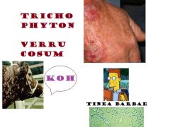
zoophilic
cows KOH demonstrate hyphae tiniae barbae --> head and neck also forearms |
|
|
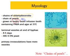
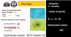
Describe Thrichophyton verrucosum microscopy
|
|
|
|
When is growth fastest for T. verrucosum?
|
|
|
|
Describe T. soudanese
|
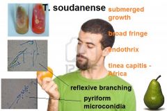
now occasional isolate would wide
tinia capitis in africa flat orange isolate mainly submered fringe slow growing endothrix |
|
|
Describe Trichophyton tonsurans
|
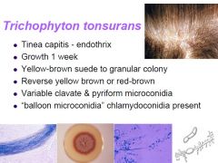
- causes tinea capitis
- endotrhix meaning spores are in the hair follicle - growth in 1 week - yellow brown colony - clavate and pyriform microconidia - balloon microconidia chlamydoconidia present |
|
|
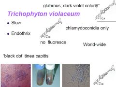
Describe T. violaceum
|
|
|
|
Constuct a table of dermatophytes
|
***
|