Reading...
![]()
Play button
![]()
Play button
![]()
Use LEFT and RIGHT arrow keys to navigate between flashcards;
Use UP and DOWN arrow keys to flip the card;
H to show hint;
A reads text to speech;
21 Cards in this Set
- Front
- Back
|
Ovulation
|
Occurs the 14 day of menstrual cycle. The ovum is release from a graafian follicle into the fallopian tube.
|
|
|
Fertilization
|
Occurs 24-36 hours after ovulation.
Fertilization occurs in the ampula of and cleavage begins. |
|
|
Cell Division Stages
|
-Gamete
-Zygote -Blastomere -Marula -Blastocyst |
|
|
Gamete
|
Female and male reproductive systems.
|
|
|
Zygote
|
Single fertilized ovum
|
|
|
Blastomere
|
Fertilized ovum at 2-cell and 4 cell stage located in the ampulla of the fallopian tube.
|
|
|
Morula
|
Mass of dividing cells located in the isthmus of the fallopian tube
|
|
|
Blastocyst
|
Organized collection of cells which implants in the uterus 7-10 days after ovulation.
The Outer lining has trophoblastic tissue which produce HCG to maintain the corpus luteum cyst. |
|
|
Blastocele
|
An inner fluid filled cavity which contain an inner cell mass which will become the yolk sac, amnion, embryonic disk.
|
|
|
Human Chorionic Gonadotropin
|
HCG is a glycoprotien which is produced by trophoblastic tissue and later by the placenta.
Detectable through maternal serum and urine. Detectable 7 to 10 days after fertilization. Doubles every 2-3 days, plateaus 8-9 weeks. Dectable in two ways Qualitative(negative and positive urine test) Quantitative (provides levels of HCG in blood stream) |
|
|
Primitive Germ Layers
|
Endoderm-(inner) becomes the GI & respiratory system.
Mesoderm-(middle)become musculoskeletal system and circulatory system Ectoderm-(outer) brain,skin, nervous system. |
|
Decidualized Endometrium
|
Decidual Basalis- developed where Blastocyst implants; matetnal contribution to the placenta
Decidual Capsularis- surrounds and cover Blastocyst Decidual Parietalis- results from hormonal influence on uninvolved endometrial tissue |
|
|
Placenta
|
Has both maternal and paternal components
Decidual component is from the Decidual Basalis Paternal component is from trophoblastic tissue |
|
|
Chorion Frondosum
|
Choronic villis developed by 5 weeks LMP.
Choronic villis with connections with decidual Basalis becomes the chorion fondosum, the fetal part of the placenta. |
|
|
Membranes
|
The chorion is the fetal contributions to the placenta proliferation.
The remainder is stretch to fit the gestation sac. The Amnion forms the inner cell mass, the remainder is stretch to fit the chorion. The amnion and chorion begins to fuse by the middle of the first trimester . Fusion is complete by 12-16 weeks |
|
|
Hemodynamic Changes
|
Trophoblastic tissue provides the developing embryo with oxygen and nutrients.
Doppler spectral wave form is high velocity low resistance flow. |
|
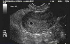
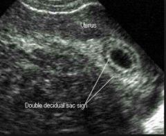
Gestational Sac
|
The first sonographic evidence that a normal intrauterine pregnancy is present. 5week LMP
Normal Songraphic Finding - sac can be round, oval or tear dropped - located in uterine fundus or mid-uterus -double decidual ring -echogenic intact borders -should be measured inner-inner -grows 1mm per day -ys should be present when MSD reaches 8mm TV. |
|
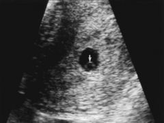
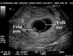
Yolk Sac
|
Yolk sac is the first structure seen within the GS.
Seen when MSD reaches 8mm TV and 20mm TA Measured inner-inner Connected to the embryo by the vitelline duct. |
|
What is considered an abnormal Yolk Sac?
|
When diameter is > 5.6mm between 5 to 10 weeks
|
|
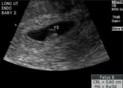
Crown Rump Length
|
The embryo should be seen sonographically when MSD reaches 16mm TV, and 25mm TA.
The embryo grows 1mm per day Most accurate measurement for dating 1st trimester pregnancy. |
|
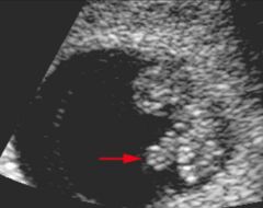
Midgut Herniation
|
The midgut herniates in the umbilical cord at around 9 weeks and return back to the abdominal cavity at 12 weeks.
The herniation is necessary for development of the abdominal vescera. |