Reading...
![]()
Play button
![]()
Play button
![]()
Use LEFT and RIGHT arrow keys to navigate between flashcards;
Use UP and DOWN arrow keys to flip the card;
H to show hint;
A reads text to speech;
69 Cards in this Set
- Front
- Back
How do Dentigerous (Follicular) Cysts appear radiographically?
|
1. Unilocular Radiolucent area associated with the crown of an unerupted tooth
2. Well-defined and often has a sclerotic border 3. Higher rate of rooth resorption (aggressive) 4. Radiolucency should be 3-4 mm |
|
|
What should your #1 differential diagnosis be if you see an impacted tooth with a radiolucency associated with the crown of an impacted tooth?
|
Dentigerous (Follicular) Cyst
|
|
Who is most commonly affected by dentigerous cysts?
|

20-30 yr White Males
Mand 3rd Molars & Max Canines |
|
|
What is the most common developmental cyst?
|
Dentigerous (Follicular) Cyst
|
|
|
What is a cyst that develops by separation of follicle from around the crown of an unerupted tooth?
|
Dentigerous (Follicular) Cyst
|
|
|
T/F
In the older classifications used in the US, the primordial cyst was considered to originate from the cystic degeneration of the enamel organ before the development of the dental hard tissue. Therefore, the primordial cyst was found in place of the tooth. |
True
Almost all Primordial cysts are Odontogenic Keratocysts (OKC), but not all OKCs are primordial cysts |
|
|
T/F
OKCs can be diagnosed using Xrays |
FALSE
OKC diagnosis is Strictly based on histopathology |
|
|
T/F
OKCs have a high reoccurence rate. |
True
Surgeons will go out of their way to make sure they eradicate the lesion completely and do a long term follow up. OKCs can reoccur 10-20 years after their initial diagnosis |
|
|
What are the pathogenic mechanisms of OKCs?
|
1. High proliferation rate of epithelium - Ki-67 staining
2. Overexpression of antiapoptotic protein - Bcl-2staining 3. Overexpression of interface protein - MMPs 2&9, TGF, IL-1a, IL-6 4. Mutations in PTCH tumor suppressor gene 5. Found in basal cell carcinomas & nevoid basal cell carcinoma syndrome 6. Also mutated in syndromic odontogenic keratocysts 6. |
|
|
What is the "not quite" 2/3 rule involved with OKCs?
|

2/3 10-40 yrs
2/3 Men >2/3 Molar ramus areas of mandible (60-80%) Grow in AP direction within the medullary cavity w/o causing bony expansion Multiple OKCs should be tested for Gorlin syndrome |
|
|
Where do the cells of origin come from for an OKC?
|
Cell rests of Dental Lamina
|
|
|
How are OKCs treated and what is their prognosis?
|
Enucleation and curettage
Other than a tendency for recurrence, the overall prognosis of an OKC is good |
|
|
How do OKCs appear radiographically?
|
1. Well-defined radiolucent lesions w/smooth, cortigated margins
2. Unilocular or Multilocular 3. Many are associated with an unerupted tooth (but #1 differential should be dentigerous cyst) 4. May appear similar to: dentigerous, radicular (at the apex), residual, lateral periodontal or globulomaxillary cyst on radiograph |
|

What is the Histological appearance of OKCs?
|

1. Thin, friable wall with little inflammatory infiltrate
2. Luminal surface shows flattened Parakeratotic Epithelial cells with a wavy/corrugated appearance 3. basal cell layer has palisaded layer of cuboidal to columnar cells, which are often hyper chromatic 4. <26% of OKCs have satellite (daughter) cysts |
|

Which of the following is a clinical feature commonly associated with the nevoid basal cell carcinoma syndrome?
a) Skeletal anomalies such as bifid rib b) Odontogenic keratocysts c) Mild ocular hypertelorism d) All of the above e) A & C only |
All of the above
|
|
What are the Major Clinical Features of the Nevoid Basal Cell Carcinoma Syndrome (Gorlin Syndrome)?
|
1. Multiple basal cell carcinomas of the skin
2. Odontogenic keratocysts 3. Epidermal cysts of the skin 4. Palmar/plantar pits 5. Calcified falx cerebri 6. Enlarged head circumference 7. Rib anomalies 8. Mild ocular hypertelorism 9. Spina bifida occulta |
|

How does Gorlin Syndrome (Nevoid Basal Cell Carcinoma Syndrome) relate to genetics?
|
Autosomal Dominant trait with high penetrance and variable expressivity.
High degree of penetrance: if the individual has the gene for a particular entity, their probabilty of expressing the phenotype is high Variable Expressivity: The gene can be expressed in a variety of ways |
|

Approximately 75% of patients that have Nevoid Basal Cell Carcinoma have multiple _______, which histologically are diagnosed as ____ that can either be at the same time or or over a span of time.
|
Jaw Cysts
OKCs |
|

Where do most Lateral Periodontal (Botryoid Odontogenic) Cysts occur?
|
2/3 occur in the Mand Canine-Premolar region
It is a cyst occuring in the lateral periodontal region Inflammatory origin or an OKC has been excluded by clinical and histological means |
|

How do you rule out Lateral radicular cysts when comparing to Lateral periodontal cysts?
|
Lateral periodontal cysts are usually asymptomatic and the associated tooth is Vital, thus ruling out the lateral radicular cyst
|
|
|
What does Botryoid mean?
|
Lateral Periodontal (Botryoid Odontogenic) Cyst
Botryoid = Cluster of Grapes |
|
|
Are Calcifying Odontogenic Cysts (COCs) true cysts?
|
YES & NO
There is a large group of these that are true cysts that behave in a more indolent factor (most COCs are cystic). At the end of the spectrum is a neoplastic group that doesn't have a cystic lumen in them and they tend to behave more aggressively (these present as solid masses) |
|
Who is affected most by COCs?
|

33 yrs
Females (Duh...COCs are not to be used on men, although this still occurs...but I digress) Max Mand Incisor-canine regions COCs associated w/odontomas occur in younger patients while the neoplastic variants appear to occur more frequently in older patients |
|
|
T/F
Neoplastic COCs make up a very Small amount of the total COCs |
True
86-98% of COCs are Non-neoplastic cysts |
|

What are the most characteristic histological features of COCs?
|
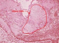
Ghost Cells, which are eosinophilic epithelial cells characterized by the loss of their nuclei. (note: ghost cells may also be found in other lesions so their presence is not an absolute positive indicator of COC)
|
|
|
What are different types of COCs?
|
Gorlin Cyst
Dentinogenic Ghost Cell Tumor Calcifying Cystic Odontogenic Tumor Calcifying Ghost Cell Odontogenic Cyst |
|
|
What is the most common developmental odontogenic cyst?
|
Dentigerous Cyst
|
|

Whatt is the difference between a lateral periapical cyst and a lateral periodontal cyst from a clinical perspective?
|
Vitality of the tooth
Lateral Periodontal Cyst is Vital |
|
|
What is the typical radiographic appearance of an adult gingival cyst?
|
There isn't one (trick question)
|
|
|
What are the cells of origin of the lateral periodontal cyst?
|
Dental Lamina
|
|

What are the features of the Nevoid Basal Cell Carcinoma Syndrom patient?
|

1. Calcification of the falx cerebri
2. OKCs 3. Hypertelorism 4. Bifid rib or other rib anomalies 5. Basal cell carcinomas 6. Frontal bossing |
|
|
What is the most common clinically significant (not the most common) Odontogenic tumor?
|
Ameloblastoma
|
|
|
What is the big significance of an OKC?
|
Reoccurrence
|
|
|
Are Dentigerous cysts and Eruption cysts true cysts?
|
True cysts: Dentigerous cysts, Eruption cysts
|
|
|
T/F
The dentigerous cyst by far represents the most common type of pathologic radiolucency |
False
|
|
|
T/F
The dentigerous cyst is the 2nd most common odontogenic cyst; the radicular cyst being the most common |
True
|
|
|
T/F
Calcifying Odontogenic Cysts may have small foci of calcified material which appear as white flecks in the cystic radiolucency |
True
|
|
|
T/F
Calcifying epithelial odontogenic tumors are always associated with unerupted teeth |
False
|
|
|
What are the sub-classifications of Odontogenic Cysts?
|
Developmental Odontogenic Cysts
Inflammatory Odontogenic Cysts |
|
|
What is the definition of a cyst?
|
A cyst is a cavity (pathologic, often fluid-filled) that is lined by epithelium
|
|
|
What makes a cyst an odontogenic cyst?
|
Cysts lined by epithelium derived from odontogenic epithelium
|
|

What is an uncommon Inflammatory Odontogenic Cyst that characteristically develops on the buccal aspect of the mandibular 1st molar in the bifurcation area?
|
Buccal Bifurcation Cyst
|
|
|
What are 3 Inflammatory cysts?
|
1. Inflammatory Odontogenic Cysts
2. Periapical or radicular cysts 3. Residual cyst |
|
What else do Buccal Bifurcation cysts commonly present with?
|
Proliferative periostitis (onion-skinned)
Pediatric patients Inflammation spreads to the apex through the bone and produces a layering effect on the inferior border of the mandible of a new reactive bone formation |
|
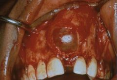
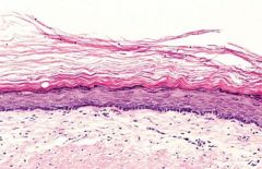
What is this a histological slide of?
|
A Primordial Cyst that is also an OKC
|
|

Do OKCs have orthokeratin or parakeratin on their surface?
|

OKCs have parakeratin (nuclei are present) epithelium on the surface
|
|

What is significant about the cells between the epithelium and connective tissue?
|
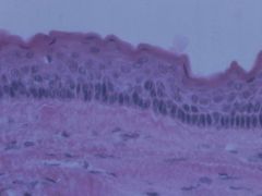
The basal cell layer is hyperchomatic and these cells are palisaded (lined up).
|
|
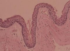
What type of epithelium is on this odontogenic cyst?
|
Orthokeratinized
|
|
|
What is the most common odontogenic tumor?
|
Odontoma (not a true neoplasm)
|
|
|
What are the cells that most often give rise to the apical periodontal cysts?
|
Rests of Malassez
|
|
|
What do odontoblasts differentiate from?
|
Mesenchymal tissue
|
|
|
What does the dental papilla become?
|
Pulpal tissue
|
|
|
Where do the rests of malassez come from?
|
Hertwig's root sheath
|
|
|
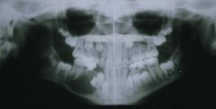
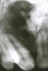
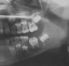
The lesion at the tip of the arrow is most likely which of the following?
a) Compound odontoma b) Complex odontoma c) Mandibular torus d) Idiopathic osteosclerosis e) pellets from a gunshot wound |
Complex odontoma
|
|
|
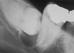
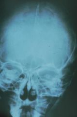
Which of the following statements is true concerning the pericoronal radiolucency shown in this projection?
a) A unicystic ameloblastoma should be considered in the differential diagnosis b) A dentigerous cyst should head the list of differential diagnoses c) Upon histologic examination the lesion could be an odontogenic keratocyst d) All of the above e) A & C |
All of the above
|
|
|
Which cyst is not an odontogenic cyst?
a) Follicular b) Primordial c) Lateral periodontal d) Incisive canal cyst e) Dentigerous |
Incisive canal cyst
|
|
|
Which cyst develops in place of a tooth?
a) Dentigerous cyst b) Fissural cyst c) Primordial cyst d) Odontogenic keratocyst e) Follicular cyst |
Primordial cyst
|
|
|
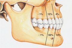
The lateral periodontal cyst is defined by its location. In which area is the lateral periodontal cyst most commonly found?
a) Mandibular 3rd molar region b) Maxillary tuberosity region c) Between the maxillary premolars d) Between the mandibular canine and first premolar e) Between the maxillary canine and first premolar |
Between the Mandibular canine and first premolar
|
|
|
Which of the following lesions is most likely to present as a multilocular radiolucency?
a) Globulomaxillary cyst b) Odontogenic myxoma c) Stafne bone cyst d) Radicular cyst e) Median palatal cyst |
Odontogenic myxoma
|
|
|
Which of the following is true of the conventional solid/multicystic ameloblastoma?
a) They occur most commonly in the posterior portion of the mandible b) They are slow growing odontogenic tumors that may cause bone expansion c) Treatment consists of marginal resection with at least a 1 cm margin d) All of the above e) A & C only |
All of the above
|
|
|
Which of the following is the most common odontogenic cyst of the jaws?
a) Dentigerous cyst b) Lateral periodontal cyst c) Primordial cyst d) Periapical or radicular cyst e) Keratocyst |
Dentigerous cyst
|
|
|
Which of the following is a cyst found in place of a tooth?
a) Dentigerous cyst b) Radicular cyst c) Odontogenic keratocyst d) Lateral periodontal cyst e) Primordial cyst |
Primordial cyst
|
|
|
Which of the following is a solitary cystlike radiolucency not necessarily contacting teeth?
a) Traumatic bone cyst b) Primordial cyst c) Both of the above |
Both of the above
|
|
|
Which of the following are true of dentigerous cysts?
a) May cause delayed eruption of a tooth b) May cause swelling c) May cause facial asymmetry d) All of the above e) A & C |
All of the above
|
|
|
T/F
Dentigerous (follicular) cysts are the second most common odontogenic cysts, after the radicular cyst and the most common developmental odontogenic cyst. |
True
|
|
|
Which of the following can appear as pericoronal radiolucencies according to your textbook?
a) Ameloblastoma b) Ameloblastic fibroma c) Adenomatoid odontogenic tumor d) All of the above e) A & C |
All of the above
|
|
|
Your textbook lists 7 distinct periapical radiolucent lesions that are the sequelae of pulpitis. Which of the following is Not included in this group of lesions?
a) Periapical granuloma b) Radicular cyst c) Abscess d) Scar e) Dentigerous cyst |
Dentigerous cyst
|
|
|
Which of the following is the predominent age predilection for odontomas?
a) 5-20 years b) 21-35 years c) 50-70 years |
5-20 years
|
|
|
Which of the following is true of adenomatoid odontogenic tumors (AOTs)?
a) Are more common in females b) Site predilection is in the mandible c) May appear as mixed radiolucent/radiopaque lesion d) All of the above e) A & C only |
A & C only
(maxillary canine region) |