![]()
![]()
![]()
Use LEFT and RIGHT arrow keys to navigate between flashcards;
Use UP and DOWN arrow keys to flip the card;
H to show hint;
A reads text to speech;
144 Cards in this Set
- Front
- Back
|
Which part of the tooth is most important forproper alignment of a bitewing radiograph, in order to reduce shape distortion? |
Crown |
|
|
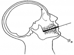
What externally visible anatomical landmarks areused to approximate the location of the apices of: (A) maxillary teeth? (B) mandibular teeth? |
(A) Ala(Nose) & Tragus (Ear)... line between them (B) Inferior Border of Mandible... ~1/2 inch above it |
|
|
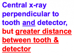
Which 2 rules of the 5 basic rules of a goodshadow image are NOT possible to achieve when using bisecting positioning for an intraoral radiograph? |
(1) Beamperpendicular to the tooth/detector (2) Detector parallel to tooth |
|
|
According to the American Academy of Oral & Maxillofacial Radiology, the goal of radiologic care is to produce high-quality images with a minimum of... |
Radiation exposure to the patient |
|
|
X-ray can damage cellular DNA by direct interaction of indirectly. What is the min mechanism of indirect degradation of DNA by X-rays? |
Free Radicals |
|
|
What is the best type of intraoral radiograph for examination of the lamina dura? |
Periapical |
|
|
Photostimulable phosphor detectors utilize “traps” to hold electrons that have been displaced from their ground states by x-rays. What portion of the electromagnetic spectrum is used to release theelectrons from these traps? |
Visible Light Portion |
|
|
Where are the mylohyoid ridge & oblique ridge in relation to the mental foramen? |
Posterior |
|
|
Arrange in order from most sensitive to leastsensitive to X-ray radiation: CT, Thyroid, Bone Marrow, Muscle |
Bone Marrow > CT > Thyroid > Muscle |
|
|
What types of x-rays occur in the cathode raytube? |
Bremsstrahlung and Characteristic |
|
|
True or False? In licensed Clinical practice,you are required to keep each radiograph for 7 years after the image is firstobtained. |
FALSE itis 7 years after patient is no longer a patient |
|
|
Correct technical term for carries that form atthe base of the tooth’s crown (near the alveolar crest) |
Root Caries or Cemental Caries |
|
|
Units of absorbed dose of radiation |
1Gray = 100 rads = 1 joule/kg |
|
|
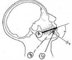
In the bisecting angle technique, imaginary linebisects between what 2 objects? |
(1) The LongAxis of the Tooth (2) Plane of the Detector |
|
|
In a cross-sectional mandibular occlusal set-up,the central ray from the BID is positioned perpendicular to the... |
Detectoror Occlusal plane |
|
|
Classical Shape of E1 Interproximal Caries Lesion |
Triangular |
|
|
Classical Shape of Buccal Surface Caries Lesion |
Round/Oval |
|
|
In a dental radiograph, the patient’s mandibular foramen should be closes to which teeth? |
Mandibular 3rd Molars |
|
|
Where are the largest medullary spaces seen indental radiography? |
Posterior Mandible |
|
|
In digital radiography, image resolution isrelated to pixel size. What controls image resolution in film radiography? |
Sizeof the silver halide crystals (grains) |
|
|
Curve of maxillary arch as usually presented ina topographical occlusal radiograph? |
|
|
|
Coherent Light Scattering |
Relative E of Incident Photo? LOWER Do Resulting Photons have enough E to Exit from Patient? NO or RARELY Approximate % of Original Incident C-ray that Undergoes this Type of Scattering? 7% |
|
|
Compton Light Scattering |
Relative E of Incident Photo? HIGHER Do Resulting Photons have enough E to Exit from Patient? YES Approximate % of Original Incident C-ray that Undergoes this Type of Scattering? 57% |
|
|
(A) mass or kVp (B) kVp (C) Object to Detector Distance (D) Alignment (E) Focal Spot Size |
|
|
What type of intraoral radiograph is the leastuseful for precisely locating supernumerary or impacted teeth? |
Bitewing |
|
|
What part of the tooth is the most important forproper alignment of a periapical radiograph, in order to reduce shapedistortion? |
Long Axis of the Root |
|
|
Change in electron binding energy as a functionof increasing distance from an atom’s nucleus? |
Decreases |
|
|
What is another accepted technical term for ”short-scalecontrast”? |
High Contrast |
|
|
What is the Cathode Ray? |
The Beam of Electrons in the X-Ray Tube |
|
|
Main purpose of the “fixing” stage ofradiographic film processing? |
To removethe unreacted silver halide crystals from the emulsion |
|
|
Who owns the dental radiographs of a patient? |
Dentist that Originally Took the X-ray |
|
|
What is another accepted name for the mylohyoidridge? |
Internal Oblique Ridge |
|
|
Two external structures on your patient’s headthat define the “Frankfort plane”? |
Orbitale (low point of inferior orbital margin) Porion (external acoustic meatus) |
|
|
According to a minimal reading assignment, PSPdetectors have a property known as wide latitude: What does wide latitude meanfor practical use of PSP detectors in Oral Radiography? |
PSPdetectors have a linear response to a wide range of X-ray exposure. It is verydifficult to under/over expose a PSP detector |
|
|
Describe the main difference between theemulsion layers in standard radiographic film vs those in screen film? |
Maindifference is that the screen film emulsion includes visible light-sensitivedrugs & chemicals that are not present in the emulsions of standard radiographicfilm |
|
|
Type of ionizing radiation that causes the mostdamage per unit volume of tissue? |
Particulate Ionizing Radiation (alpha particles; beta particles; cathode rays) |
|
|
Define Ionizing? |
Have enough energy to displace electrons in tissue |
|
|
Compton scattering: |
High energy X-ray displaces an electron from a tissue atom. C-ray’s path is divertedby the X-ray still has a relatively high energy (high enough to continue intothe tissue & penetrate to the detectors) |
|
|
FMX |
Full Mouth Series |
|
|
BID |
Beam Indicating Device |
|
|
mAs |
milli ampere-seconds |
|
|
HVL |
Half Value Layer |
|
|
3 Advantages of Digital Radiography Compared to Film-Based Radiography? |
(1) Less radiation exposure to the patient (2) less environmental impact (less waste/chemicals) (3) requires less storage space |
|
|
Main difference in the x-ray that initiate Compton scattering v. Coherent Scattering? |
Theenergy (wavelength) of the X-rays High Energy >>> Compton Scattering Low energy >>> Coherent Scattering |
|
|
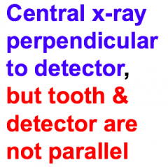
What condition causes “image foreshortening” inan oral radiograph? |
(1) central C-ray is perpendicular to the detector, BUT... (2) the tooth(object) and the detector are not parallel to each other |
|
|
5 steps of the PSP process for obtaining aradiographic image? |
(1) X-ray beam displaces valance electron fromsensor shell/orbit (2) Displaced electron is captured in fluoride “trap” (3) Scanning laser Light displaces trapped electrons (4) Electrons fall back to ground state, giving off visible light (5) Photo detector records light |
|
|
Who was Otto Walkhoff? |
Otto Walkhoff is credited with being the firstdentist to try Roentgen’s new discovery (X-rays) to form an image |
|
|
Current vs Early Units: Exposure |
Current: C/kg Early: Roentgen (R) |
|
|
Current vs Early Units: Dose |
Current: Gray Early: Rad |
|
|
Current vs Early Units: Equivalent Dose |
Current: Sievert (Sv) Early: rem |
|
|
List Conditions that can look like cariouslesions on a radiograph |
(1) Mach Band Effect (2) Cervical Burnout (3) Non-Metallic Restorations (4) Toothbrush Abrasion |
|
|
What is the geometry of the collimator openingused in panoramic radiograph? |
Slit |
|
|
3 normal anatomic structures that result in doublereal images on a panoramic radiograph |
(1) Hard Palate (2) Hyoid Bone (3) Spine |
|
|
3 factors that determine whether an atom will beionized by radiation? |
(1) Atomic Number (2) Location of Electron being Displaced (3) Energy of Incident Radiation |
|
|
Name 3 types of electromagnetic ionizingradiation |
(1) X-rays (2) Gamma Rays (3) UV rays (some) |
|
|
Ionizing radiation can damage somatic cells. What characteristic of somatic cells determines their relative sensitivity toionizing radiation? |
Mitotic Rate |
|
|
The quantity of X-rays produced by an X-raymachine varies... |
Directly & Linearly with: a) Exposure Time b) Current |
|
|
The penetrating power of X-rays produced by anX-ray machine varies... |
Directly and Non-Linearly with: a) Voltage |
|
|
3 anatomical conditions that are commonchallenges to correct placement of bitewing radiograph of the premolars |
(1) Tori (2) Shallow Maxillary Palate (3) High Muscle Attachments |
|
|
Difference between absorbed dose & effectivedose in ionizing radiation biophysics? |
AbsorbedDose: the amount of E absorbed by the tissue (units: Grays or joules/Kgof tissue) Effective Dose: takes the type of tissueinto account by using a tissue weighting factor
|
|
|
According to the reading assignment why shouldthe cost of a radiograph be included in the cost of the clinician’s diagnosticevaluation? |
So thatthere is no question that the dentist owns the radiographs |
|
|
The contrast of a radiograph is affected most by what variable of the X-ray machine? |
kVp |
|
|
Medullary Spaces in the mandible are __________ than in the maxilla. |
Larger |
|
|
Medullary Spaces in the anterior mandible are __________ than in the posterior mandible. |
Smaller |
|
|
True or False? The American Academy of Oral 7 MaxillofacialRadiology regulates the maximum, cumulative absorbed does allowed per dentalpatient per year in the US. |
False |
|
|
What is the feature of the panoramic radiographthat indicates that the patient’s chin was too high during the exposure? |
Theteeth in the radiograph are aligned more in a straight line (left to right) oreven in a slight frown |
|
|
Occlusal carious lesions in posterior teethoften are not visible on intraoral radiographs until they are radiographicclass 3 lesions. Why are they usually not visible before this? |
Becausethe large amount of enamel that the X-ray would have to go through (BL direction) to get to the detector |
|
|
In the electromagnetic spectrum, what is the specific type of ionizing radiation that has higher enerdy than X-rays. |
Gamma Rays |
|
|
Advantage of the bisecting angle techniquecompared to the parallel technique for a periapical radiograph? |
Thedetector (film) can be placed closer to the teeth (sharper images) |
|
|
True or False? The US FDA requires that Cray machines in adental clinic be inspected every 3 years? |
False. This is a NYS regulation. |
|
|
Follow Up for a failed Inspection in NYS... |
Every 60 days OR LESS |
|
|
FDA Regulations |
continue to be revised and slightly updated (important to know when an X-ray machine was FDA approved) |
|
|
Legal Risk Management |
|
|
|
Approximate number of electron volts required togenerate a free radical in tissue? |
~ 10 eV |
|
|
Dense White Line Close to Tooth? |
Lamina Densa |
|
|
Cephalometric radiographs are used most oftenfor which dental specialty? |
Orthodontics |
|
|
In a radiograph, the image of the incisive foramen is high or low density? |
High Density |
|
|
A bitewing radiograph is the best way to detectwhat general type of caries? |
Interproximal |
|
|
What kind of restorative materials are mostradiolucent? |
Resin or Resin Composites |
|
|
Principle Quantum Numbers |
K = 1 L = 2 M = 3 N = 4 |
|
|
Maximum Electrons Per Shell |
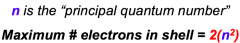
K = 2 L = 8 M = 18 N = 32 |
|
|
For healthy teeth, how does the radiographicappearance of a young person’s periodontal ligament space differ from theradiographic appearance of an elderly person’s periodontal ligament space? |
PDL space on a young, healthy person isthicker than that on an elderly/healthy person |
|
|
In a periapical radiograph, what is the appearance of Trabeculae |
Radiopaque network (may exhibit step-ladder pattern) |
|
|
In a periapical radiograph, what is the appearance of Medullary Spaces |
Radiolucent Blocks between the trabeculae |
|
|
On a panoramic radiograph, where is the nasalseptum, relative to the image of the nasal spine? |
nasal septum is superior to the nasal spine |
|
|
Another accepted name for the external obliqueridge? |
Oblique Ridge |
|
|
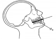
And on a panoramic or periapical technique for aperiapical radiograph, how would you adjust the X-ray equipment in order to reduce the penumbra? |
Move the X-ray source further away from the patient |
|
|
True or False? Screen film is a type of digital detector? |
False. |
|
|
|
|
|
|
|
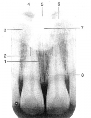
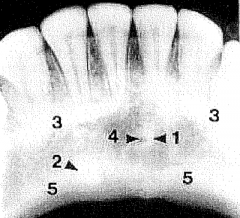
1. Radiolucent line indicated by the two white arrows? 2. Radiolucent on both side of radiolucent line? 3. Radiolucent areas in mesial surface of the crowns? |
1. mid-palatine suture 2. incisive foramen 3. composite-resin restorations |
|
|
|
|
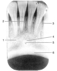
1. Identify the radiolucent structure in the upper right area. 2. Describe the radiograph in terms of area, restorations, and apparent periodontal health. |
1. Maxillary SInus 2. This is a periapical radiograph of the patients left posterior maxilla, showing the 1st & 2nd molars and both premolars (cropped). There are two restorations on the 1st molar. The periodontal health of the patient is fair to good. Bone height is even, although a bit low, and the PDLs are close to the roots of the teeth. |
|
|
|
|
|
|
|
|
|
|
|
|
|
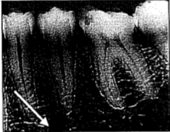
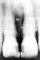
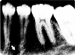
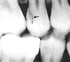
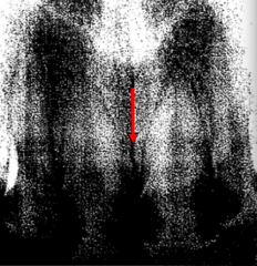
What is the radiolucent area indicated by the white arrow? |
Mental Foramen Landmarks: Near the apices of the mandibular premolars |
|
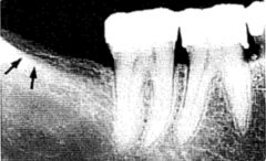
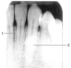
1. What structure is indicated by the black arrows? 2. Describe radiograph by type and location. |
1. Oblique Ridge or External Oblique Ridge 2. The is a periapical radiograph of the patients right posterior mandible. |
|
|
|
|
Type Area Notable Features Description |
A Periapical Radiograph of the mandibular posterior left premolar/molar area [tooth # 19-22]showing a MO amalgam restoration on 2nd premolar [#20] and endodontic treatment with a full crown on 1st molar [#19]. Add sentence describing any carious lesions and there location/severity. |
|
|
4 Disadvantages of Digital Radiography |
(1) System Cost (2) Detector Cost and Unpredictable Longevity (3) Relatively New Technology (several competing systems) (4) Digital Format requires Computer, etc. |
|

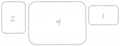
Size of the Detectors Shown? |
|
|
|
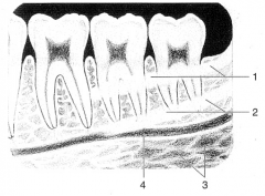
Radiographic Features of Cancellous Bone vs. Cortical Bone |
Cancellous Bone: both radiopaque/radiolucent features:
|
|
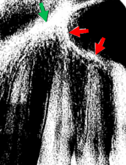
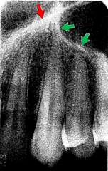
1. What is the Green arrow pointing at? 2. What is the Radiolucent area that the Red arrows are pointing at? |
1. Inferior Border of the Nasal Fossa 2. Floor of the Maxillary Sinus |
|
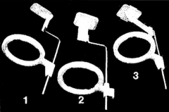
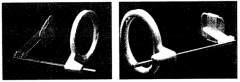
Name the specific type of radiograph obtained with the devices pictured. |
1. Anterior Periapical 2. Posterior Periapical 3. Bitewing |
|
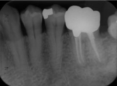
Describe |
This is a periapical radiograph of the patients left posterior mandible, showing both premolars and the 1st & 2nd molars. Bone Height is good, but the PDL space is widening at the mesial surface of the 2nd molar. Carious lesions are not visible in the radiograph. |
|
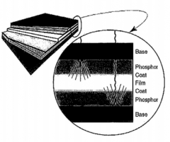
What kind of detector schematic is shown in this drawing? |
screen film |
|
Name the radiographic projection- be specific. |
Anterior Maxillary Occlusal |
|
Name the radiographic projection- be specific. |
Cross-Sectional Mandibular Occlusal |
|
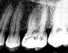
A) What is the radiographic classification of this lesion? B) What is the operative classification of this lesion? |
A) Class III B) D1 or D2 |
|
|
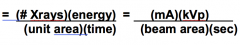
X-ray is set at 90 kVp, 15 mA, 2.5 mm aluminum filtration, and .2 sec exposure time with a distance of 8 inches. If you change 10 mA, what would be the new exposure time if you don't want to change the image density? |
Density = mA x s (15 mA) x (.2 sec) = (10 mA) x (X sec) X = .3 sec |
|
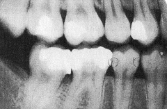
1) type of x-ray? 2) side of mouth? 3) teeth with restorations? 4) tooth with caries? |
1) Bitewing 2) Right 3) #2, #3, #5, #30, #31 4) Mandibular 2nd Premolar |
|
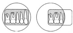
Which is set up wrong for a bitewing? Why? |
Left. Teeth would be positioned behind the detector, and not between the detector and source. |
|
1) What radiographic technique is indicated? 2) What radiographic projection is indicated? |
1) bisecting technique 2) anterior mandibular occlusal (mandibular symphysis is included) |
|
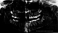
Why is this a bad panoramic radiograph? |
Condyles are on different levels and not the same size. The dentition is not all in focus. |
|
1) What structure is the arrow pointing to? 2) Describe the type and location. 3) Identify condition indicated by large radiolucent areas. |
1) Median Palatine Suture 2) This is a periapical radiograph of the patients maxillary incisors. 3) The large radiolucent areas indicate rampant caries. |
|
|
If a radiograph was produced using a source to film distance of 8 inches and an exposure time of 1 sec, what would be the correct exposure time to get the same image with a source to film distance of 16 inches? |
![4 sec
[C] or [T] can be used in place of E.](https://images.cram.com/images/upload-flashcards/43/26/21/24432621_m.png)
4 sec [C] or [T] can be used in place of E. |
|
1) Red Arrow? 2) Green Arrow? |
1) Floor of the Nasal Cavity 2) Floor of the Maxillary Sinus |
|
What is the radiographic term used to describe the problem in this image? |
Cone-Cut or Partial Image |
|
|
Most Common Cause of Malpractice Suits is the Failure of the Dentist to use radiographs in the diagnosis or management of.... |
1) pain 2) infection 3) swelling |
|
|
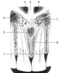
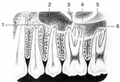
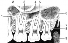
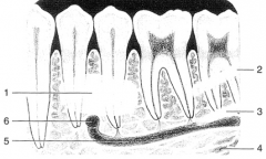
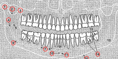
1. Condyle 2. Sigmoid Notch 3. Coronoid Process 4. Mandibular Foramen 6. Mandibular Canal 7. Mental Foramen 10. Lingual Foramen 11. Genial Tubercles 12. Inferior Border of Mandible 14. Mylohyoid Ridge |
|
Draw the lines demonstrating the bisecting technique and explain what they are. |
A) Plane of the Detector B) Bisecting Line C) Long Axis of Central Incisors |
|
|
What is the main advantage of CMOS/APS over CCD for dental radiography? |
More Rapid Processing |
|
|
What X-ray detection mechanism is common to these techniques: CCD, PSP, & CMOS/APS |
Detectors Electrons are displaced from their ground state |
|
Identify (1), (3),(4), & (5) |
(1) Genial Tubercles (3) Mental Ridges (4) Lingual Foramen (5) Inferior Border of the Mandible |
|
|
X-Ray Interactions with Matter |
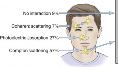
1. None 2. Coherent Scattering 3. Photoelectric Effect 4. Compton Effect |
|
|
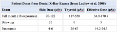
Typical Equivalent Doses of Dental X-Rays? |
|
|
|
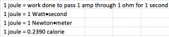
What is a Joule? |
|
|
|
Wave Theory |

c = velocity of "light" v = frequency l = wavelength |
|
|
Quantum Theory |
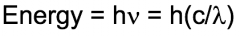
h = planck's constant |
|
|
Ionizing Effect on Biomolecules |
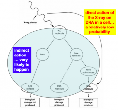
1) Direct 2) Indirect (via free radical formation) |
|
|
Cells sensitivity to ionizing radiation damage... |
Directly proportional to reproductive capacity. Inversely proportional to cells degree of differentiation.
|
|
|
Interactions associate with oral radiology... |
cathode rays interacting with targets = X-rays X-rays interacting with your patient = scattering and radiographic |
|
|
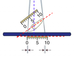
Image Foreshortening |
|
|
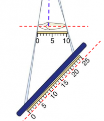
Image Elongation |
|
|
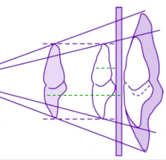
Image Enlargement |
|
|
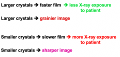
Large or Small Crystals? |
|
|
|
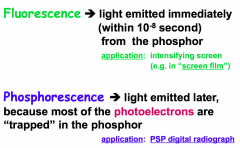
Fluorescence vs Phosphorescence |
|
|
|
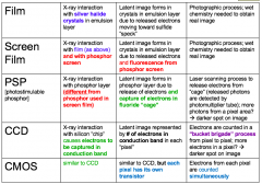
Radiographic Detection Mechanisms: Summary |
|
|
|
Intensity |
Intensity = quality x quantity quantity: determined by mAs quality is penetrating ability (X-ray energy) Inverse Square Law: Intensely is inversely related to distance SQUARED |
|
|
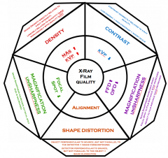
X-Ray Variables |
|
|
|
Dose and Equivalent Dose: X-Ray |
Equivalent Dose (Sv) = Dose (Gy) |
|
|
Dose and Equivalent Dose: Alpha Particle |
Equivalent Dose (Sv) > Dose (Gy) |