![]()
![]()
![]()
Use LEFT and RIGHT arrow keys to navigate between flashcards;
Use UP and DOWN arrow keys to flip the card;
H to show hint;
A reads text to speech;
71 Cards in this Set
- Front
- Back
|
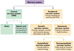
Describe the general organisation of the nervous system . |
|
|
|
If a neuron is entirely containedwithin the brain and/or spinalcord, what kind of neurone is it? |
A CNS neurone |
|
|
If any part of a neurone (dendrites, axon or cell body) projects outside of the brain and spinal cord, what kind of neurone is it? |
A PNS neurone |
|
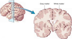
What forms the grey and white matter? |
Grey matter = cell bodies White matter = axon tracks (myelin) |
|
|
Do you have any control over the function of the sympathetic and parasympathetic nervous systems. |
No |
|
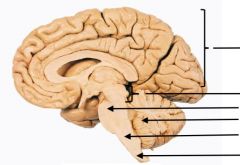
Label this brain from top to bottom |
Forebrain Midbrain Pons Cerebellum Medulla Spinal cord |
|
|
What do the pons, cerebellum and medulla form? |
The hindbrain |
|
|
The medulla oblongata keeps you alive-you wouldn't survive for long without it. It's the bit of the brain that houses the cardiovascular and respiratory centres. The medulla monitors and gets information from the periphery on things ...... |
blood pressure, blood CO2, heart rate (and then autonomically produces the correct response) |
|
|
What kind of cells can be found in the cerebellum? |
Purkinje cells. |
|
|
What are the roles of the cerebellum? |
Maintenance of balance and posture Coordination of voluntary movement Motor learning Cognitive functions (e.g language) |
|
|
What is the role of the midbrain? |
Relay auditory and visual information between top and bottom of brain. Controls reflex eye movement |
|
|
What are the two bumps in the midbrain concerned with? |
Reflex eye movement (e.g if you hear something out the corner of your eye, your eyes move towards it.) This is more common in birds and amphibia. |
|
|
In neuroanatomy, what is a nucleus? |
A group of cell bodies doing the same job. |
|
|
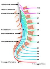
How many spinal nerves are there normally? |
31 |
|
|
What vertebral level does the spinal cord end at? |
L1/L2 |
|
|
What is the caudal equina? |
The area where there is no spinal cord and the nerves just hang. |
|
|
As we develop after three months, the spinal cord and vertebral don't grow at the same rates any more. Which grows at a slower rate? |
The spinal cord |
|
|
By the time you are born, your spinal cord end is high up your back. How does this affect how nerves leave the vertebra? |
They leave at more of a slant |
|
|
What are cranial nerves? |
Those nerves which arise from the brain and brain stem rather than the spinal cord. |
|
|
The spinal cord is segmented - how do nerves enter and leave? |
As rootlets |
|
|
The outer part of the cerebrum isthe.... |
Cerebral cortex |
|
|
The cerebral cortex is throwninto several ridges and grooves. What are they known as? |
Ridges= gyri Grooves =sulci |
|
|
What are deeper grooves known as? |
Fissures |
|
|
The gyri and sulci of the cerebral cortex increase the surface areaof the brain to approx. 2,500 cm2- what does this allow? |
Much more neural material to be contained within the skull. |
|
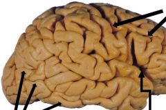
Label in a clockwise way |
Sulci Fissure Gyri |
|
|
The cerebrum is divided into lobes. Some functions are associated with individual lobes but... |
No function is located to only one lobe and no lobe is associated with just one function. |
|
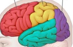
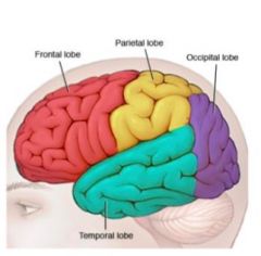
Label this! |
Red= frontal lobe Yellow = parietal lobe Green= temporal lobe Purple= occipital lobe |
|
What runs the length of the spinal cord? |
The central canal |
|
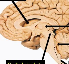
Label this starting from top left then move in a clockwise way |
Lateral ventricle 3rd ventricle 4th ventricle Central canal Cerebral aqueduct |
|
|
What is INSIDE the ventricles? |
Cerebral spinal fluid (CSF) |
|
|
What makes the choroid plexus? |
Specialised ependymal cells |
|
|
What are the functions of the CSF? |
• Buoyancy • Protection • Removal of waste products |
|
|
What happens to CSF? |
It is reabsorbed back into the venous blood stream |
|
|
What happens if CSF is not absorbed back into the venous blood stream? |
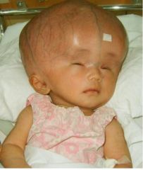
Hydrocephalus. |
|
|
Ventricles expand as they get more CSF within them, so the cortex is pushed out. How is hydrocephalus is often treatable? |
If a stent is put in and the XS CSF is drained. |
|
|
The CNS has 3 outer layers (meninges). What are these three outer layers and what is their common role ? |
The pia mater (innermost layer) The arachnoid mater (middle layer) The dura mater (outer layer) Protect the CNS |
|
|
What is the pia mater like? |
Fine and delicate |
|
|
What is the arachnoid mater like? |
Silky, web-like |
|
|
What is the dura mater like? |
Tough, hardy |
|
|
The CSF leaves the ventricles of the brain and exits into the space between which two layers? What is this place called? |
The pia mater and the arachnoid mater The sub arachnoid space |
|
|
The brain has a consistency of jelly as it has no collagen. Why is the dura mater important? |
It helps protect the brain and give it some stability. |
|
|
The pia mater is a fine, vascular membrane that allows entry of what into the CSF? |
Blood vessels |
|
|
Why does the pia mater help contain the CSF? |
It is impermeable |
|
|
What is the arachnoid mater closely associated with? |
The dura mater |
|
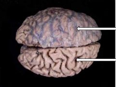
What is present in the top section of the brain that is missing in the bottom section? |
Arachnoid mater |
|
|
Why does the subarachnoid space hang loosely and is not attached to the dura? |
Hangs loosely because If dura was attached, overtime when you bend over it would tear. |
|
|
Surrounding the brain, what is the dura mater closely attached to? |
Periosteum of the cranium (the connective tissue surrounding the skull) |
|
|
In the skull, the dura mater is composed of two layers which separatein places to form what? |
Sinuses carrying venous blood. |
|
|
Which ventricle does the CSF leave the brain from? |
The fourth ventricle |
|
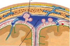
Label this starting from the top left and moving clockwise |
Arachnoid granulations Dura mater Arachnoid mater Sub-arachnoid space Pia mater Falx cerebri Superior sagittal sinus |
|
|
After leaving the sub-arachnoid space, where does the CSF go? |
Into thesuperior sagittal sinus |
|
|
What mediates the transfer of CSF from the subarachnoid space to the superior sagittal sinus? |
Arachnoid granulations |
|
|
What is the superior sagittal sinus? |
A blood vessel that carries venous blood from brain to heart |
|
|
What would examples be of the anatomical division of the CNS? |
Spinal cord, hindbrain, midbrain, forebrain |
|
|
Is the CNS hollow? |
Yes |
|
|
The central canal and ventricles are filled with.... |
CSF |
|
|
If a clinician wants to take a sample of cerebrospinal fluid, what is the most convenient (and safest) place to obtain such asample? |
CSF is commonly taken by lumbar puncture from thelumbar cistern. Since the spinal cord ends in adults at the level of the intervertebral disk between L1 and L2, a needle is inserted below this. Typically the space between L3/L4 orL4/L5 is used. |
|
|
Epidural anaesthetics are often given for pain relief, particularly during childbirth. Theyare injected into the epidural space. Where do you think this space is, in relation to the duramater ? |
The epidural space is the area between the dura mater and the vertebral wall (ie just outside the dura). |
|
|
Why is anaesthetic not administered subdurally? |
Anaesthetic is not administered subdurally as this could put it in the sub- arachnnoid space where it could travel in the CSF to anesthetise inappropriate nerves with potentially life-threatening consequences. |
|
|
What connects the brain to the PNS? |
The spinal cord |
|

What are some functions of the frontal lobe? |
Executive functions: thinking, planning, organising and problem solving, emotions and behavioural control, personality |
|

What are some functions of the parietal lobe? |
Perception, making sense of the world, arithmetic, spelling |
|

What is the function of the occipital lobe? |
Vision |
|
What are some functions of the temporal lobe? |
Memory, understanding, language |
|
|
What is the cerebrum? |
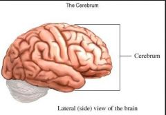
The section of the forebrain that makes us human ( thinking, reason, emotion, voluntary action) |
|
|
What is the corpus callosum? |
A network of myelinated cells that connects the left and right cerebral hemispheres |
|
|
What is the cerebellum? |
"little brain" controls and coordinates the movement of our muscles alcohol affects this part of the brain |
|
|
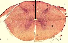
Anatomically, what is the main difference between the brain and the spinal cord? |
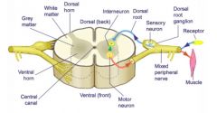
In the brain, grey matter is found towards the outside and white matter is found on the inside In the spinal cord, white matter is on the outside, grey matter on inside N.B in the spine, sensory neurones enter from back (dorsal) and motor neurones exit from the front (ventral) |
|
|
The spinal cord can be broken down into 4 regions. What are these regions? |
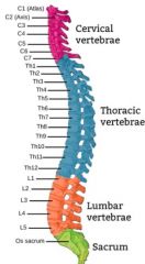
Cervical region Thoracic region Lumbar region Sacral region |
|
|
What is the dorsal root ganglion? |
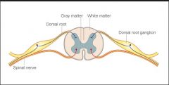
Sensory neurones enter the spinal cord from the back of the spine. The sensory neurone cell bodies lie in the dorsal root ganglia, and their axons extend into the spinal cord. |
|
|
There are normally 31 spinal nev |
8 cervical 12 thoracic 5 lumbar 5 sacral 1 coccygeal |