![]()
![]()
![]()
Use LEFT and RIGHT arrow keys to navigate between flashcards;
Use UP and DOWN arrow keys to flip the card;
H to show hint;
A reads text to speech;
153 Cards in this Set
- Front
- Back
|
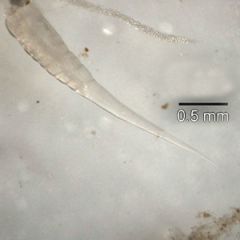
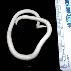
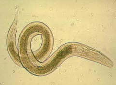
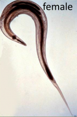
Enterobius vermicularis adult |
|
|
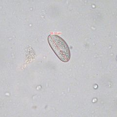
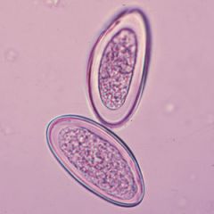
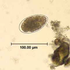
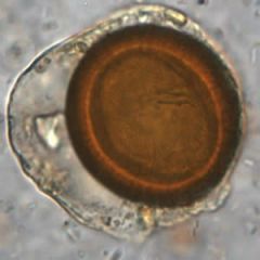
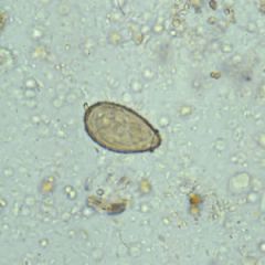
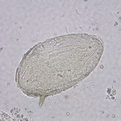
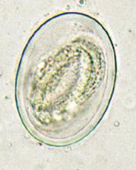
Enterobius vermicularis egg 50 - 60 by 20 - 30 um, thick, double-layeredshell, flattened on one side. |
|
|
Enterobius vermicularis egg 50 - 60 by 20 - 30 um, thick, double-layeredshell, flattened on one side. |
|
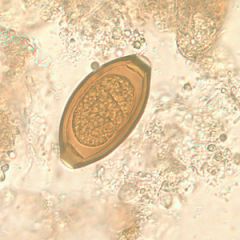
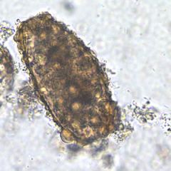
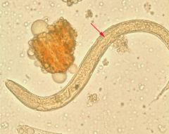
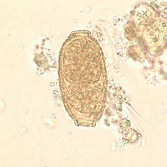
what is this?
what is the pathology related to? |
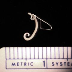
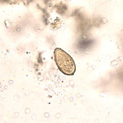
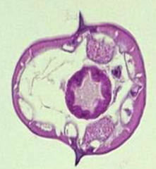
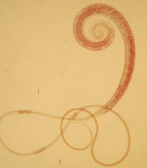
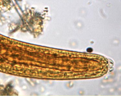
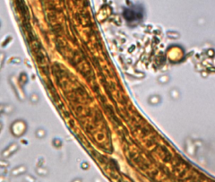
Trichuris trichiura adult Whipworm: Adults live up to 8 years threaded in intestine. Pathology related to damage to mucosa and an allergic response. |
|
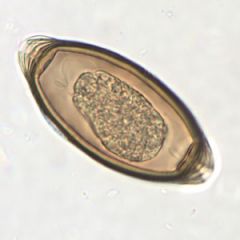
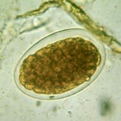
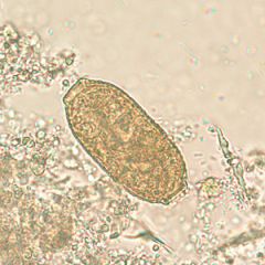
what is this?
what other parasite is commonly found with it? |
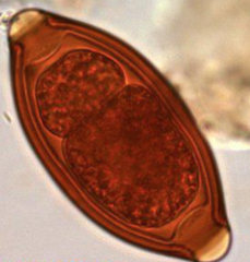
Trichuris trichiura egg: whipworm Frequently found along with Ascaris lumbricoides |
|
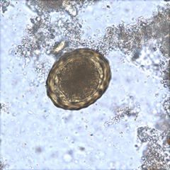
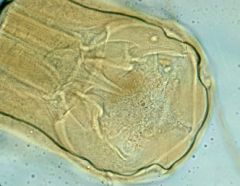
Common name? What is the infective stage of this parasite? What is the diagnostic stage? |
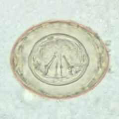
Trichuris trichiura egg Whipworm Mature eggs are the infective stage, they mature in the environment. The immature egg is diagnostic. |
|
|
Ascaris lumbricoides adult Adults very large(6 – 12 inches in length). Live in the small intestine(do not attach). |
|
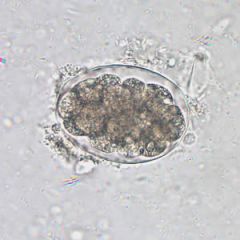
Whats this?
Sizes? |
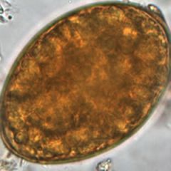
Ascaris lumbricoides egg fertilized 45 - 75 by 35 - 50 um, oval,thick - shelled, bile-stained, central zygote; may be mammillated(outer albuminoid coat present)or decorticated (outer coat absent). |
|
Whats this?
Sizes? |
Ascaris lumbricoides egg unfertilized 85 - 95 by 43 - 47 um,thin shell, internal contents are a mass of highly refractive granules; may be irregularmammillated or decorticated. |
|
|
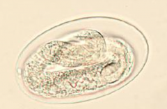
Hookworm egg Identical for both genera 60-75 μm by 35-40 μm; oval. Smooth, thin shell. |
|

|
Hookworm (Ancylostoma duodenale) adult RARELY SEEN |
|
|
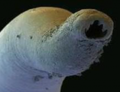
Hookworm (Necator americanus) adult Cutting plates RARELY SEEN |
|

|
Hookworm egg |
|
|
Hookworm egg |
|
|
Hookworm egg |
|
|
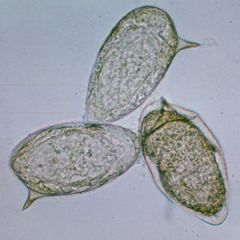
Trichostrongylus egg Lab diagnosis is by: Egg: 75-95 μm by 40-50 μm; tapered atone end (“chicken-egg” shape), thin shell. Egg larger and more pointy thanhookworm egg |
|
|
Trichostrongylus egg Lab diagnosis is by: Egg: 75-95 μm by 40-50 μm; tapered atone end (“chicken-egg” shape), thin shell. Egg larger and more pointy thanhookworm egg |
|
|
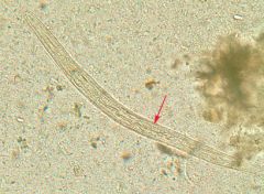
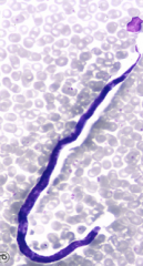
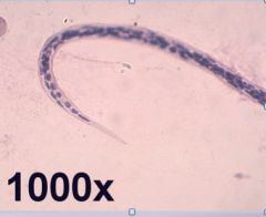
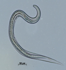
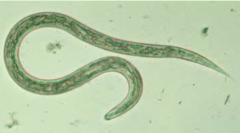
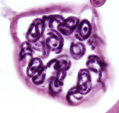
Strongyloides stercoralis Rhabditiform "threadworm" |
|
|
Strongyloides stercoralis Rhabditiform "threadworm" |
|
|
Strongyloides stercoralis Rhabditiform "threadworm" |
|
|
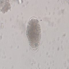
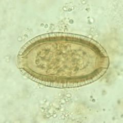
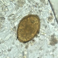
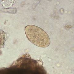
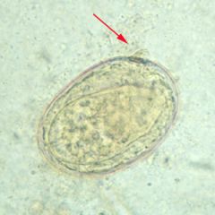
Capillaria philippinensis egg UNEMBRYONATED 35 - 45 by 21 umPolar plugs (may be hard to see),striated shell, also more rectangular than T. trichiura. |
|
|
Capillaria philippinensis egg 35 - 45 by 21 um Polar plugs (may be hard to see), striated shell, also more rectangular than T. trichiura. |
|
|
Capillaria philippinensis egg |
|
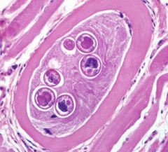
id
which infected animal meat transmits it? |
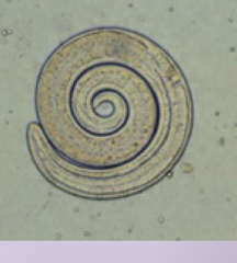
Trichinella spiralis PORK... or bear! carnivore.. |
|
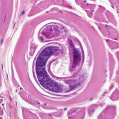
id
what is the infective stage what is the diagnostic stage |
Trichinella spiralis infective stage: encysted larvae ingested diagnostic stage: larvae in striated muscle |
|
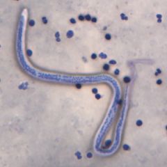
ID
Morphology? Periodicity? Location? |
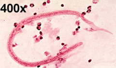
Wuchereria bancrofti Morphology of microfilaria:sheathed, discrete nuclei,nuclei do not extend to the tip of the tailPeriodicity: nocturnal-collect specimenbetween 10pm-2am Location of microfilaria:blood |
|
ID
Morphology? Periodicity? Location? |
Wuchereria bancrofti Morphology of microfilaria: sheathed, discrete nuclei, nuclei do not extend to the tip of the tail Periodicity: nocturnal-collect specimen between 10pm-2am Location of microfilaria: blood |
|

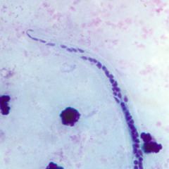
#1 |
#1: Wuchereria bancrofti (lymphatic)Sheath,no nuclei in the tip of the tail |
|
|
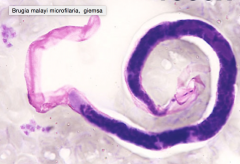
Brugia malayi
Morphology of microfilaria:sheathed, Two terminal nuclei separated with asignificant gap,column of nuclei is compact Periodicity: nocturnal-collect specimenbetween 10pm-2am Location of microfilaria:blood |
|
|
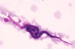
Brugia malayi |
|
2 |
#2: Brugia malayi (lymphatic)Sheath, 2 distinct nuclei in the tip of the tail |
|
ID
where do these sometimes migrate to, and then cause psychological problems? |
Loa loa Travel to the eye! |
|
ID
Periodicity? |
Loa loa Microfilariae showdiurnal periodicity:Peripheral blood during day, lung, etc.at nightDiagnosis is best made from blood collected duringthe mid-day (10 AM-2 PM) |
|

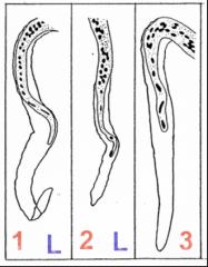
#3 What is the vector? Diagnostic stage? |
#3: Loa loa FLY! Infective larvae are injectedinto host. Females producemicrofilariae(diagnostic stage) thatcirculate in peripheral blood. (tissueand “eye worm”)Sheath, nuclei extending to the tip of the tail |
|
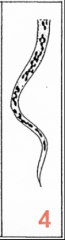
4 (Found in SKIN) |
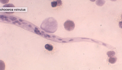
Onchocerca volvulus No sheath, no nuclei in the tip of thetail |
|
ID
morphology periodicity location |
Onchocerca volvulus Morphology of microfilaria:unsheathed, nuclei donot extend to tip of tail,column of nucleiis moderately compact Periodicity: nonperiodic Location of microfilaria:subcutaneous tissue |
|
|
Onchocerca volvulus unsheathed, nuclei don't go to tip of tail, non periodic, in subcutaneous tissue |
|
#6 Vector? |
#6: Mansonella ozzardi Unsheathed, nuclei don't go to tip, compact nuclei, located in the BLOOD, non periodic Biting fly vector |
|
|
Mansonella ozzardi Unsheathed, nuclei don't go to tip, compact nuclei, located in the BLOOD, non periodicBiting fly vector |
|

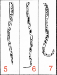
5 |
#5: Mansonella perstans Unsheathed, nuclei go to tip, compacted column of nuclei, non periodic, located in the blood. |
|
|
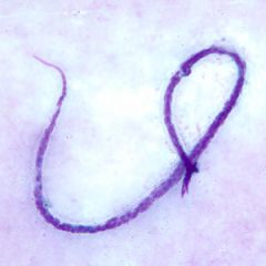
Mansonella perstans No sheath, nuclei extending to the tip ofthe tail |
|
|
Mansonella perstans No sheath, nuclei extending to the tip ofthe tail |
|

#7 |
#7: Mansonella streptocerca Vector is the fly. HOOKED tail. In the skin. OCCURS in Africa only |
|
|
Mansonella streptocerca hooked tail |
|
|
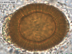
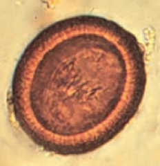
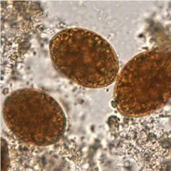
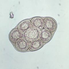
Taenia egg |
|
|
Taenia egg |
|
|
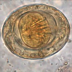
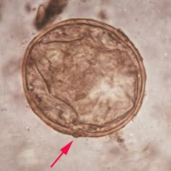
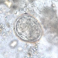
Taenia egg The egg (most common diagnostic stage) is bile stained, round, 30 – 43 um., has a thick cell wall with radial striations. Inside the embryonated egg is a six hooked oncosphere. |
|
ID
Diagnostic stage? |
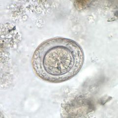
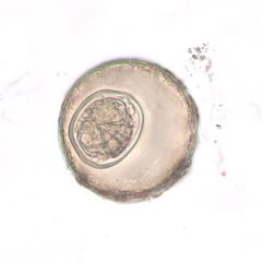
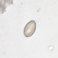
Hymenolepis nana egg Eggs are the diagnostic stage They are oval 30 to 50 µm (Smaller than Hymenolepis diminuta) |
|
What is this? common name? unique lifecycle? |
Hymenolepis nana egg dwarf tapeworm! most common of all cestode infections flour beetle intermediate, can skip and be autoinfective, human to human transfer |
|
ID
whats on the inner membrane? |
Hymenolepis nana egg On the inner membrane are two poles, from which 4 to 8polar filaments spread out between the two membranes. |
|
|
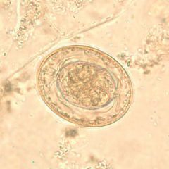
Hymenolepis diminuta egg Eggs are round or slightly oval, size 70 - 85 µmwith striated outer membrane and a thin inner membrane. The space between themembranes is smooth or faintly granular. Withoutpolar filaments.The oncosphere has six hooks. |
|
|
Hymenolepis diminuta egg |
|
intermediate host? |
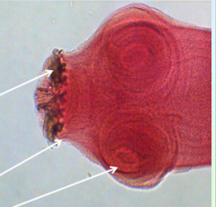
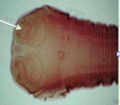
Diphyllobothrium latum egg Two intermediate hosts! copepod and fish The scolex has two sucking grooves (instead of suckers/hooklets) |
|
what's unique about this structure for the group? |
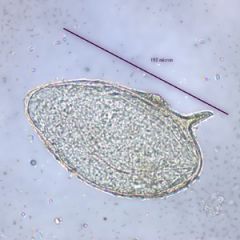
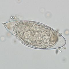
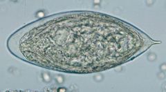
Diphyllobothrium latum egg Unembryonated, operculated, bile stained, 58– 76 um by 40 – 50 um. There is a knoblike structure on the end opposite the operculum.The operculum is unique for thetapeworms (cestodes) We will see them again in the flukes (trematodes) |
|
ID what pathologies does it cause |
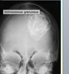
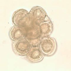
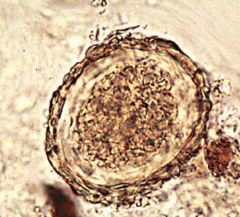
Echinococcus granulosus hydatid cysts, can be in bone |
|
id?
common name? |
Dipylidium caninum egg packet Called the dog tapeworm.Children are most often infectedby ingesting fleas containing the larval stage. |
|
id?
vector? |
Dipylidium caninum egg packet flea vector |
|
id 1st & 2nd intermediate hosts? |
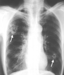
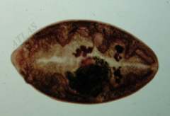
Paragonimus westermani egg in sputum Snails are the first intermediate host and crustaceansare the second intermediate host |
|
ID how do humans get infected |
Paragonimus westermani egg in sputum Acquired through ingestion of metacercariae inraw, pickled, or undercooked freshwater crabs or crayfish |
|
ID known as..... |
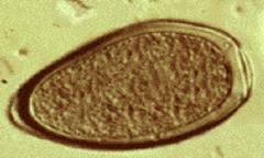
Fasciolopsis buski/hepatica egg Known as the giant intestinal fluke. |
|

ID
Hosts? How do humans get it? |
Fasciolopsis buski/hepatica egg First intermediate host is the snail;second intermediate host is freshwater vegetation Humans get it by ingesting infected vegetation (bamboo shoots and water chestnuts) |
|
ID
Known as.... |
Clonorchis/Opisthorchiss egg Known as the Chinese liver fluke. |
|
ID characteristics... |
Clonorchis/Opisthorchis egg 29 – 35 um, embryonated,flask shaped, operculated with prominent shoulders, and a knob at the end opposite theoperculum. |
|
|
Opisthorchis/Clonorchis egg |
|
ID
where's it found in the world and the body? |
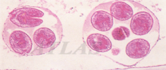
Schistosoma mansoni egg Found primarily in Africa, South America,West Indies, and Puerto Rico. It livesmainly in the veins of the large intestine. |
|
|
Schistosoma mansoni egg |
|
|
Schistosoma mansoni egg |
|
ID
Where's it found in the world and the body? |
Schistosoma japonicum egg Found primarily in the Far East. (Japan,China, Philippines). It lives mainly in the veins of the small intestine. |
|
ID Does it have a spine? |
Schistosoma japonicum egg Has a small curved spine (can be difficult to see) |
|
|
Schistosoma japonicum egg |
|
ID Where's it found in the world and the body? |
Schistosoma haematobium egg
Found primarily in the Nile Valley,Mideast, and East Africa. It livesmainly in the veins of the bladder. |
|
|
Schistosoma haematobium egg |
|
|
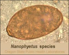
Nanophyetus egg |
|
|
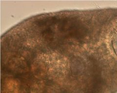
Nanophyetus adult |
|
|
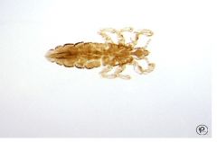
Pediculus humanus adult |
|
|
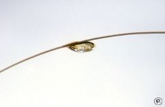
Pediculus humanus nit |
|
|
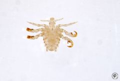
Phthirus pubis adult |
|
|
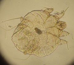
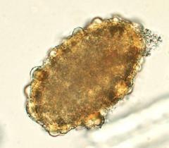
Sarcoptes scabei adult |
|
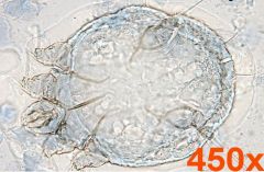
|
Sarcoptes scabei adult |
|

|
Sarcoptes scabei adult |
|
|
Common name: pinworm Scientific name: _________________ Most common symptom: How is it transmitted? |
Enterobius vermicularis anal itching fecal-oral |
|
|
With pinworm, is it the male or female that causes pathology? How do they do it? |
ENTEROBIUS VERMICULARIS Female lives in intestine, transits to anus atnight, deposits eggs in perianal area. |
|
|
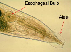
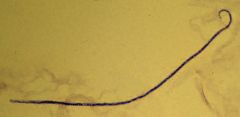
Enterobius vermicularis with alae 8-13mm |
|
|
Enterobius vermicularis female adult |
|
|
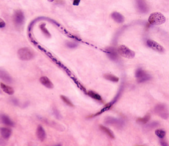
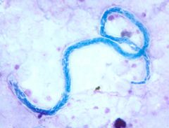
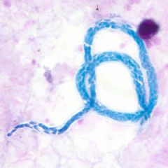
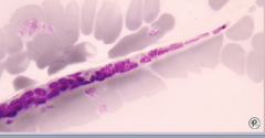
Enterobius vermicularis in histology |
|
|
What is the scotch tape prep used to detect? |
Pinworm! Enterobius vermicularis |
|
|
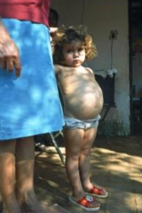
Which parasite caused the prolapsed anus? |
Whipworm |
|
|
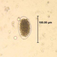
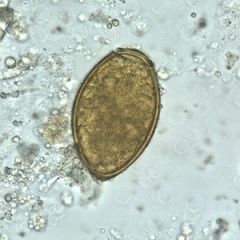
Trichuris trichiura 50 - 55 by 22 - 24 umBarrel-shaped, bile stained,thick shell, mucoid plugsat each end,unembryonated. |
|
|
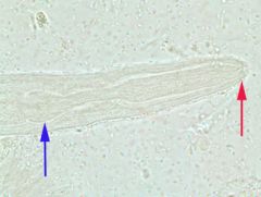
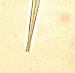
Trichuris trichiura Slender, thread likeanterior portion(“whip”). Thick, posterior portion(“handle”). |
|
|
Ascaris lumbricoides infection |
|
|
Ascaris lumbricoides egg fertilized |
|
|
Ascaris lumbricoides egg unfertilized |
|
|
Ascaris lumbricoides egg decorticated |
|
|
What are the most common humanhelminthic infections? |
1. Ascaris 2. Hookworm |
|
|
Whats the most common symptom of hookworm infection? |
Iron deficiency anemia (caused by blood loss at the siteof intestinal attachment of the adult worms) is the mostcommon symptom of hookworm infection, and can beaccompanied by cardiac complications. Gastrointestinaland nutritional/metabolic symptoms can also occur. |
|
|
How do you make the diagnoses of hookworm infection? |
Diagnose by identification of eggs in feces (adultsremain attached to intestinal mucosa) andhypochromic, microcytic anemia. |
|
|
Which parasite causes an intense desire to eat dirt (loss of iron)? |
Hookworm! |
|
|
What's the infective form of the hookworm? |
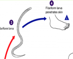
Filariform |
|
|
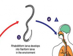
What is the route that hookworm takes in the body?! |
1. Filariformlarva penetrate the skin 2. travels to the heart (via the lymph system) and then to the lungs 3. Theyare then coughed up 4. swallowed 5. mature in the intestine. |
|
|
What are the two hookworm species? |
1. Necator americanus (“New World”) 2. Ancyclostoma duodenale ("Old World") Found in the Middle East, N. Africa, S. Europe |
|
|
Hookworm (Necator americanus) adult Cutting plates RARELY SEEN |
|
|
Hookworm (Ancylostoma duodenale) Mouth has 2 pairs of teeth. RARELY SEEN |
|
|
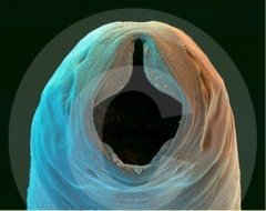
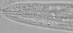
Hookworm Rhabditiform larvae Long buccal canal (mouth), genital primordium small (or not present). Need to know this to distinguish from Strongyloides stercoralis |
|
|
Hookworm Filariform larvae: (identical for both genera). Infective stage: RARELY SEEN |
|
|
Hookwormegg embryonated Hookwormegg, embryonated |
|
|
What is the infective form of Trichostrongylus? |
Found in small intestine.Eggs are shed into soilfrom herbivorous animals. Humans acquire by ingestinginfective filariform larvae orpossibly skin penetration fromLarvae on plant material. |
|
|
Which parasite has a similar life cycle as Strongyloides stercoralis ? What's different about it? |
Life cycle similar to hookwormexcept has an auto-infectivecycle (infective filariform larvain the host invades internalmucosa or perianal skin). |
|
|
How do you diagnose Strongyloides |
Rhabditiform larvae in feces. |
|

|
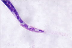
Strongyloides stercoralis Short buccal canal |
|
|
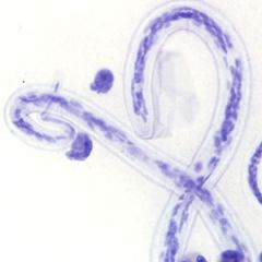
Strongyloides stercoralis Prominent genital primordium |
|
|
Strongyloides stercoralis Notched tail |
|
|
What is the infective stage of Capillaria philippinensis |
Humans get infection from eatinglarvae (infective stage) from infectedinsufficiently cooked fish (fresh water). |
|
|
Which of the nematodes requires only one host? |
Capillaria hepatica |
|
|
Which parasite migrates around the body, but mainly lives in the small intestine? |
Ascaris lumbricoides |
|
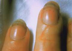
Splinter hemorrhages beneath the nails |
Caused by Trichinella spiralis Trichinosis from ingesting infected meat PORK |
|
|
Trichinellalarvae |
|
|
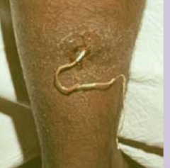
Dracunculus medinesis Causes Dracunculosis: Guinea Worm Disease |
|
|
which parasite uses the copepod as an intermediate host? |
Dracunculus medinesis humans drink water with infected copepods in it! |
|
|
NOTE: filarial worms transmitted by insect vectors includes.... |
Lymphatic (elephantiasis) -Wuchereria bancrofti -Brugia malayi Loa Loa (African eye worm) Onchocerca volvulus (river blindness) |
|
|
two lymphatic filaria: |
Wuchereria bancrofti Brugia malayi Second most common mosquito borne disease.Wuchereria bancrofti isencountered in tropical areas worldwide; Brugia malayi islimited to Asia |
|
|
Sheathed filarial worms: Transmitted by?? |
Wuchereria bancrofti - mosquito Brugia malayi - mosquito |
|
ID |
Wuchereria bancrofti Morphology of microfilaria: sheathed, discrete nuclei, nuclei do not extend to the tip of the tail Periodicity: nocturnal-collect specimen between 10pm-2am Location of microfilaria: blood |
|
|
Loa Loa |
|
|
Onchocerca volvulus in tissue |
|
|
Tapeworm: Taenia saginata found in ___________ meat. Taenia solium found in __________ meat. |
T. saginata=beef T. solium=pork |
|
|
What is the major difference in the life cycles of the two tapeworms? |
Major difference in the life cycles isthat humans canalso be the intermediate hostfor T. solium. |
|
|
Laboratory diagnosis of adult tapeworminfection: |

Finding the egg, scolex, or proglottid(s)in feces.The egg (most common diagnostic stage) isbile stained, round, 30 – 43 um., has a thick cell wall with radialstriations. Inside the embryonated eggis a sixhooked oncosphere. |
|
|
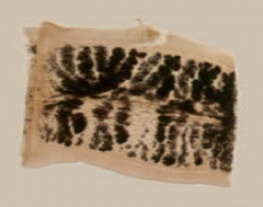
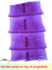
T.solium: 7 –13 uterine branches on one side |
|
|
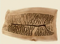
T. saginata: 15 – 20 uterine branches on one side. |
|
|
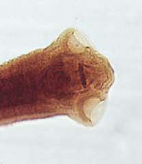
T. solium: 4suckers and a rostellum with a double row of hooklets. |
|
|
T.saginata: 4suckers. |
|
|
Which parasite is found in flour beetles? |
Hymenolepis nana |
|
|
Hymenolepis nanaadult |
|
|
RAT tapeworm? |
Hymenolepis diminuta It is acquired by ingesting fleas thatcontain the infective cysticercoid |
|
|
Which parasite has two intermediate hosts? |
Diphyllobothrium latum egg Two intermediate hosts! copepod and fish |
|
|
Diphyllobothrium latum wider than it is long... |
|
|
Known as hydatid cyst disease. . . |
Echinococcus granulosus |
|
|
Tapeworms are ______odes. Flukes are __________odes. |
tapeworms are chested flukes are trematodes |
|
|
What is common about all the trematode's life cycles? |
•Eggsmust reach water to mature •Allhave snail species as the first intermediate host |
|
|
Also known as the lungfluke! Paragonimus westermani |
|
|
Paragonimus westermani adult |
|
ID |
Paragonimus westermani –Size:80-118um X 48-60 um–Oval,flattened operculum, slightshoulders, unembryonated, thickenedarea opposite the operculum |
|
|
Known as the giant intestinal fluke. |
Fasciolopsis buski Known as the giantintestinalfluke. |
|
|
Known as the sheep liver fluke. In sheep, it is known as liver rot. |
Fasciola hepatica |
|
|
What do Clonorchis feed on? |
bile |
|
|
which of the flukes is not hermaphroditic |
Schistosoma species Bloodflukes =live in the veins |
|
|
schistosomaldermatitis is called |
swimmer's itch |
|
|
Head lice: Body lice: Pubic lice: |
HeadLice: (Pediculushumanus capitis). Body Lice: (Pediculushumanus corporis) Pubic Lice: (Phthirus pubis) |
|
|
What is the primary human bot fly. |
Dermatobia hominis |
|
which parasite is this |
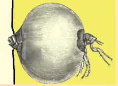
Tunga penetrans |
|
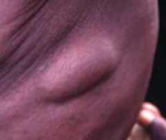
What causes these nodules? |
Onchocerca volvulus microfilariae are found in subcutaneous |
|
|
which of the mansonella sp. is not found in the blood? |
mansonella streptocerca |
|
|
which of the mansonella spp. is only found in the Americas |
Mansonella ozzardi |