![]()
![]()
![]()
Use LEFT and RIGHT arrow keys to navigate between flashcards;
Use UP and DOWN arrow keys to flip the card;
H to show hint;
A reads text to speech;
102 Cards in this Set
- Front
- Back
|
Pathogenic flagellates |
G. lamblia, D. fragilis, T. vaginalis |
|
|
Only flagellate to inhabit small intestine |
G. lamblia |
|
|
Flagellate cysts that are uninuclated |
Chilomastix mesnili, Retortamonas intestinalis |
|
|
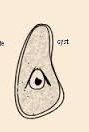
Characteristic shape of C. mesnili cyst |
lemon or nipple-shaped |
|
|
G. lamblia cyst features |
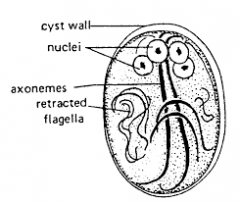
with axoneme, parabasal bodies, and remnants of flagella |
|
|
C. mesnili cyst other features |

Curved fibril alongside cytostome referred to as Sheperd's crook |
|
|
Enteromonas hominis cyst other feature |
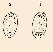
Bipolar arrangement of nucleus, resembles Endolimax nana cyst |
|
|
Retortamonas intestinalis cyst other features |
Cystostome extends above nucleus with bird's beak appearance and resembles C. mesnili cyst |
|
|
Motility of G. lamblia trophozoite |
Falling leaf-like |
|
|
Number of nuclei for G. lamblia cyst |
4 |
|
|
Number of nuclei for E. hominis |
2 or 4 |
|
|
Flagellates with 2 nuclei in their trophozoite form |
G. lamblia and D. fragilis (80%) |
|
|
Number of flagella for G. lamblia trophozoite |
8 |
|
|
Number of flagella for C. mesnili trophozoite |
4 (3 anterior and 1 cytostome) |
|
|
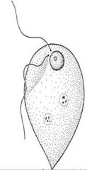
Number of flagella for Enteromonas hominis trophozoite |
4 (3 anterior and 1 posterior_ |
|
|
Number of flagella for Retortamonas intestinalis trophozoite |
2 (1 anterior and 1 posterior) |
|
|
Number of Dientamoeba fragilis trophozoite flagella |
None |
|
|
Number of P. hominis trophozoite flagella |
4 (3 posterior and 1 in undulating membrane) |
|
|
Characteristic appearance of G. lamblia trophozoite |
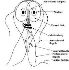
Old man w/ glasses appearance; prominent sucking-disc and axostyle; bilaterally symmetrical |
|
|
Characteristic motility of C. mesnili |
Cork-screw or rotary |
|
|
Characteristic appearance of C. mesnili trophozoite |
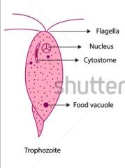
Prominent cytostome with spiral croove |
|
|
Enteromonas hominis characteristic appearance |
One side flattened
|
|
|
Flagellates with jerky movement |
E. hominis, and R. intestinalis |
|
|
D. fragilis trophozoite characteristic feature |
Multiple hyaline, leaf-like pseudopodia; nucleus has fragmented tetrakaryosome |
|
|
Pentatrichomonas hominis trophozoite characteristic appearance |
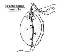
Long undulating membrane; axostyle; and fine, scattered granules |
|
|
R. intestinalis trophozoite distinguishing feature |
Cytostome extend 1/2 length of the body |
|
|
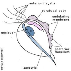
General characteristics of Trichomonads |
exist only in trophozoite forms, axostyle extends beyond their bodies, and equipped with anterior tuft of flagella and undulating membrane |
|
|
T. tenax axostyle and undulating membrane characteristics |
Thick axostyle with undulating membrane almost as long as costa |
|
|
P. hominis axostyle and undulating membrane characteristics |
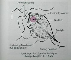
Semi-rigid axosytyle with undulating membrane as long as costa |
|
|
T. vaginalis axostyle and undulating membrane characteristics |
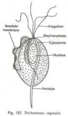
Axostyle is split into fibrils and Undulating membrane <2/3 of costa |
|
|
T. vaginalis cytoplasmic inclusions |
Large amount of siderophil granules |
|
|
Giardia lamblia disease caused |
lambliasis, or traveler's diarrhea and has been associated with Gay bowel syndrome |
|
|
Flagellate that also causes trouble with children in daycare |
G. lamblia |
|
|
Dientamoeba fragilis disease caused |
intermittend diarrhea |
|
|
T. vaginalis disease caused |
Non-specific vaginitis, urethritis, or prostatitis |
|
|
T. vaginalis vaginitis signs and symptoms |
foul-smelling yellowish/greenish vaginal discharge accompanied by intense itchiness in vaginal area |
|
|
Present forms in Leishmania |
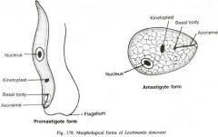
Amastigote and Promastigote |
|
|
Present forms in T. brucei complex |
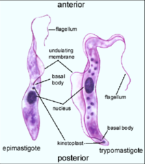
Epimastigote and Trypomastigote |
|
|
Present forms in T. cruzi |
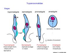
Amastigote, Promastigote, Epimastigote, Trypomastigote |
|
|
Diagnostic stage in man (intracellular) for Leishmania spp. |
Amastigote |
|
|
Diagnostic stage in man (extracellular) |
Trypomastigote |
|
|
"Habitat" of amastigotes
|
inside endothelial cells and phagocytes because they cannot be degraded |
|
|
Stage of Leishmania that occurs in sandfly vector and is recovered from clulture |
Promastigote |
|
|
Infective stage form in Leishmaniosis |
Promastigote |
|
|
Stage in hemoflagellate life cycle that appears in tse tse fly in T. brucei complex |
Epimastigote |
|
|
Diagnostic stage forms in T. brucei complex |
Trypomastigote |
|
|
Infective stage for T. brucei complex |
Metacystic trypomastigote |
|
|
Shape of trypomastigote |
C, S, or U-shaped |
|
|
Forms present in man in T. cruzi infections |
Amastigote, promastigote, epimastigote, trypomastigote |
|
|
Stages in T. cruzi infection present in bugs |
Promastigote, epimastigote, and Metacystic trypomastigote |
|
|
Stages in T. cruzi infection present in man |
Amastigote to Trypomastigote |
|
|
Leishmania spp. with Phlebotomos papataci sandfly vector |
L. tropica, and L. donovani |
|
|
L. brazilliensis sandfly vector |
Phlebotomos intermedius |
|
|
L. tropica habitat |
Skin |
|
|
L. braziliensis habitat |
skin and mucus membranes |
|
|
L. donovani habitat |
visceral organs (liver, spleen, and lymph nodes) |
|
|
MOT of Leishmania spp. |
Skin inoculation of promastigote by sandfly vector |
|
|
Cutaneous leishmaniasis |
Local or metastatic skin lesions with elevated margins, which are painless but leave ugly scar on healing |
|
|
Muco-cutaneous leishmaniasis description |
Appear like cutaneous leishmaniasis but becomes a leprosy-like lesion called tapir nose |
|
|
Visceral leishmaniasis description |
fever, organomegaly, and anemia |
|
|
Two kinds of cutaneous leishmaniasis |
old world and new world |
|
|
Old world cutaneous leishmaniasis causative agents and characteristic lesion |
L. tropica and L. major causes "Oriental Sore" |
|
|
New world cutaneous leishmaniasis causative agent and characteristic lesion |
L. mexicana causes "Chiclero ulcer" |
|
|
Visceral Leishmaniasis aka |
Kala-Azar fever |
|
|
Kala-Azar meaning |
means darkening of skin |
|
|
Causative agent of Muco-cutaneous Leishmaniasis |
L. braziliensis |
|
|
Trypanosoma gambiense vector |
Glossina palpalis, G. tachinoides |
|
|
T. rhodensiense vector |
Glossina morsitans, G. swynnertoni |
|
|
Brucei complex |
T. gambiense and T. rhodesiense |
|
|
T. cruzi vector |
Reduviid, Triatoma, Panstrongylus spp. |
|
|
Aka assassin bugs |
Reduviid |
|
|
Habitat of Brucei complex during febrile stage |
Blood, Lymph |
|
|
T. cruzi amastigote habitat |
Endothelial and muscle cells |
|
|
Habitat of Brucei complex during sleeping stage |
CSF |
|
|
T. cruzi trypomastigote habitat in febrile stage |
Blood |
|
|
T. cruzi trypomastigote habitat in sleeping stage |
CSF |
|
|
MOT of Brucei complex |
Skin inoculation of metatrypomastigote by tse tse fly |
|
|
MOT of T. cruzi |
contamination of bite site and conjunctiva by bug feces |
|
|
Old world or African Trypanosomiasis |
Gambian (chronic) and Rhodesian (acute) |
|
|
Gambian aka |
West African trypanosomiasis |
|
|
Rhodesian aka |
East african tryponasomiasis |
|
|
New World or American Trypanosomiasis aka |
Chaga's Disease |
|
|
Clinical manifestations of Old world Trypanosomiasis |
Trypanosome chancre, Invasion of blood, Invasion of lymph nodes, Invasion of CNS |
|
|
The Winterbottom sign is seen in? |
Trypanosomiasis invasion of lymph nodes especially in the cervical lymph nodes |
|
|
Kerandel sign is seen in? |
Sleeping stage or Invasion of CNS of old world trypanosomes |
|
|
2 important signs for acute New World trypanosomiasis |
Romanas and Chagoma |
|
|
Romanas sign definition |
unilateral conjunctivitis or orbital edema |
|
|
Chagoma sign definition |
primary lesions that appear at site of inoculation few hours after bite |
|
|
Chronic Chaga's disease signs and symptoms |
Organomegaly of the heart, colon, and esophagus |
|
|
Chronic Chaga's disease time frame |
last for 20 years or more |
|
|
Chaga's disease different types |
acute, chronic, and congenital |
|
|
Largest intestinal protozoan |
Balantidium coli |
|
|
Balantidium coli trophozoites also secretes? |
Hyaluronidase |
|
|
Shape of ulcer for B. coli |
Wide neck with rounded base |
|
|
Habitat of B. coli |
Large intestine |
|
|
Size of B. coli cyst |
45 x 64 um |
|
|
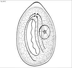
Balantidium coli cyst appearance |
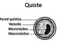
Has a kidney or bean-shaped macronucleus, spherical micronucleus, contractile vacuoles, and retracted cilia |
|
|
Purpose of B. coli macronucleus |
for vegetations |
|
|
Purpose of B. coli micronucleus |
Reproduction |
|
|
B. coli trophozoite size |
50x70um |
|
|
Morphology of B. coli trophozoite |
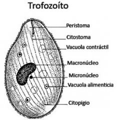
Equipped with cell mouth (cytostome), and cell anus (cytopyge), provided with 2 contractile vacuoles, and with micronucleus at concavity of macronucleus |
|
|
B. coli characteristic movement |
directional tumbling |