Reading...
![]()
Play button
![]()
Play button
![]()
Use LEFT and RIGHT arrow keys to navigate between flashcards;
Use UP and DOWN arrow keys to flip the card;
H to show hint;
A reads text to speech;
25 Cards in this Set
- Front
- Back
|
Describe Type I muscles fibers
|
Slow-twitch (red) fibers
rich in Mitochondria & Oxidative enzymes |
|
|
Describe Type II muscle fibers
|
Fast-twitch (white) fibers
Poor in Mitochondria |
|
|
What determines the type of fiber a myocyte contains?
|
Motor neuron
|
|
|
Define Neurogenic Atrophy
|
disease of the Anterior Horn cell or its axon
|
|
|
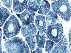
Describe the pathology seen in Neurogenic Atrophy
|
Initial: individual atrophic angular fibers
Later: Groups of atrophic angular fibers |
|
|
Neurogenic Atrophy
|
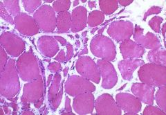
What is seen here?
|
|
|
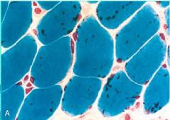
What helps in diagnosis Neurogenic Atrophy?
|
Fiber typing = after reinnervation, a cluster of Type I fibers are adjacent to a cluster of Type II fibers
|
|
|
Fiber-typing used to diagnose Neurogenic Atrophy
|
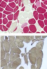
What is seen here?
|
|
|
What diseases can cause Neurogenic Atrophy?
|
1. ALS
2. Vasculitis 3. Trauma 4. Nerve compression (spinal cord disease) |
|
|
List 3 Inflammatory Myopathies
|
1. Dermatomyositis
2. Polymyositis 3. Inclusion Body Myositis |
|
|
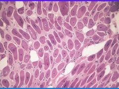
Dermatomyositis
-perifascicular pattern of injury |
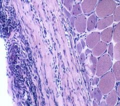
What is seen here?
|
|
|
What is the clinical presentation of Dermatomyositis?
|
Rash + weakness + muscle pain
*has an association with Cancer (Lung CA) |
|
|
What is the immune target of Dermatomyositis? What is it mediated by?
|
Capillaries
Antibodies + Complement |
|
|
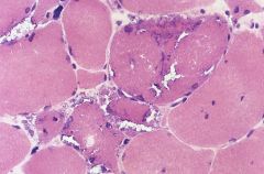
Polymyositis = lack of skin involvement
-Endomysial process -inflammation tends to occur within the meat of muscle -nuclei are also sometimes not at the periphery |
What is seen here?
|
|
|
Inclusion Body Myositis
-rimmed vacuoles that may contain Beta-amyloid & tau within |
What is seen here?
|
|
|
Describe the clinical features of Inclusion Body Myositis
|
1. occurs in Distal muscles first
2. Older subjects 3. Resistant to therapy = not responsive to steroids 4. Rimmed vacuoles |
|
|
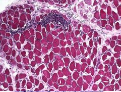
pathology of Dystrophies
-fiber size variation -increased central nuclei -degeneration, necrosis -regeneration -fibrosis |
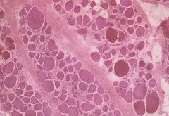
What is seen here?
|
|
|
Describe the pathology of Dystrophy
|
1. Fiber size variation
2. increased central nuclei 3. degeneration, necrosis 4. regeneration 5. fibrosis |
|
|
Dystrophy characterized by X-linked inheritance with a mutation in the dystrophin gene
|
Duchenne & Becker Muscular Dystophy
|
|
|
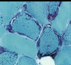
Central Core Disease (congenital myopathy)
-central pallor -disorganized sarcomeres with deficiency of ATPase |
What is seen here?
|
|
|
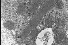
Nemaline Myopathy (congenital myopathy)
-Rod-like inclusions derived from the Z-disc |
What is seen here?
|
|
|
Myotubular (Centronuclear) Myopathy
-desmin, loss of myofibrillar material (EM) |
What is seen here?
|
|
|
Mitochondrial Myopathy
-aggregates of abnormal mitochondria -Ragged red fibers -Cytochrome oxidase negative fibers |
What is seen here?
|
|
|
-aggregates of abnormal mitochondria
-Ragged red fibers -Cytochrome Oxidase negative fibers |
Mitochondrial Myopathy
|
|
|
Mitochondrial Myopathy
Proximal weakness -other neurological symptoms -lactic acidosis -Cardiomyopathy |
What disease is this? What does it cause?
|