Reading...
![]()
Play button
![]()
Play button
![]()
Use LEFT and RIGHT arrow keys to navigate between flashcards;
Use UP and DOWN arrow keys to flip the card;
H to show hint;
A reads text to speech;
31 Cards in this Set
- Front
- Back
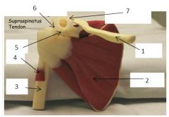
Posterior Scapula
|
1 = Acromion
2 = Humerus 3 = Teres Minor 4 = Infraspinatus 5 = Spine of Scapula 6 = Supraspinatus |
|
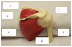
Anterior Scapula
|
1 = Clavicle
2 = Subscapularis 3 = Humerus 4 = Biceps head 5 = Coracoid 6 = Coracoacromial Ligament 7 = Acromio-clavicular Ligament |
|
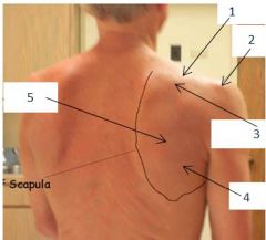
Edge of scapula is labeled
|
1 = Supraspinatus
2 = Acromian 3 = Spine of Scapula 4 = Teres Minor 5 = Infraspinatus |
|
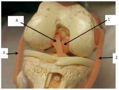
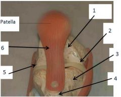
Right knee ligaments (viewed from the front with patella removed)
|
1 = Posterior Cruciate Ligament
2 = Medial Cruciate Ligament 3 = Lateral Cruciate Ligament 4 = Anterior Cruciate Ligament |
|
Right Knee from front
|
1 = Femur
2 = Medial Joint Line 3 = Tibia 4 = Tibial Tuberosity 5 = Lateral Joint Line 6 = Patellar Tendon |
|
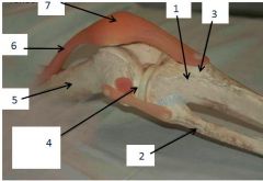
Lateral Right Knee
|
1 = Tibia
2 = Fibula 3 = Tibial Tuberosity 4 = Lateral Joint Line 5 = Femur 6 = Quadriceps Tendon 7 = Patella |
|
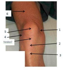
Right Knee from front
|
1 =Medial Joint Line
2 = Anterior Tibial Tuberosity 3 = Tibia Patellar Tendon 4 = Lateral Joint Line 5 = Patella 6 = Quadriceps Muscle |
|
|
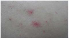
Roth Spots - pale centered hemorrhage, caused by multiple disease processes, including bacterial endocarditis
|
|
|
Emboli/Infarcts - the small fleck is a plaque (platelet/fibrin/cholesterol embolus) resulting in an infarct (the grey area to the right and above the plaque)
|
|

|
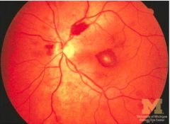
Cotton wool spots - caused by microinfarcts - exploded ganglion cells extrude their axoplasm into the retina
DDX: HTN, DM, HIV, severe anemia or thrombocytopenia, hypercoaguable states, connective tissue disorders, viruses |
|
|
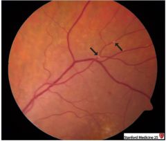
Arteriovenous Nicking - at AV crossing points (arrow), arteries indent and displace veins
Chronic HTN thickens and stiffens arteries *Arteries are narrower than veins in the retina |
|
|
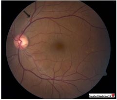
Optic disc edema - optic disc is elevated and it's surface is covered by cotton wool spots (damaged axons) and flame hemorrhages (damaged vessels)
DDX (the four I's): increased intracranial pressure (papilledema), infarction, inflammation, infiltration (cancer) |
|
|
Optic disc edema - optic disc is elevated and it's surface is covered by cotton wool spots (damaged axons) and flame hemorrhages (damaged vessels)
DDX (the four I's): increased intracranial pressure (papilledema), infarction, inflammation, infiltration (cancer) |
|
|
Optic disc edema - optic disc is elevated and it's surface is covered by cotton wool spots (damaged axons) and flame hemorrhages (damaged vessels)
DDX (the four I's): increased intracranial pressure (papilledema), infarction, inflammation, infiltration (cancer) |
|
|
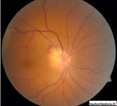
Pathological Optic Cupping
Cup to Disc ratio should be <0.5; in this picture it is 0.8 Indicates Glaucoma |
|
|
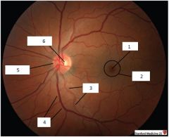
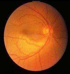
1 = Fovea
2 = Macula 3 = Arteries (skinnier) 4 = Veins (more prominent) 5 = optic disc 6= optic cup |
|
|
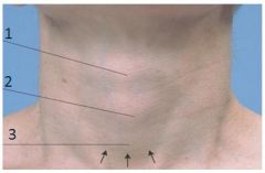
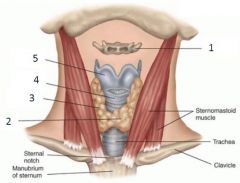
1 = Thyroid cartilage
2 = Cricoid cartilage 3 = Thyroid gland |
|
|
1=hyoid bone
2=thyroid isthmus 3=thyroid lobe 4=cricoid cartilage 5=thyroid cartilage |
|
|
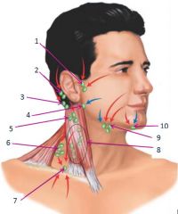
1= pre-auricular
2= posterior auricular 3= occipital 4= tonsillar 5= superficial cervical 6= posterior cervical 7= supraclavicular 8= deep cervical 9= submandibular 10= submental |
|
|
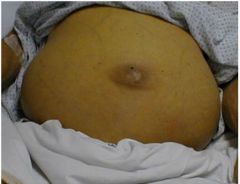
Ascites - sign of advanced liver failure
|
|

|
Scleral icterus - sign of advanced liver disease
|
|
|
Spider - sign of advanced liver disease
|
|
|
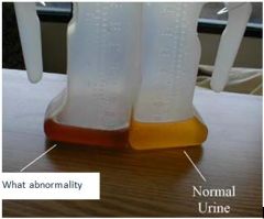
Bilirubinuria - sign of advanced liver disease
|
|
|
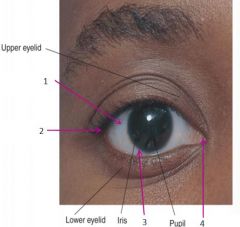
1= sclera
2= Lateral Canthus 3= Limbus 4= Medial Canthus |
|
|
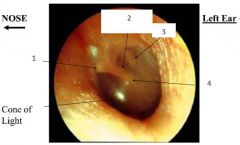
1= short process of malleus
2= long process of malleus 3= incus 4= umbo |
|
|
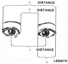
A = outer canthal distance
B = inner canthal distance C = interpupillary distance D = palpebral fissure length |
|
|
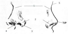
1 = Nasal Bridge
2 = Ala nasae 3 = Naris 4 = Columella 5 = Naison |
|
|
Papilledema - sign of increased intracranial pressure. NOT the same as optic disc edema
= disc hyperemia possibly with dilated disc capillaries. Nerve fiber layer (NFL) opacification and swelling. Blurred disc margin(s). |
|
|
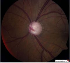
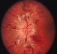
Severe papilledema
More severe developed papilledema will have elevation of the disc and opacified NFL obscuring disc vessels. There may be dilated veins, disc splinter hemorrhages, cotton wool spots, or hard exudates. With severe disc leakage a macular star may be seen, this is more prominent on the disc side of the macula. |
|
|
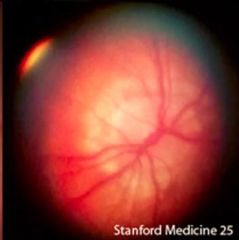
Central retinal artery occlusion - this picture looks really normal but whatever you say Dr. Reynolds
"cherry red spot" with surrounding pale retina is characteristic look |
|
|
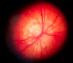
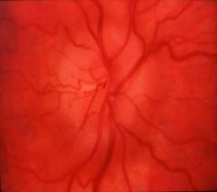
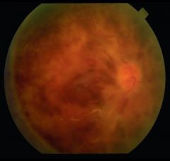
Central vein artery occlusion - BLOOD AND THUNDER
|