Reading...
![]()
Play button
![]()
Play button
![]()
Use LEFT and RIGHT arrow keys to navigate between flashcards;
Use UP and DOWN arrow keys to flip the card;
H to show hint;
A reads text to speech;
321 Cards in this Set
- Front
- Back
|
What does the spinal cord allow for?
|
*Information to travel to the brain from spinal cord or brain to spinal nerve
*Neural connection for reflexes to occur (acts as control center to a certain extent) |
|
|
What is an example of what the spinal cord can do?
|
Includes ability to withdraw from a painful stimulus
|
|
|
What part of the brain is the lower brain or subcortical level
|
All of it except for the cerebral cortex
|
|
|
What does the lower brain control?
|
Unconcious bodily function
|
|
|
What are examples of what the brain or subcortical level control?
|
Respiratory rate
Equilibrium Emotional Patterns |
|
|
What is the higher brain or cortical level?
|
Cerebral cortex
|
|
|
The higher brain or cortical level is the _______ storehouse
|
Memory
|
|
|
What does the higher brain work in combination with?
|
Subcortical brain
|
|
|
When the higher brain and subcortical brain work together what do they produce?
|
Precision in our actions and allow for thinking and use of our past experience and memories
|
|
|
How many synapses does a monosynapse have?
|
1
|
|
Complete the table
|
|
|
|
For an example "Walking down the street and step on an uneven peice of sidewalk, and your ankle starts to roll ("inversion"). Muscles on lateral side of leg (fibrous longus brevis) experience a sudden stretch (also called the "muscle stretch reflex")" what are the sensory receptors for that?
|
Muscle spindles infibularis detect change in length, there will be a "dynamic response" in which primary endings of muscle spindle will send increased rate of impulses along afferent neurons
|
|
|
For an example "Walking down the street and step on an uneven peice of sidewalk, and your ankle starts to roll ("inversion"). Muscles on lateral side of leg (fibrous longus brevis) experience a sudden stretch (also called the "muscle stretch reflex")"
What is the sensory neurons? |
Endings in the muscle spindles send signal along type 1a fibers
(Large diameters Transmit signals @ ~70-120 m/sec) AKA Aα fibers (largest/fastest neurons) |
|
|
For an example "Walking down the street and step on an uneven piece of sidewalk, and your ankle starts to roll ("inversion"). Muscles on lateral side of leg (fibrous longus brevis) experience a sudden stretch (also called the "muscle stretch reflex")"
What is the area of spinal and sensory receptor synapses? |
Branch of 1a fibers, reach directly to anterior horn of spinal cord
|
|
|
For an example "Walking down the street and step on an uneven peice of sidewalk, and your ankle starts to roll ("inversion"). Muscles on lateral side of leg (fibrous longus brevis) experience a sudden stretch (also called the "muscle stretch reflex")"
What are the interneurons and motor neurons sensory neurons that synapse with the spinal cord |
After it synapses, motor neuron innervates same muscle being stretch (agonist).
There is a monosynaptic reflex, no interneuron between the sensory and motor neuron |
|
|
For an example "Walking down the street and step on an uneven piece of sidewalk, and your ankle starts to roll ("inversion"). Muscles on lateral side of leg (fibrous longus brevis) experience a sudden stretch (also called the "muscle stretch reflex")"
What is the type of motor fibers that leave spinal cord to innervate muscle |
Motor neuron has cell body in the anterior horn of the spinal cord. The motor neurons are Aα fibers (large and fast neurons).
The monosynaptic reflex occurs between Aα (1a) afferent nerve/Aα motor nerve Additional synapse with Aγ fibers that communicate with infrafusal muscle fibers (within muscle spindles) of the agonist |
|
|
For an example "Walking down the street and step on an uneven piece of sidewalk, and your ankle starts to roll ("inversion"). Muscles on lateral side of leg (fibrous longus brevis) experience a sudden stretch (also called the "muscle stretch reflex")"
What is the effect in the muscle (bot hthe extrafusal and intrafusal motor fibers) |
Contraction of the same msucle that was lengthened (agonist, in this case, the fibularis)
The A α motor neurons supply the extrafusal muscle fibers (AKA skeletal muscle fibers) Infrafusal muscle fibers recieve message on the slower Aγ fibers which causes them to contract too and ensure the muscle spindle itself maintains the correct "tune" to detect changes in the whole muscle length |
|
|
Which of the following are "efferent" in nature, as opposed to "afferent"?
A) Somatic nervous system B) Parasympathetic nervous system C) Sympathetic nervous system D) All of the Above E) Only B and C |
D
|
|
|
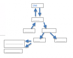
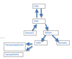
What does the CNS include
|
Brain and spinal cord
|
|
|
What does the CNS do?
|
Integrative and control centers
|
|
|
What does the PNS include?
|
Crainal nerves
Spinal nerves |
|
|
The PNS is the communication lines between the ______ and ____
|
*CNS
*Rest of the body |
|
|
What is the sensory (efferent) division of the PNS inlcude
|
Somatic sensory nerve fibers
Visceral sensory nerve fibers |
|
|
What does the sensory (afferent) divison of the PNS do?
|
Conduct impulses from receptors t othe CNS
|
|
|
What does the motor (efferent) divison include?
|
Motor nerve fibers
|
|
|
What does the motor (efferent) divison of the PNS do?
|
Conducts impulses form the CNS to effectors (muscles and glands)
|
|
|
What two branches can the motor (efferent) divison of the PNS be divided into?
|
Autonomic Nervous System (ANS)
Somatic Nervous System |
|
|
Is the somatic nervous system for voluntary or involuntary movement
|
Voluntary
(Somatic motor) |
|
|
The somatic nervous system conducts impulses from the _____ to the _____
|
CNS
Skeletal muscles |
|
|
/Is the autonomic nervous system (ANS) involuntary or voluntary
|
Involuntary
(Visceral motor) |
|
|
The autonomic nervous system (ANS) conducts impulses from the _____ to _____
|
CNS
Cardia muscles, smooth muscles, glands |
|
|
What two branches can the autonomic nervous system (ANS) be divided into?
|
The sympathetic divison
Parasympathetic division |
|
|
What does the symapthetic divison of the autonomic nervous system do?
|
Mobilizes body systems during emergency situations
|
|
|
What does the parasympathetic division of the ANS do?
|
*Conserves energy
*Promotes nonemergency functions |
|
|
Is the autonomic nervous system output or input
|
Output
|
|
|
What is the functions of the spinal cord>
|
Two-way highway
Reflex control |
|
|
What is the subcortical part of?
|
All but the cerebral cortex
|
|
|
What is the subcortical used for?
|
Unconcious control (Autonomic control)
|
|
|
What is considered the cortical?
|
Cerebral cortex
|
|
|
What is the cortical used for
|
Thinking and memory integration
Consciouns |
|
|
Does the perception of a reflex in a brain come before and after the detection and response in the spinal cord?
|
After
|
|
|
When there is a stretch reflex, where is the muscle stretch reflex found in?
|
Lateral muscles (fibularis group)
|
|
|
Where are the stretch reflex sensed at first?
|
Muscle spindles
|
|
|
During a stretch refelx, muscle spindles fire what type of neurons? What are they like?
|
1a neurons
Large (fast) |
|
|
During a stretch reflex, the 1a neurons synapse with a what type of motor neuron? What is it like?
|
α motor neuron (efferent)
Large, fast neurons |
|
|
α motor neurons are efferent or afferent?
|
Efferent
|
|
|
What muscles do the α motor neurons synapse with?
|
Fibularis muscle
|
|
|
When the α motor neurons synapse with the fibularis muscles, is the fibularis muscles agonists or antagonists?
|
Agonist
|
|
|
When the α motor neuron synapses with the fibularis muscles, is that monosynaptic?
|
Yes
|
|
|
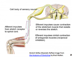
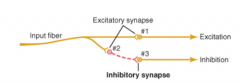
In a stretch reflex, apart from synapsing with an α motor neuron, what else can the type 1a neuron synapse with?
|
Inhibitory interneuron which then synapses with antagonist muscle α motor neuron
Inhibits the antagonist |
|
|
What the name mechanism which inhibits the antagonist muscle and excits the agnoist muscle
|
Reciprocal inhibition
|
|
|
What is the entire mechanism of the stretch reflex
|
|
|
|
What is reciprocal inhibition?
|
Antagonist muscle is being inhibited while the lateral muscles are contracted (excited)
|
|
|
What is the relative length of the preganglionic neurons of a parasympathetic system?
|
Long
|
|
|
Where are the preganglionic neurons of the parasympathetic neurons coming from? (Soma)
|
Soma in brain stem and spinal cord at S2 - S4
|
|
|
Are post-ganglionic neurons myelinated or unmyelinated?
|
Unmyelinated
|
|
|
Are pre-ganglionic neurons myelinated or unmyelinated
|
Myelinated
|
|
|
What is the relative length of post-ganglionic neurons in the parasympathetic system
|
Short
|
|
|
What is the relative length of the pre-ganglionic neurons of the sympathetic system
|
Short
|
|
|
Where is the soma of the pre-ganglionic neurons of the sympathetic system found?
|
In the spinal cord at ~ T1-L2
|
|
|
What is the relative length of post-ganglionic neurons of the sympathetic system?
|
Long
|
|
|
What neurotransmitters are released by the pre-ganglionic neurons of the parasympathetic system?
|
Acetylcholine (ACH)
|
|
|
What neurotransmitters are released by the postganglionic neurons of the parasympathetic system
|
Acetylcholine (ACH)
|
|
|
What neurotransmitters are transmitted by the preganglionic neurons of the sympathetic system.
|
Acetylcholine (ACH)
|
|
|
What neurotransmitters are released by the postganglionic neurons
|
Norepinephrine (NE) or epinephrine (Epi) or Acetylcholine (ACH)
|
|
|
What are the types of receptors found on postganglionic neurons of the parasympathetic system?
|
Nicotinic cholinergic receptors
|
|
|
What are the types of receptors found in target organs of the parasympathetic system?
|
Muscarinic cholinergic receptors
|
|
|
What are the types of receptors found on the postganglionic neurons of the sympathetic system
|
Nicotinic cholinergic
|
|
|
What are the types of receptors found on the target organs of the sympathetic system
|
Adrenergic receptors are found effector organs (α1, α2, β1, β2, β3) for Norepinephrine or epinephrine
Muscarinic cholinergic receptor (sweat glands, piloerector muscles in skin) for ACH |
|
|
What is the speed of the parasympathetic system?
|
Rapid (becuase of the extra amount of myelinated fibers)
|
|
|
What is the duration of response of the parasympathetic system
|
Quick because ACH is quickly broken down at the neuroeffector junction by acetylcholinesterase, so duration of effect is short
|
|
|
What is the speed of the sympathetic system
|
Slower (because less myelination)
|
|
|
What is the duration of response for the sympathetic system?
|
Prolonged effect
|
|
|
Why does the sympathetic system have a prolonged effect?
|
Because of blood borne epinephrine and norepinephrine from the adrenal gland. Also norepinephrine and epinephrine is slower to be broken down at neuroeffectorj junction and have locally longer effectors
|
|
|
What does the tone of the parasympathetic and sympathetic?
|
Refers to way in which sympathetic and parasympathetic systems are always sending signals to their effector organs at a base level. Therefore, either system can be increased or decreased from these base (or basal) levels) allowing for quick and dramatic changes in the body (IE you can turn up the sympathetic while simultaneously turning down the parasympathetic, which produces a big effect on organs which are innervated by both).
|
|
|
The muscles spindles are found in ________ fibers
|
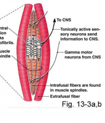
Infrafusal
(Inside skeletal muscles -> The spindles are wrapped around) |
|
|
What is the different between the extrafusal and the intrafusal fibers?
|
The extrafusal msucle fibers are regular old muscle fibers while the intrafusal fibers are the inside and have muscle spindles wrapped aroudit.
|
|
|
What affaerent information is sent to the CNS from the muscle spindles?
|
Length
Rate of change in length |
|
|
How are the muscle spindles activated>
|
Stretched apart
|
|
|
What afferent information does the golgi tendon organs send to the CNS?
|
*Tension
*Rate of change in tension |
|
|
What are the tendons of the golgi tendon organ like?
|
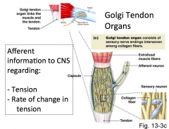
They are stretch tendons
And the sensors will get "squished" which creates the action potentials |
|
|
Are α motor neurons myelinated or unmyelinated?
|
Myelinated
So they are fast |
|
|
Why is your withdrawl reflex when you burn yourself so quick?
|
Not going to the brain
Not controlled by the cortex reflex Monosynaptic Myelinated vs not myelinated |
|
|
What are the various aspects of a sensory-motor response that affects time?
|
1) The complexity of the receptor
2) Fiber type/size and the distance traveled 3) Number of syanpases |
|
|
What does the complexity of the receptor mean?
|
How long it takes to o from the stimulus to the response (How long to be changed to action postential)
|
|
|
Is the visual system the least complex?
|
No
(It is the most complex) |
|
|
What are somatic senses?
|
Body sense
|
|
|
What different types of somatic sense (as opposed to "special senses") can you think of?
|
Proprioception (joint positon sense)
Visceral (Organs -> Not near as sensitive as sensitive area of skin) Deep tissue (If punched in the arm, not just skin, joints, bone level, etc will feel it) Surface of body (skin) |
|
|
A (or I-III) neurons are _________ size?
|
large to medium
|
|
|
A ( or I-III) neurons are myelinated or unmyelinated?
|
Myelinated
|
|
|
Are A (or I-III) neurons fast or slow?
|
Fast
|
|
|
C ( or IV) neurons are _______ size?
|
Small
|
|
|
C (or IV) nerons are myelinated or unmyelinated
|
unmyelianted
|
|
|
Are the C (or IV) neurons fast or slow?
|
Slow
|
|
|
What are the two size classfication systems of neuronal fibers?
|
Blue line represents myelinated vs unmyelinated
|
|
|
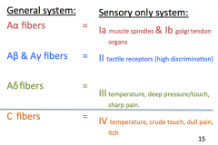
What are the large and myelinated neurons in the general system?
|
Aα, Aβ/Aγ and Aδ fibers
|
|
|
What are the large and myelinated neurons of the sensory system
|
1a/1b, II, III
|
|
|
What is the 1a
|
Muscle spindles
|
|
|
What is the 1b neuron
|
Golgi tendon organs
|
|
|
What is the II neuron
|
Tactile receptors (high discrimination)
|
|
|
What is the III sensory neuron for?
|
Temperature, deep pressure/touch, sharp pain
|
|
|
What is the C fibers of the general system as a sensory neuron
|
IV
|
|
|
What is the IV sesnory neuron for?
|
Temperature, crude touch, dull pain, itch
|
|
|
What is the size of the type I & II sensory nerves ?
|
Large
|
|
|
Is the type I & II sensory nerves fast or slow?
|
Fast
|
|
|
Is the type I & II myelinated or unmyelinated?
|
Myelinated
|
|
|
what is the type I & II sensory neurons used for
|
Critical sensations (joint position, tactile)
|
|
|
what size the the type III neurons?
|
Medium
|
|
|
Is the type III sensory neuron fast or slow?
|
Fast
|
|
|
Are the type III sensory neurons myelinated or unmyelinated
|
Myelinated
|
|
|
WhAre the type III sensory neurons, what are they for?
|
Free nerve endings (sharp pains) some temp
|
|
|
What are the type IV sensory neurons' size?
|
Small
|
|
|
What is the type IV sensory neuron's speed?
|
Slow
|
|
|
Is the type IV sensory neuorn myelinated or unmyelinated?
|
Unmyelinated
|
|
|
What sort of the sensations is the type IV sensory neuron used for?
|
Tickle, slow aching pain, some temp
|
|
|
For the motor neuronal fibers, which are the largest and fastest?
|
Type Aα neurons
[Skeletal muscle fibers (extrafusal) ] |
|
|
For the motor neuronal fibers, what is the smaller and not as fast fibers?
|
Type Aγ neurons
[Muscle spindle (intrafusal) fibers ] |
|
|
for the motor nuronal fibers, which one is the smallest and slowest?
|
Type C neurons (unmyelinated)
[Autonomic post-ganglionic nerves] |
|
|
How do we detect touch, temperature, pain etc?
|
Stimulus is converted to action potentials that reach specific portions of the brain
|
|
|
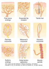
What are the somato-sesnory receptors?
|
Mechano receptors [Only response to mechanical pressure]
Thermoreceptors Chemoreceptors Nociceptors (pain) [communicate to brain about pain] Electromagnetic Receptors (Vision |
|
|
What are the main three somato-sesnory receptors?
|
Mechanoreceptors
Thermoreceptors Chemoreceptors |
|
|
What are all the different receptors (picture)
|
|
|
|
How do sensory receptors convert "sensations" into action potentials?
|
Typically, open ion channels and change the membrane potential. When threshold is reached, action potentials move along the neuron membrane
|
|
|
How does the temperature receptor work?
|
The temperature change causes the conformational change
|
|
|
What is the most common precursor to the action potential that allows for the sensory receptors to convert the "sensaitons" into the action potentials
|
*At membrane needed a depolarization caused by the stimulus
* Ion channels open to allow all potential change *In mechanoreceptors, mechanical signals cause ion channels to open |
|
|
How does our physiology differ for touch, position, pain and temperature sensations
|
The receptor types vary to translate different stimuli into action potentials
The exact type of receptor differs |
|
|
Are all sensations perceived in the brain?
|
Yes
Brain tells you where it should hurt Actual pain is just a perception in the brain |
|
|
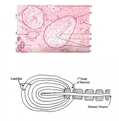
What does the pacinian corpuscle look like?
|
|
|
|
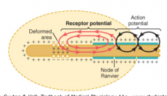
How does the pacinian corpuscle work?
|
When pushes down on it, causes the ion channels to open and if it was to just stay there, there would nbe no change and ion channels woudl no longer open
It only allows ion movement went there is a change on the receptor |
|
|
How does the deformation of the pacinian corpuscle
|
There is a change in the pressure
|
|
|
If you are wearing a watch or ring, is it changing the deformation of the corpuscles under it right now?
|
No, but you changed the deformation when you first put it on; afterwards, it does not change at all...or else you would constantly get signals telling you that you had your watch on, etc
|
|
|
Have an image of the working of the pacinian corpuscle
|
|
|
|
What does the fusiform gyrus do?
|
Helps you recognize faces (IE damage to it limits someon's ability to recognize faces)
|
|
|
What is the amygdala do?
|
Gauges emotional significance of what you look at
(Limbic system) |
|
|
Explanation for phenomena when someone can recognize their mother but thinks they are an imposter
|
Fusiform gyrus is intact, but neurons to the amygalda are severed, so they can recognize their mother but don't feel any emotional connection to the face so they assume the person is an imposter.
|
|
|
What is the vidence that Ramachandran uses to back up his theory
|
Patient's ANS is not stimulated when neural informaiton about mother's face is presented (eg the expected galvanic skin response does not occur when they see their mother-Hands begin to sweat)
Also they do recognize their mother's voice on the phone, suggesting the connection between auditory recognition areas and the amygdala are likely intact |
|
|
What is learned paralysis
|
Repeated bouts in which CNS commands arm to move with no response due to the severed peripheral nerves
|
|
|
How is a mirror box used to releive the pain of a "paralyzed" phantom arm
|
Visual informaiton from move of healthy arm perceived from the reflection in mirror helps to change learned paralysis. Practicing with the mirror allows the neural connection to be rewired and the phantom limb and pain disappears
|
|
|
Describe synthesia?
|
People can have numbers visualized in different colors (or musical tunes, letters, months of the year, etc)
|
|
|
What did Dr. Ramachandran explain is the neurological basis of this bility?
|
The color and number areas in the fusiform gyrus are next to each other so it allows the unique genes to cross wire.
|
|
|
How does Dr. Ramachandran believe synthesia is related to creativity?
|
People with synthesia have a greater ability to produce metaphors and are creative due to the interconections (which are trimed down a lot) in areas of the brain
|
|
|
Why do people assign the same shapes to the names "Bubba and Kiki"
|
Because auditory and visual corticies send information between each other in the angular gyrus and the information is interrelated (sound "kiki" seems to match in our brain with sharp edges of one shape and sound for "Bubba" match the round edges in other shape)
|
|
|
Why is the cerebral cortex dependent on the thalamus
|
Almost all sensory pathways (with the exception of olfaction) travel through the thamalus before reaching the cortex. If the portion of the thalamus, that communicates with a specific part of the cortex is damaged, that area of the cortex does not function either. There are numerous afferent and efferent connections between the cortex and thalamus
|
|
|
What does the primary motor do?
|
Sends signals to cause the muscles to contract
|
|
|
What do the secondary supplementary motor and premotor areas do?
|
Work with the basal ganglia to create functional patterns of movements
|
|
|
What does the primary sensory areas do?
|
Receives signals from the sensory receptors for the somatosensory, visual and auditory senses
|
|
|
What does the supplementar y and premotor areas and secondary sensory do?
|
Provide "patterns" for motor activity (interperetes incoming stimulus)
|
|
|
What is the function of the Wernicke's Area
|
Interpret language
Important for cognition and intellectual function |
|
|
What is the angular gyrus used for
|
Feeds visual information from words into the Wernicke's area
Helps to reading ability-The interpretation of words received via visual input |
|
|
What are the two main functions of the prefrontal association area?
|
1) Thinking and planning complex motor activities
2) Thinking in general and possibly storing short term or "working" memory (thinking about ideas as they come into your head) 3) Related to personality 4) Ability to connection actions to consequences |
|
|
Is the frontal lobe the last or first to develop
|
Last (As late as 25)
|
|
|
What is the main action of Broca's area?
|
Plans motor output necessary to speak using words or phrases
|
|
|
What are the main functions of the limbic association area?
|
1) Behavior
2) Emotions 3) Motivation |
|
|
Where is the face recognition portion of the brain?
|
Inferior surface of the brain: medial occipital and temporal lobes
|
|
|
Which hemisphere is the most developed Wernicke's area in the right-handed individuals?
|
Left
|
|
|
If the dominant Wnicke's area was damged what would be the functions that would be lost?
|
Lose function to process intellectual functions of the brain
Almost complete lack of intelligences (as our culture tends to define it); including the ability to read, perform math, think logically) |
|
|
What is the function of the corpus callosum?
|
Communicate between two hemispheres
|
|
|
What are some functions of the non-Dominant Wernicke's area
|
1) Interprete music
2) Visual patterns 3) Spatial relationships 4) Body language 5) Voice intonation 6) Body experiences from the limbs and hands |
|
|
What are some functions that are LOST in patients following a prefrontal lobotomy?
|
1) Problem solving
2) Mutli-tasking 3) Ambition 4) Learning 5) Sequential tasks 6) Staying focused 7) Appropriate sexual activity / excretion 8) Maintain mood |
|
|
What is the "holistic theory" of thoughts
|
Thoughts are patterns of stimulation of various areas of the brain including the cerebral cortex, thalamus, limbic system and brain stem
|
|
|
Define short term memory
|
Memories that last for seconds or a couple of minutes (remember 7 lucrative pieces of inforamtion for the duration of time you are thinking about it (seconds to minutes)
|
|
|
Define intermediate term memory
|
Memories that last for days to weeks then fade
Changes to the pre or postsynaptic neuron that helps the memory trace remain activated (minutes to weeks) |
|
|
Define the long term memory
|
Stored for a lifetime
Structural changes in the pre or postsynaptic neurons next to the memory trace to remain activated (years to lifetime) |
|
|
Where is the hippocampus
|
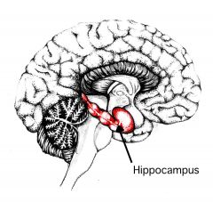
Medial portion of the temporal lobe
|
|
|
What effect does the removal of hippocampus have on memory?
|
Removal of both hippocampi make it impossible to create intermediate or long term memory
|
|
|
Why is the hypothalamus described by Guyton and Hull as "one of the most important of the control pathways of the limbic system?"
|
Sends and receives information from all portions of the limbic system. In addition, it sends information to the brain stem and beyond to the ANS, sends into the thalamus and cortex and it sends information to the pituitary galnd (which in turn secretes hormones with the sympathetic effect).
|
|
|
When you have an example voluntary motor movement of seeing an apple on the table and wanting to pick it up, what is the first thing that happens with light and the sensory impulse?
|
The light reflecting from the apple is translated into a sensory impulse, which has multiple synapses and areas in the brain that will be involved with translation and interpretation of those sensory impulses
|
|
|
When you have an example voluntary motor movement of seeing an apple on the table and wanting to pick it up, after the light is translated into sensory impulses, the reaching and grasping is a pattern of _________. Where will the signal for the action likely begin in? Where is that area found? And this forms the "motor image" which is what? This can directly stimulate the appropriate areas in the ______
|
*Movement.
* Premotor area *Anterior ot the primary motor cortex *"Motor image" is where the pattern is broken down into its individual parts. From here the signal for each pattern of motion, which includes activity in one muscle of the shoulder, arm and hand *Primary motor cortex (or the message could first be sent to the basal ganglia and thalamus followed by the primary motor cortex |
|
|
When you have an example voluntary motor movement of seeing an apple on the table and wanting to pick it up, after it goes through the primary motor cortex, what else may be stimulated to produce the "background" muscle activity? What is that "background" muscle activity?
|
*Supplementary motor area
*Body positioning or hand and eye positioning that would assist my ability to reach up and grasp the apple |
|
|
When you have an example voluntary motor movement of seeing an apple on the table and wanting to pick it up, after the signal is sent down to muscles themveles what are they sent down on?
|
Pyramidal tract
(Also known as the corticospinal tract; literally meaning from the cortex to the spinal cord) |
|
|
When you have an example voluntary motor movement of seeing an apple on the table and wanting to pick it up, after the signals are sent down to muscles via the pyramidal tract, the impulses that are concernerd with helping the arm reach out and the fingers to grasp the apple will synapse with the ________
|
Interneurons in the spinal cord
and some alpha motor neurons directly (IE neurons that will lead to deltoid muscle of the shoulder, extesory muscles of the hand, flexor muscles of the elbow, etc |
|
|
When you have an example voluntary motor movement of seeing an apple on the table and wanting to pick it up, what happens in the cerebellum?
|
Place where sensory and motor information is compared and adjustments are made. The cerebellum receives informatio nabout the desired motor patern from the motor cortex and then compares this to the sensory informaiton it is receing. Helps to produces smooth coordinated movements. In this example, if my arm was not reaching far enough, the cerebellum would be able to excite motor pathways to produce more muscle contraction in the muscles the extend the arm, so that my reach pattern was more accurate
|
|
|
When you have an example voluntary motor movement of seeing an apple on the table and wanting to pick it up, finally, many complex unconciously controlled motor actions of the body would require input from what?
|
Basal ganglia
The simple action of reach for the apple may not, but the writing of the alphabet, throwing a football, hammering nail, etc would include connections between the motor cortex and the basal ganglia (as well as connecting with the thalamus) |
|
|
Instead of having impulses synapse with interneurons in the spinal cord and some alpha motor neurons, some fibers from the motor cortex could use a different but similar pathway to the motor neurons in the spinal cord called the corticorubrospinal system. What does that do?
|
First, fibers travel form the interconnection from the red nucleus to the cerebellum and back as well, allowing influence by the cerebellum on these signals. From the midbrain, the fibers continue down the spinal cord via the rubospinal tract, which crosses to the opposite side of the body in the lower medulla (just as the corticospinal tract did). These fibers lead to interneurons, and directly to alpha motor neurons which innervate muscles relevant to the movement
|
|
|
Some portions of the basal ganglia allow for "unconcious thinking" which is what?
|
Reacting to a situation based on previous knowledge or memory.
|
|
|
What else can the basal ganglia do?
|
*Ability to produce movements in the correct scale (large vs small) and with the right timing, but much of the data behind this assertion has been deduced based on patients with brain damage and has not been proven
|
|
|
Do all of the areas of our skin have the same "2-point-discrimination" ability?
|
No
|
|
|
Is the 2 point discrimination of the pads on our finger very good or not?
|
Very good
|
|
|
What might allow for better 2-point discriminatin on some areas of skin compared to others?
|
*Size of receptor field per afferent neuron
*Receptor density *"Lateral inhibition" (or surround inhibition) enhances discrimination *Convergence of first order neurons onto second order neurons ` |
|
|
What is receptor density about?
|
How much receptors there are
|
|
|
If there is a higher receptor density, is there a better chance for two point discrimination?
|
Yes
|
|
|
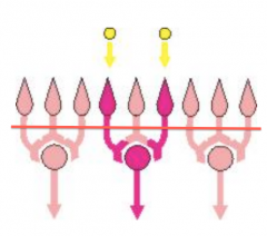
The receptor field is the area tat ______ neuron will association with
|
One
|
|

Which has better two point discrimination
|
The Left one
|
|

For the receptor field on the left, there are _______ receptive fields that stimulated by the _______ points of stimulation
|
Two
Two |
|

For the receptor field on the right side, there is ______ receptive field that is stimulated by the _______ points of stimulation the same distance apart as in the left one
|
One
Two |
|
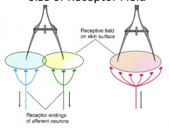
How many points can be felt for the receptor field on the left?
|
Two
|
|

How many points can be felt for the receptor field on the right
|
One
|
|

Why can the brain only perceive one point for the left sided receptor field
|
Because the receptor field encompasses both points, so the brain can only perceive one point
|
|
|
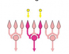
What is lateral inhibition?
|
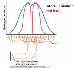
Inhibition to the sides by same neuron that is stimulated, so by time signal at cortex can differentiate one from another because it puts next to it are always going to be inhibited on either side
|
|
|
The lateral inhibition helps to identify one or two signals?
|
Two
|
|
|
For the lateral inhibition, as neurons are moving from the primary and synpasing onto different layers like the medullla, ast the different areas that are synpasing, have lateral branches that come off that are _______ (inibitory or excitator) so the neurons right next to it have what?
|
*Inhibitory
*Neurons right next to it get stimulated by negative inhibitory stimulus |
|
|
What is an image of convergence?
|
|
|
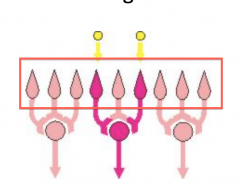
In the convergence diagram above, what are the things that are boxed?
|
1st order neuron (actual sensory receptors)
Neuron with the receptive field Each point is under its ow receptor field at the levelo f the 1st order neuron |
|
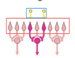
In the diagram above of convergence, what are the things boxed in blue?
|
The points of the paper clip
|
|
What is everything under this line?
|
These are second order neurons; they converage into one stimulus, so it makes it seem like there is only one receptive field that is stimulated
|
|
|
Does convergence lead to worse or better two point discrimination?
|
Worse
Because two neurons converge onto 1 neuron and only shows 1 signal It starts with the paper clip reaching two different receptive fields but then at the end, after the 2nd order neurons they only hit one |
|
|
Does lateral inhibition help make two point discrimination better or worse?
|
Better
Because there is no cross talk and it is easier for the signal to be discriminated |
|
|
When you increase intensity of the axonal stimulation what is the physiological change that occurs?
|
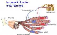
There is an increase number of motor units that is recruited
|
|
|
For this image, when you increase the intensitity of the stimulus, what happens?
|
IE at 6 mA -> Only small motor units are being activated because there is only a small force
at 20 mA there is a bigger stimulus and therefore a higher stimulation and an increase in force output because more motor units are recruited to meet threshold of both axons (increase intensity)) |
|
|
When you increase the frequency of axonal stimulation is there an increase in motor unit recruitment?
|
No
|
|
|
When you increase the frequency of axonal stimulation, is there an increase in frequency of action potentials in the recruited muscle?
|
Yes
|
|
|
Why is there an increase in the frequency of action potentials in recruited muscles?
|
1) Ca++ in sarcoplasm
2) No slack in elastic components 3) Increased temperature affects the enzymes |
|
|
Why is there increase Ca++ in sarcoplasm with an increase frequency of axonal stimulation?
|
There is a temporal summation i nthe axon so see muscle summation
Each time the action potential to the muscle release Ca++ muscle twitch duration of time because the AP is fast but Ca++ released and is also taken back up; not all of the calcium is taken back up so there is an accumulation of the ca++ in the sarcoplasm which allows for an increase in force |
|
|
What are the 4 F's of the ANS
|
Fight
Flight Feed **** |
|
|
Why do we say parasympathetic or sympathetic branch "dominates"
|
Because you're getting stimulation or "tone" from both the sympathetic and also the parasymapthetic all the time. They are not "on" and "off" switches, they are "tuning dials" that can be moved seperately
|
|
|
In the rest and digestion systme, does the parasympathetic or the sympathetic dominate?
|
Parasympathetic
|
|
|
In the fight or flight system, does the sympathetic or parasympathetic system dominate?
|
Sympathetic
|
|
|
What is the neurotransmitter released by the alpha motor neuron?
|
Acetylcholine at the neuromusculat junctiion
|
|
|
What type of fiber is the postganglionic neuron?
|
C fiber
|
|
|
Is the postganglionic neuron of the sympathetic branch myelinated?
|
No
|
|
|
What is the neuron size of the sympathetic postganglionic neuron?
|
Small (C)
|
|
|
What is the neuron size for the alpha motor neuron?
|
Large (Aα)
|
|
|
Which of the followin are ways that sympathetic postganglionic neurons are different than α-motor neurons?
A. Neurotransmitters released B. Myelination C. Neuron size D. All of the above E. Only A and C |
D
|
|
|
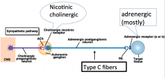
What do the postganglionic and preganglionic sympathetic neurotransmitters look like?
|
|
|
|
When a neuron is named "cholinergic" it is naming its what?
|
neurotransmitter
|
|
|
How is the term "adrenergic" related to norepinephrine or epinephrine?
|
Named for the adrenal gland
Adrenaline and noradrenaline are the British names for these neurotransmitters |
|
|
Most postganglionic sympathetic neurons are _______
|
Adrenergic
|
|
|
Are all postganglionic sympathetic neurons adrenergic?
|
No, there are a few that are cholinergic
|
|
|
Where are the cholinergic postganglionic sympathetic neurons found?
|
*Nerves to sweat glands
*Piloerector muscles in skin (Arrector Pili Muscle) *Some blood vessels |
|
|
Cells of the adrenal gland originated embryologically as _____ tissues
|
Nervous
|
|
|
Cels f the adrenal gland are ridimentary ______ neurons
|
Postganglionic
|
|
|
What do the adrenal glands secrete into the blood?
|
Neurotransmitters
|
|
|
How does the adrenal gnd work?
|
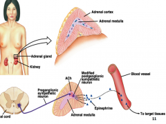
The adrenal medulla releases the epinephrine (similar to the postganglionic sympathetic neuron; however, the postganglionic neurons mostly release norepinephrine)
It is a modified postganglionic neuron and is now just a cell in the adrenal gland, it releases its neurontransmitters into the blood intead of just into the next neuron so there is a wider effect? |
|
|
What does the sympathetic system have a wider effect than the parasympathetic system?
|
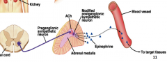
Because the sympathetic system's postganglionic neurons synapse with the adrenal gland. The adrenal medulla is a modified nerve cells that release their neurotransmitters into the blood instead of just into the next neuron so there is a wider effect present.
|
|
|
Which neurotransmitter is released in the greatest quantity from the adrenal gland
|
*Epinephrien (E, Epi) - 80%
-But some norepinephrine (NE) - 20% |
|
|
Which neurotransmitter is released in the greatest quantity from the postganglionic neurons in the symapthetic system?
|
-Mostly norepinephrine (NE)
-Epinephrine -Just a few acetylcholine |
|
|
Which of the following is true of the parasympathetic nervous system?
A. Short preganglionic neurons B. Synpase in ganglia close to spinal cord C. Uses mainly cholinergic neurons D. None of the above E. Only B and C |
C
|
|
|
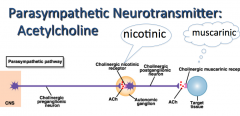
What does the parasympathetic branch of the ANS look like?
|
|
|
|
Do the preganglionic neurons of the parasymapthetic travel close to or far away from the organs?
|
Close to
Almost to the organ before they synapse |
|
|
The sympatetic pathways use what neurotransmitters?
|
Acetylcholine and norepinephrine
|
|
|
The parasympathetic pathways use what neurotransmitters?
|
Acetylcholine
|
|
|
Which of the following receptor types are typically G protein coupled receptors?
A. Nicotinic cholinergic B. Muscarinic cholinergic C. Adrenergic D. All of the above E. Only B and C |
E
|
|
|
Where can the G protein coupled receptors of a parasympathetic or sympathetic branch be found?
|
On the target organs
|
|
|
Muscarinic cholinergic and adrenergic recetpros are both G protein coupled receptors that open ion channe
|
G protein coupled receptors that open ion channels directly or produce second messengers
|
|
|
What is an image of a G-protein opening ion channels
|
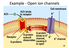
The beta and gamma units activate the ion channel
|
|
|
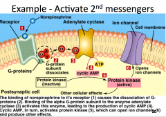
What is an image of a G-protein activating 2nd messengers
|
|
|
|
Which of the following are true regarding activation and duration of response?
A. Sympathetic system has the quickest effect B. Parasympathetic system has the longest duration effect C. Sympathetic system has the longest duration effect D. Both A and C E. Both A and B |
C
|
|
|
The sympathetic system has a slower or faster effect?
|
Slower
|
|
|
Why does the sympathetic system have a slower effect but the body still needs it to work fast?
|
Because it is about the balance of the two; there are dials; giving the oportunity for the sympathetic, it wants more sympathetic and can just turn down the parasymapthetic response so now there is more sympathetic response
Can have one response by removing the opposing stimulus |
|
|
Why does the parasympathetic response the quickest response time
|
There are long presynaptic neurons that are myelinated and fast
|
|
|
Why does the sympathetic branch have the longest duration response
|
Medulla of the adrenal glands release Epi into blodstream, which takes time to clear
|
|
|
For the vasodilator theory, when there is an increase rate of metabolism in tissue, there is an ______ numberof metabolites. What are metabolites?
|
*Increase
*Vasodilator substances |
|
|
What are examples of vasodilator substances (or metabolities)
|
*Adenosine
*Carbon dioxide *Adenosine phosphate compounds *Histamine *K+ ions *H+ ions |
|
|
In the vasodilatory theory, when the vasodilator substances leak into the tissues and reachassociated blood vessels which are ___________ what happens to the vessels?
|
*Precapillary sphincters, metarterioles and arterioles
*Dilation (relax vascular smooth muscle) |
|
|
In the oxygen lack theory, when there is increase metabolism in the tissue, that does what to the oxygen delivered by local blood vessels
|
It uses up the oxygen delivered by the local blood vessels
|
|
|
For the oxygen lack theory, the smooth muscle in the precapillary sphincters also requires what to contract (vasoconstrict)
|
Oxygen
|
|
|
When tissues use up the oxygen being delivered the blood flow ______ due to _______
|
Increase
Vasodialation (relatxation of vascular smooth muscle) |
|
|
When oxygen is delivered it allows for vessels to _______. And this mechanism can continue cyclically to meet ________ demands of the tissues
|
*Vasoconstrict
*Oxygen |
|
|
What is autoregulation
|
A system that allows for the maintenance of blood flow (L/min) despite changes in blood pressure, due to adjustment in the resistance applied by the blood vessels (vasodilation or vasoconstriction)
|
|
|
What are the two competing theories of autoregulation?
|
1) Metabolic theory
2) Myogenic theory |
|
|
For the metabolic theory, it states that when there is an increase in blood flow it is caused by an _____ in blood pressure that will do what?
|
*Increase
*Supply lots of oxygen and remove any building up metabolities *Cause smooth muscle in the blood vessels to contract (vasocontract) *And will decrease the blood flow to the appropriate rate to meet the demand of the tissues |
|
|
The myogenic theory states that when there is an increase in blood pressure what will that cause for the smooth muscle of the blood of the blood vessels? And what would happen because of that?
|
*They will be stretched
*Mechanical stretching leads to opening of mechanically gated ion channels that allows postiive ions (Ca++) to enter the smooth msucle and cause depolarization and contraction |
|
|
The myogenic theory shows that an increase in blood pressure will produce what?
|
A concomitant vasoconstriction, regulating the flow of blood throughout the vesseld
|
|
|
What are the four metabolic factors for cerebral blood flow regulation?
|
1) Carbon dioxide concentration
2) Hydrogen ion concnetration 3) Oxygen concnetration 4) Substances released from astrocytes |
|
|
How does carbon dioxide concentration contribute to CBF regulation. When there is an increase in [CO2] what happens?
|
There is an increase of cerebral blood flow because of vasodilation (actually caused by the conversion of CO2 -> Carbonic acid and H+ ion release)
|
|
|
Does an increased [CO2] cause vasoconstriction or vasodialatin?
|
Vasodilation
|
|
|
Why does an increase in [CO2] cause a vasodilation?
|
Conversion of the CO2 to carbonic acid and H+ ion release
|
|
|
Does an increase in [H+] cause an increase or decrease cerebral blood flow?
|
Increase
|
|
|
Does an increase in [H+] cause vasoconstriction or vasodilation?
|
Vasodilation
|
|
|
Does low oxygen cause low or high blood flow to the brain?
|
Low blood flow to the brain
|
|
|
Why does low oxygen cause low blood flow to the brain
|
Because the vessels are vasodilated
|
|
|
For substances that are released from astrocytes, the excitation of glutaminergic neurons (neurons that release glutamate) are connected to astrocytes seem to cause an increase in blood flow in where?
|
Adjacent vessels
|
|
|
Astrocytes do what with metabolites?
|
They release metabolities that produce vasodilation in the adjacent vessels
|
|
|
Do neurons rely completely on aerobic or anaerobic metabolism?
|
Aerobic metabolism
(Metabolism that uses O2 to produce energy. |
|
|
How do neurons produce energy?
|
On a second-by-second basis, if deprived for even 10 seconds, the ATP runs out, ion pumps stop working and the neuron can't produce action potentials
|
|
|
For brain energy what is required?
|
Glucose and oxygen
|
|
|
What is not required for glucose uptake?
|
Insulin
|
|
|
What is required for glucose uptake
|
A steady concentration of glucose in the blood is required along with oxygen
|
|
|
If blood glucose levels drop for more than a few minutes what happens to energy?
|
It will not be produced and the neurons will stop functioning
|
|
|
What are the four key facts to the clinical presentation of concussions?
|
1) Consciousness and orientation
2) Memory 3) Attention, concentration and behavior 4) Speech and language |
|
|
What does a damage to cerebral hemispheres of brain stem do the consciousness and orientation?
|
It leads to a loss of consciousness and idsorientation (IE where are you? What day is it? etc)
|
|
|
Damage to the hippocampus may lead to what?
|
Amnesia (anterograde or retrograde)
Memory of new events occurring since the concussion, or memory of events occurring before the concussion may be affected |
|
|
Frontal lobe damage may lead to difficulty with what?
|
Paying attention to the task at hand, or can lead to a change in the person's typical behavior or personality
|
|
|
Damage to the Brocca's area in the frontal lobe or wernicke's area of the temporal lobe, can lead to what?
|
Different visions of aphasia (difficulty with word generation)
|
|
|
What are the two types of brain injury?
|
Inertial
Impact |
|
|
What is inertial brain injury caused by?
|
Sudden acceleration or deceleration of the head and lead to "diffuse axonal injury"
|
|
|
For an inertial brain injury what does the suden stretch of the axon lead to?
|
Damage in the cytoskeleton in which cell organelles (being constantly transportd from the cell body to the axon terminal) build up in the damaged area of the axon and lead to axonal sweeling and eventual speration
|
|
|
For an impact brain jury upon contact what happens to a subset of blood vessels?
|
They are damaged and allow blood to leak into the brain cavity
|
|
|
Is there any free space in the cranium following an impact brain injury?
|
No
|
|
|
Since there is no free space in the crainium after an impact brain injury, what does that cause?
|
Brain is deformed by the hematoma leading to further damage to vessels and neurons
|
|
|
Diffuse Axonal Injury (DAI) does what to the axons?
|
Stretching
Shearing |
|
|
What does a DAI follow?
|
A sudden acceleration or declaration of the brain in the cranium
|
|
|
What can a diffuse axonal injury cause to the axon intially? What does it eventually lead to?
|
Subtle damage to the axon initally
Leads to axon separation and eventual neuron death It is believed that the stretch of shearing of the axon can occur, in which cell organelles build up at the site of damage and lead to eventual axon separation |
|
|
For a diffuse axonal injury what will happen to the neurotransmitters
|
They will be disrupted in the damaged neural circuits
|
|
|
In areas where blood flow has been damaged by the DAI, the associated neurons will quickly stop _______ due to lack of ________ production, which requires both ______ and ____ in the neuron
|
*neurotransmission
*ATP *Glucose *Oxygen |
|
|
Do you need a loss of consciousness to be considered a concussion?
|
No
|
|
|
Is a concussion a transient neurological dysfunction resulting from a hit to the head which resolves within 5-7 days of injury?
|
No
|
|
|
Concussions are induced by _______ forces
|
Biomechanical
|
|
|
What is a secondary brain injury?
|
*Begins shortly after impact and continues for hours or days
*Intracellular and extracellular derangements |
|
|
What are examples of secondary brain injury?
|
*Transient massive depolarization
*Ionic shifts *Neurotransmitter release and neuronal excitation *Cell energy failure *Membrane disruption *Cellular edema (swelling) *Cerebral edema (brain swelling) |
|
|
What is a diffuse axonal injury?
|
A type of brain injury caused by shearing forces that occur between different parts of the brain as a result of rotational acceleration (occurring over many points of the brain and can't specify just one)
|
|
|
Where are neurotransmitters and vessicles created in the neuron?
|
In the cell body
|
|
|
What happens after the neurotransmitter is created in the cell body?
|
It is transported down the axon
|
|
|
After a neurotransmitter is released from the presynaptic terminal and attaches to the receptor in the synaptic cleft, and is seperated, what happens?
|
It is reuptaken to be recycled and the vessicles without neurotransmitter transmitted back to the cell body
|
|
|
What happens to the neuron to allow for uncontrollable depolarization with Na+ flooding in?
|
Mechanical stretching
|
|
|
How can mechanical stretching of the brain occur?
|
*Impact
*The cranium stops suddenly *The brain is in CSF so it won't stop moving and just slosh around *Causes a sudden stretch |
|
|
Because of mechanical stretching, what can come flooding in?
|
*Ca++ comes from the axon and release of neurotransmitters and causes a bunch of neurotransmitters in the synaptic cleft
|
|

What is happening in 1?
|
There is an action potential that is coming down that allows for a depolarization and then Ca++ come flooding into the axon
|
|

What is happening in 2?
|
There is the neurotransmitter of glutamate that is released into the synapteic cleft and then Na+ is going going in and the K+ is coming out because of the excess of glutamate.
|
|

What is happening in 3?
|
There is a K+ efflux
|
|
|
How are we supposed to get back to homeostasis with the giant amount of K+ built up outside of the neuron?
|
Through the Na+/K+ ATPase pump which is ATP driven
|
|
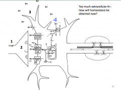
What is happening in 4?
|
Increased membrane pumping
|
|

What kind of cells can remove the extracellular K+?
|
Glial cells
|
|
|
Is the Na+/K+ ATPase pump moving against or with the gradient?
|
Against
|
|
|
What supplies the energy for the Na+/K+ pump?
|
ATP
|
|
|
How is ATP delivered to the brain?
|
Blood (glucose)
|
|
|
What happens if we don't get enough glucose to the brain?
|
No energy to drive
*Clean up excess K+ *Inability to transmit signals *Mechanism for glial cells have an increase energy demand to clean up excess K+ -> So even less energy for that |
|
|
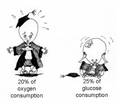
The brain comprises ____% of total body weight
|
2
|
|
|
The brain comprises ____% of resting cardiac output?
|
15%
|
|
|
How much percent of the brain is for oxygen consumption
How much of the brain is for glucose consumption |
|
|
|
Under normal conditions, Cerebral Book Flow (CBF) is tightly coupled to...
A) Neuronal activity B) Metabolic waste byproducts C) Cerebral artery blood pressure D) Both A and B E) None of the above |
D
|
|
|
Cerebral blood flow is "autoregulated" extremely well between MAP of?
|
60-140
|
|

Where is the autroregulation of the CBF on the graph?
|
It is between the two regions that CBF is very consisten
Act to increase Blood pressure: exercise, stress, etc. |
|
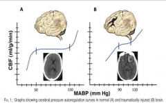
What is the brain on the left showing
|
After a neuronal stretching, there is increase in blood pressure
There is a decrease in the autoregulation range and decreased blood flow, if there is exercise it decreases the amount of oxygen and increases the demand for oxygen which will cause light headedness and fatigue |
|
|
What are the symptoms of reduced CBF?
|
*Fatigue
*Persisting Symptoms *Exertion-based symptoms (Headaches) |
|
|
What is is the fatigue that is caused by the reduced CBF?
|
*Increased anaerobic glycolysis
*Production of lactate that is converted int lactic acid *Acid depresses neuronal activity *The decrease neuoronal activity means feeling more tired |
|
|
hat are the persisting symptoms that can be caused by reduced CBF?
|
Dimished blood flow results in low influx of nutrients and oxygen as well as a lack of wast eremoval
|
|
|
What is the exercition-based symptoms (headache) caused by reduced CBF?
|
Brain is unable to maintain stable intracranial pressurei n the face of fluctuating systemic arterial pressure (dysautoregulation)
|
|
|
Immediately after injury, the demand for glucose _____, cerebral blood flow _____ and oxygen metabolism _______
|
*Increases
*Decreases *Decreases |
|
|
What are the physiological problems of concussions
|
*Massive axonal depolarization
*Flood of excitatory neurotransmitter release *Incessant neuronal firing *Calcium efflux *Potassium efflux *Increased neuronal energy demand *Excess calcium reduces oxygen metabolism *Limited supply of glucose to the brain *High energy demand *Oxygen debt (energy crisis!) *Neuronal exhaustion |
|
|
What are signs/symptoms of concussions?
|
*Headache
*Nausea *Vomitting *Balance problems *Dizziness *Fatigue *Sensitivity to light/noise *Numbness/tingling *Irritable *SAd *mental "fogginess" *Feeling slow *Difficulty concentrating *Difficulty remembering *Confusion |