![]()
![]()
![]()
Use LEFT and RIGHT arrow keys to navigate between flashcards;
Use UP and DOWN arrow keys to flip the card;
H to show hint;
A reads text to speech;
43 Cards in this Set
- Front
- Back
|
stimulus-latency response |
The lag between the stimulus and initiation of the action potential is called latency. When thestimulus strength is increased above threshold, the time to reach threshold, the latency, decreases. |
|
|
upstroke |
rapid depolarization |
|
|
overshoot |
hyperpolarization |
|
|
AP sequential steps |
1- Depolarization -> Na+ Channel opens -> increase gNa -> shift of Em towards ENa2- Na+ channel inactivation and K+ channel opens -> decrease gNa and increase gK3- K+ Channel Closes |
|
|
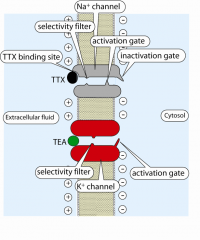
Na/K channel inhibiting agents |
|
|
|
V-Snare |
synaptobrevin |
|
|
T-Snare |
syntaxin is on pre-synaptic membrane |
|
|
Curare |
block acetylcholine receptors D-tubucurareis used surgically. |
|
|
Succinylcholine |
Non competitive AchR activator keeps the channelopen, succinylcholine produces paralysisby depolarization block |
|
|
Neostigmine,physostigmine |
physostigmine inhibit acetylcholinesterase and potentiates acetylcholine action in persons with weakNMJ responses(myasthenia gravis) |
|
|
sarin and VX |
effects on autonomic nervous system (meant to use in warfare but now arebanned) SalivationLacrimationUrinationDefecationEmesis |
|
|
Myasthenia gravis |
muscle weakness •Acquired autoimmunedestruction of acetylcholine receptors •SNMG – sero negative myasthenia gravis haveantibodies against MUSK – muscle specific kinase - involved in formation of the NMJ |
|
|
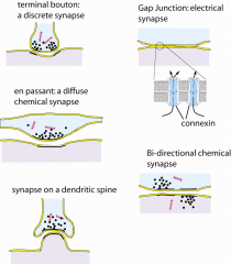
Different types of synapses |
|
|
|
Two types of synaptic vesicles |
small synaptic vesicles and large dense core vesicles |
|
|
Spatial and temporal summation |
simultaneous EPSP & IPSP input summation repetitive EPSP or IPSP |
|
|
Isometric vs. isotonic |
constant length vs. constant tension (hence, the lifting of afterload can occur) |
|
|
Pennation |
Pennationallows more force to be developed from a muscle that will still fit into thebody – but the trade-off is slowervelocity of contraction Muscle pennation increases force but reduces velocity |
|
|
Dystrophin & dystroglycan |
Special proteins span the sarcolemma atthe costameresand transfer force from the cytoskeletonto the extracellular matrix |
|
|
Costamere |
The costamere is a structural-functional component of striated muscle cells which connects the sarcomere of the muscle to the cell membrane. |
|
|
Varicosity |
bulged areas in nerve cells where neurotransmitters are accumulated within |
|
|
Connexin |
A channel protein that is embedded within gap junction |
|
|
Dense bodies |
A point where contractile fibers are anchored at. When the structure is located at the cell membrane, it is called the membrane dense area or attachment plaque. |
|
|
Caveolae |
Thesurface membrane is coveredwith small invaginations calledcaveolae. These may serve aslocal sources of Ca2+ foractivation of the muscle. |
|
|
MLCK / MLCP |
myosin light chain kinase (contraction)/phosphatase (relaxation) |
|
|
Duty cycle |
the part of the time that the muscle is activated, increases withintensity because the period of activation decreases less than the period of rest. This describes the situationwhen we increase the speed of a repetitive action such as walking. higherintensity exercise: higher duty cycle less time for metabolism to recover theresting state At the highest intensity of exercise, there is no rest phase |
|
|
ATP / creatine phosphate intracellular concentration |
5mM / 15-20mM |
|
|
Muscle fiber typing |
Type 1: slow oxidative Type 2a: fast oxidative Type 2b: fast glycolyic |
|
|
Endomysium / perimysium / epimysium |
surrounds single muscle fiber / a primary bundle of fibers (fascicle) / the whole muscle fiber |
|
|
Two types of fatigue |
high intensity / short duration = build up of Pi + H+ medium intensity / long duration = depletion of glycogen |
|
|
Tonic vs phasic muscle |
constant activation vs. activation by trigger |
|
|
phospholamban |
activates SERCA upon phosphorylation |
|
|
lipid rafts |
•formedby sphingomyelinand cholesterol. •Lipidrafts: •Segregatesignaling mechanisms •Segregatemolecules. |
|
|
Covalent attachment to the membrane |
GPI (glycosylphosphophatidylinositol), farnesyl, myristyl, palmitosyl |
|
|
Total body water composition |
14 + 28 = 42 plasma + ISF + ICF = TBW |
|
|
Agents for water composition |
Deuterium oxide = all Inulin = ECF Tracer = plasma protein (plasma) |
|
|
ionophore |
•Ionophores aremolecules that allow ions to cross membranes. •Ionophores areof two types: carriers and channel formers. |
|
|
NCX |
Na+/Ca2+ exchanger Three sodium in / one calcium out; hence, one net positive charge enters the cell Can reverse its direction upon physiological change in cell potential. eg. depolarization of the heart |
|
|
[NaCl] physiological Isotonic level |
0.9% 5% D-glucose |
|
|
Four types of G coupled metabotropic receptors |
Gs = epi binds b1 or b2 -> ↑ [cAMP] -> activates PKA Gi = epi binds a2 -> ↓[cAMP] = epi binds M2 receptor -> alpha inhibits adenylyl cyclase and beta-gamma activates K channel Gq = epi binds a1 -> alpha activates PLC -> ↑ IP3 -> ↑[Ca2+] = epi binds a1 -> alpha activates PLC -> ↑ DAG -> DAG activates PKC G12 = Gα12-GTP -> activates GEF -> activate small G proteins |
|
|
GPCR involvement in photo-transduction in the retina |
Gαt-GTP -> activates cGMP phosphodiesterase -> ↓ [cGMP] -> ↓ gNa -> hyperpolarization |
|
|
Vitamin D |
can act as a steroidal hormone |
|
|
Ion permeability at resting potential |
PCl > PK > PNa |
|
|
phospholamban |
inhibitor of SERCA; stops inhibition upon phosphorylation by PKA or PKG |