Reading...
![]()
Play button
![]()
Play button
![]()
Use LEFT and RIGHT arrow keys to navigate between flashcards;
Use UP and DOWN arrow keys to flip the card;
H to show hint;
A reads text to speech;
61 Cards in this Set
- Front
- Back
- 3rd side (hint)
|
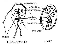
Identify this and the structures
|
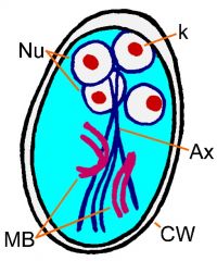
Giardia cyst
|
|
|
|
Identify this and structures
|
|
Giardia Cyst, notice the structures
|
|
|
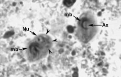
What are these and where would you find them?
|
|
Giardia cysts
|
|
|

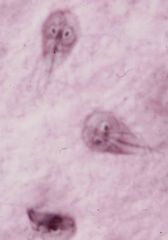
Giardia trophozoite.
|
|
|

What are these?
|
giardia trophozoite
|
|
|

|
|
|
|
|

giardia cysts
|
|
|
|

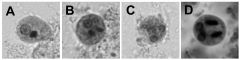
Various stages of E. histolytica cysts. A) An early precyst with a single nucleus. B and D) A cyst at the 2 nucleus stage. C) A mature cyst with 4 nuclei and no chromatoid bodies. Note how the nuclei decrease in size with each subsequent division. A-C are stained with trichrome and D is stained with iron hematoxylin.
|
|
|

|
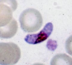
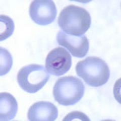
crescent-shaped gametocytes
P falciparum |
|
|
|
Which plasmodium has a crescent shaped gametocyte?
|
P. Falciparum
|
|
|
What is this?
|

P falciparum ring stage. These are early trophozoitesmay appear to have two nuclei
|
|
|
|
Which Plasmodium have Schüffner’s dots?
|
vivax and ovale
|
|
|
|
What is the halo around the falciparum gametocyte known as?
|
Laveran's bib
|
|
|
|
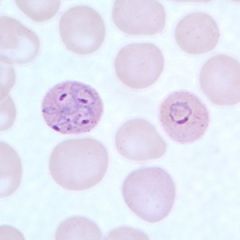
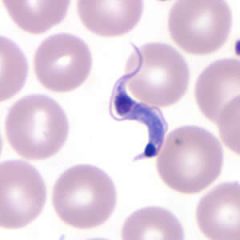
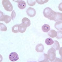
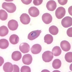
Plasmodium vivax
|
may have irregular ameboid appearance
reddish granules called Schueffner granules |
|
|
|
What are Plasmodium parasites with 2 or more nuclei known as?
|
schizont
|
|
|

|

vivax schizont
|
|
|

what is this?
|
Ring-form of P. vivax have a thick cytoplasm with a single, large chromatin dot. The cytoplasm becomes amoeboid, Schüffner's dots appear as the trophozoites mature. Infected RBCs are often larger
|
|
|
|
What are the two types of falciparum in circulation?
|
ring stage and gametocytes
|
|
|
|
What is a distinguishing feature of vivax trophozoit and schizont infection of eurythrocytes?
|
Schueffner's dots will be reddish
|
|
|
|
What are plasmodium parasites with more thatn one nucleus called?
|
schizont
|
|
|
|
trophozoite of P malariae
|
compact and deeply staining, no enlargement of the host cell
|
|
|
|
Trophozoite of vivax
|
ameboid and host cell is enlarged. Schnueffer's dots
|
|
|
|
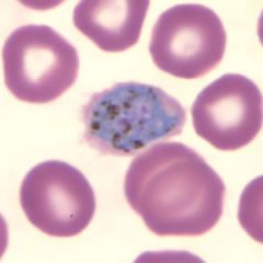
Schizont of malariae
|
rosette structure. 8-12 nuclei
|
|
|
|
Schizont of vivax
|
enlarged and has Schnueffer's dotscan contain up to 32 nuclei/merozoites
|
|
|
|
Gametocyte of malariae
|
fills entire normal sized erythrocyte
|
|
|
|
Gametocyte of vivax
|
nearly fills entire enlarged host and Schnuffer's dots present
|
|
|

|
|
|
|
|

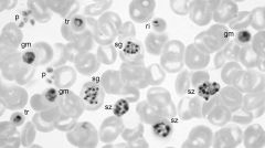
Composite showing various morphological forms of P. malariae. ri = ring; tr = trophozoites; sz = schizont; sg = segmenter; gm= gametocyte; p = platelets
|
|
|
|

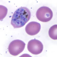
Schizonts of P. ovale in thin blood smears
|
|
|

|

Composite showing various morphological forms of P. malariae. ri = ring; tr = trophozoites; sz = schizont; sg = segmenter; gm= gametocyte; p = platelets
|
|
|

|
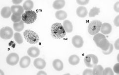
Composite showing various morphological forms of P. vivax. ri = ring; et = early trophozoite; lt = late trophozoite; sz = schizont; gm= gametocyte; p = platelets
|
|
|
|
malariae schizonts
|
are usually compact not enlaged
|
|
|
|
distinguish between gametocytes of ovale and vivax
|
vivax larger.
|
|
|

|
Schizonts of P. ovale tend to be smaller and contain fewer merozoites (average 8). Elongation to an oval shape Schüffner's dots can be observed with proper staining.
|
|
|
vivax schizont
|

ovale schizont
|
|
|
|

ovale rings
|
|
|
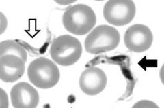
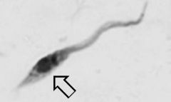
T. cruzi
|
African Trypanosome
|
|
|
|

|
|
|
|
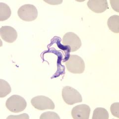
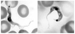
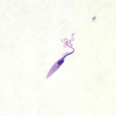
trypomastigote of African Trypanosome spp
|
|
|
|
which Trypanosome spp has a trypmastigote dividing cycle?
|
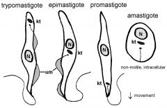
African Trypanosome. T. Cruzi does not. In T cruzi, this is the epimastigote form
|
|
|
|
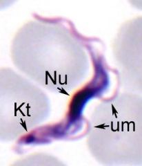
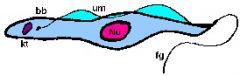
trypomastigote: kinetoplast at anterior end. nucleus in middle
|
|
|
|
|
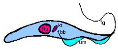
Epimastigote: kinetoplast in middle next to nucleus. This is the replicating form of T cruzi. has shorter um
|
|
|
|
amastigote
|
|
|
|
where are the trypomastigotes of the African trypanosomes found?
|
in the blood. this is the replicating form. in the lab these are all obtained from rats or mice
|
|
|
|
trypomastigotes of African Trypanosome. notice the replicating form
|
|
|
|
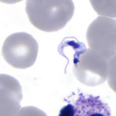
T. gambiense and T. cruzi blood-stage trypomastigotes. notice that the Tg has a more prominate um.
The kinetopplastid seems to be larger in diameter than body or T cruzi also more often c shaped |
|
|
|
What forms of T cruzi are in humans
|
Trypomastigote and amastigote. Amastigote is intracellular and replicate in cell
|
|
|
|
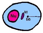
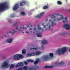
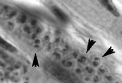
T cruzi amastigotes in tissue. Dont forget to identify the interior kinetoplast
|
|
|
|
T cruzi in tissue. Notice the nuclei and kinetoplast
|
|
|
|
In lab, where is the epimastigote of T cruzi from?
|
culture smear
|
|
|
|
typical epimastigpte T cruzi. found in vector. replicating form. kintoplast appears as bar
|
|
|
|
In lab what are the specimens from bone marrow and liver?
|
amastigotes of Leishmania
|
|
|
|
Are the amastigotes of T cruzi and Leishmania distinguishable?
|
No, must know what kind of tissue and cells. Leishmania only infect phagocytes
|
|
|
|
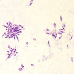
Leishmania promastigotes from culture. not found in human tissue
|
|
|
|
distinguishing T cruzi epimastigotes from Leishmania promatigote
|
Promastigote does not have um and are fatter also not found in human tissue
|
|
|
|
T cruzi trypomastigote
|
|
|
|
T cruzi epimastigote
|
|
|
|
P. malariae ring
|
|
|
|
ovale rings
|
|
|
|
ovale trophozoite
|
|
|
|
ovale trophozoite. notice the fringe
|
|