![]()
![]()
![]()
Use LEFT and RIGHT arrow keys to navigate between flashcards;
Use UP and DOWN arrow keys to flip the card;
H to show hint;
A reads text to speech;
211 Cards in this Set
- Front
- Back
|
Which of the following are NOT one of the 4 basic types of tissue in the human body? A) Integumentary B) Connective C) Nervous D) Osseous E) Muscular F) Epithelial |
A and D |
|
|
How many separate bones are found in the adult human body? |
206 |
|
|
Which system maintains the acid-base balance in the body? |
Urinary |
|
|
The two divisions of the human skeleton are: |
Axial and Appendicular |
|
|
Which portion of the long bones is responsible for the production of red blood cells? |
Spongy or Cancellous |
|
|
What type of tissue covers the ends of the long bones? |
Hyaline or Articular Cartilage |
|
|
The narrow space between the inner and outer table of the flat bones in the cranium is called the: |
Diploe |
|
|
What is the primary center for endochondral ossification in long bones? |
Diaphysis (shaft) |
|
|
What is the name of the secondary growth centers of endochondral ossification found in long bones? |
Epiphyses |
|
|
The aspect of long bones where bone growth in length occurs is termed: |
Metaphysis |
|
|
A skull suture has the structural classification of a __________ joint. |
Fibrous |
|
|
The symphysis pubis has the structural classification of a __________ joint |
Cartilaginous |
|
|
Which specific joint is the only true syndesmosis, amphiarthrodial, fibrous joint? |
Distal Tibiofibular Joint |
|
|
Sternum is classified as a |
Flat Bone |
|
|
Femur is classified as a |
Long Bone |
|
|
Tarsal Bones are classified as |
Short Bones |
|
|
Pelvic Bones are classified as |
Irregular Bones |
|
|
Scapulae are classified as |
Flat Bones |
|
|
Humerus are classified as |
Long Bones |
|
|
Vertebrae are classified as |
Irregular Bones |
|
|
Calvarium are classified as |
Flat Bones |
|
|
The three structural classifications of joints are synovial, cartilaginous, and: |
Fibrous |
|
|
The First Carpometacarpal Joint has a ____________ movement |
Sellar (Saddle) |
|
|
The Intercarpal Joint has a ____________ movement |
Plane (Gliding) |
|
|
The Hip Joint has a ____________ movement |
Spheroidal (Ball and Socket) |
|
|
The Proximal Radioulnar Joint has a ____________ movement |
Trochoidal (Pivot) |
|
|
The Interpahlangeal Joint has a ____________ movement |
Gingylmus (Hinge) |
|
|
The Fourth Metacarpophalangeal Joint has a ____________ movement |
Ellipsoidal (Condyloid) |
|
|
The Knee Joint has a ____________ movement |
Bicondylar |
|
|
The Wrist Joint has a ____________ movement |
Ellipsoidal (Condyloid) |
|
|
The Joint between C1 and C2 has a ____________ movement |
Trochoidal (Pivot) |
|
|
The Ankle Joint has a ____________ movement |
Sellar (Saddle) |
|
|
Which plane divides the body into equal anterior and posterior parts? |
Midcoronal |
|
|
T or F: The term radiographs and image receptor refer to the same thing. |
False |
|
|
A longitudinal plane that divides the body into right and left parts is the: |
Sagittal Plane |
|
|
Near the source or beginning |
Proximal |
|
|
Away from head of the body |
Caudad |
|
|
Inside of something |
Interior |
|
|
Increasing the angle of a joint |
Extension |
|
|
Outward stress of the foot |
Eversion |
|
|
Movement of an extremity away from the midline |
Abduction |
|
|
Turning palm downward |
Pronation |
|
|
A backward movement |
Retraction |
|
|
To move around in the form of a circle |
Circumduction |
|
|
Toward the center |
Medial |
|
|
Away from the source or beginning |
Distal |
|
|
On the opposite side of the body |
Contralateral |
|
|
Lying down in any position |
Recumbent |
|
|
Head lower than the feet position |
Trendelenburg |
|
|
Upright position, palms forward |
Anatomic Position |
|
|
Top of the foot |
Dorsum Pedis |
|
|
Frankfort horizontal plane |
Base plane of skull |
|
|
A plane at right angle to the longitudinal plane |
Horizontal plane |
|
|
Head higher than feet position |
Fowler's Position |
|
|
Palm of hand |
Palmar |
|
|
Sole of foot |
Plantar |
|
|
Front half of body |
Anterior |
|
|
A plane that divides body into anterior and posterior halves |
Midcoronal Plane |
|
|
A recumbent position with knees and hips flexed with support for legs |
Lithotomy Position |
|
|
The direction or path of the central ry of the x-ray beam defines the positioning term: |
Projection |
|
|
A patient is placed in a recumbent position facing downward. The left side of the body is turned 30 degrees toward the image receptor. Which specific position has been performed? |
LAO or Lateral Anterior Oblique |
|
|
A patient is placed into a recumbent position facing downward. The x-ray tube is directed horizontally and enters the left side and exits the right side of the body. An image receptor is placed against the right side of the patient. Which position has been performed? |
Ventral Decubitus |
|
|
A patient is erect with her back to the image receptor. The central ray enters the anterior aspect and exits the posterior aspect of the body. Which projection has been performed? |
Anteroposterior |
|
|
A patient is lying down facing upward with the posterior surface of the body against the image receptor. The right side of the body is turned 45 degrees toward the image receptor. The x-ray tube is directed vertically and enters the anterior surface of the body. Which position has been performed? |
RPO or Right Posterior Oblique |
|
|
An elbow projection is taken with the posterior surface placed against the image receptor. The elbow is rotated 20 degrees outwardly. Which specific projection has been performed? |
AP oblique with lateral rotation |
|
|
A specific projection of the foot in which the central ray enters the anterior surface and exits the posterior surface is termed: |
Dorsoplantar |
|
|
A patient is placed in a recumbent position with the body tilted so that the head is higher than the feet. The image receptor is under the patient and the x-ray tube is above the patient. Which is the general position of the patient? |
Fowler's |
|
|
The anterior surface of the right knee of the patient is facing the image receptor. The anterior aspect of the knee and lower leg is rotated 15 degrees toward the midline. Which specific projection has been performed? |
PA oblique with medial rotation |
|
|
What is the name of the projection in which the central ray merely skims a body part? |
Tangential |
|
|
What is the name of the specific projection in which the central ray enters the left side of the chest and exits the opposite side? |
Transthoracic |
|
|
What is the specific projection that enters the posterior aspect of the skull and exits the acanthion? |
Parietoacanthial |
|
|
Which one of the following is an example of an axial projection? A) Transthoracic Lateral B) Mediolateral Ankle C) AP chest with 20 degree cephalic angle D) AP abdomen with 30 degree rotation to the left |
C) AP chest with 20 degree cephalic angle |
|
|
Which one of the following positioning terms is no longer considered valid in the US? A) Radiographic View B) Radiographic Position C) Radiographic Projection D) Semi-axial Projection |
|
|
|
Proximal is opposite of |
Distal |
|
|
Cephalad is opposite of |
Caudad |
|
|
Ipsilateral is opposite of |
Contralateral |
|
|
Medial is opposite of |
Lateral |
|
|
Superficial is opposite of |
Deep |
|
|
Internal is opposite of |
External |
|
|
Lordosis is opposite of |
Kyphosis |
|
|
AP is opposite of |
PA |
|
|
Superior is opposite of |
Inferior |
|
|
Dorsoplantar is opposite of |
Plantodorsal |
|
|
Which one of the following is NOT one of the four primary image quality factors? A) Density B) Contrast C) Kilovoltage (kV) D) Detail E) Distortion |
C) Kilovoltage (kV) |
|
|
The amount of blackness on a processed radiograph is called: |
Density |
|
|
Which exposure factor primarily controls radiographic density? |
mAs |
|
|
T or F: For an underexposed radiograph, the mAs must be increased by a factor of four to produce a visible change in radiographic density. |
False |
|
|
A radiograph of the knee reveals that it is overexposed and must be repeated. The original technique used 10 mAs. What change will improve the image during the repeat exposure? |
Decreasing to 5 mAs |
|
|
The primary controlling factor for radiographic contrast is: |
kV |
|
|
Chest radiography requires long-scale contrast. Which set of exposure factors will produce this? A) 50 kV, 20 mAs B) 65 kV, 15 mAs C) 110 kV, 2 mAs D) 80 kV, 5 mAs |
C) 110 kV, 2 mAs |
|
|
Which one of the following sets of exposure factors will produce the highest (short-scale) radiographic contrast? A) 60 kV, 30 mAs B) 80 kV, 20 mAs C) 96 kV, 5 mAs D) 120 kV, 2 mAs |
A) 60 kV, 30 mAs |
|
|
T or F: Kilovoltage is a secondary controlling factor for radiographic density. |
True |
|
|
T or F: A low-kilovoltage technique (50 kV) produces a long-scale contrast image. |
False |
|
|
A radiograph of the elbow reveals that it is overexposed. The technologist wants to adjust kV rather than mAs for the repeat exposure. This is contrary to common practice. The original analog exposure factors were 70 kV and 5 mAs. Which of the following kV settings would reduce radiographic density by one-half? A) 80 kV and 5 mAs B) 66 kV and 5 mAs C) 60 kV and 5 mAs D) 56 kV and 5 mAs |
C) 60 kV and 5 mAs |
|
|
Which of the following techniques or devices will reduce the amount of scatter radiation striking the IR? A) Collimation B) Lower kV C) Grids D) All of the above |
D) All of the above |
|
|
T or F: Recorded detail or spatial resolution is optimal with a long object image receptor distance (OID) and a short SID. |
False |
|
|
Which of the following factors best controls involuntary cardiac motion artifact? A) Careful instructions given to the patient B) High kV technique C) Practicing with patient when to hold breath D) Shortening the exposure time |
D) Shortening the exposure time |
|
|
The technologist is asked to produce a high-quality image of the carpal (wrist) bones. the emergency room physician suspects that the patient has a very small fracture of one of the bones. Which one of following sets of technical factors will produce an image with the highest degree of radiographic resolution? A) 1.0-mm focal spot and 30-inch (77cm) SID B) 2.0-mm focal spot and 36-inch (92cm) SID C) 0.5-mm focal spot and 40-inch (102cm) SID D) 0.3-mm focal spot and 40-inch (102cm) SID |
D) 0.3-mm focal spot and 40-inch (102cm) SID |
|
|
Rather than rely on the anode heel effect, what can be used to equalize density of specific anatomy? |
Compensating filter |
|
|
Which type of compensating filter is recommended for an AP projection of the shoulder? |
Boomerang |
|
|
Which type of compensation filter is recommended for an axiolateral hip projection? |
Wedge |
|
|
Which type of grid cutoff is created if the CR and the face of the grid are not perpendicular to each other? |
Off-level |
|
|
Which one of the following projections requires the use of a grid? A) PA hand B) Axial calcaneus (heel) C) AP abdomen D) AP elbow |
C) AP abdomen |
|
|
The misrepresentation of an object's size or shape projected on a radiograph is called: |
Distortion |
|
|
Which of the following sets of factors MINIMIZES radiographic distortion to the greatest degree? A) 40-inch (102cm) SID and 8-inch(20-cm) OID B) 44-inch (113cm) SID and 6-inch (15cm) OID C) 72-inch (183cm) SID and 3-inch (7.5cm) OID D) 60-inch (154cm) SID and 4-inch (10cm) OID |
C) 72-inch (183cm) SID and 3-inch (7.5cm) OID |
|
|
T or F: To best use the anode heel effect, the thinner aspect of the anatomic part should be placed under the cathode aspect of the x-ray tube. |
False |
|
|
The best method to reduce distortion of the joints of the hand is to keep the fingers __________ to the IR. |
Parallel |
|
|
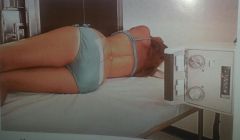
Right lateral decubitus with PA projection |
|
|
T or F: Changes in kV have little impact on patient dose with digital imaging. |
False |
|
|
T or F: kV and mAs do not have the same direct effect on image quality with digital imaging as they do with IR-screen imaging. |
True |
|
|
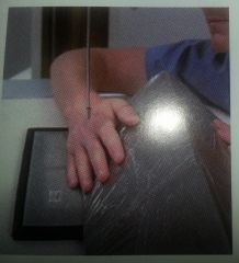
PA oblique lateral rotation |
|

|
LPO upright with AP projection |
|

|
RAO upright with PA projection |
|

|
RAO recumbent with PA projection |
|
|
AP oblique internal rotation |
|
|
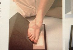
Axial of foot or Dorsalplantar projection |
|
|
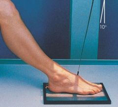
PA axial calcaneous or Plantardorsal projection |
|
|
Left lateral decubitus with AP projection |
|
|
The amount of blackness seen on a processed radiograph is called: |
Density |
|
|
T or F: When placing radiographs of the hand on the view box, the digits should be pointing upward. |
FALSE |
|
|
Misrepresentation of object size or shape as projected onto radiographic recording data is the general definition for: |
Disortion |
|
|
What change will improve recorded resolution? |
Decrease OID |
|
|
Which place specifically divides the body into equal right and left halves? |
Midsagittal |
|
|
Radiographers can control voluntary motion by: |
use shorter exposure time use faster IR give good instructions to patient |
|
|
A radiograph of the abdomen demonstrates involuntary motion due to bowel peristalsis (involuntary contractions). Which of the following factors will best eliminate this problem with the repeat exposure? |
decrease exposure time and make appropriate changes to the mA to compensate |
|
|
Caudad |
away from the head end of the body |
|
|
Proximal |
near the source or beginning |
|
|
Extension |
increases the angle from flexed to straightened position |
|
|
Radial Deviation |
towrd the radial side of the wrist |
|
|
Inversion |
inward stress movement |
|
|
Dorsiflexion of the foot |
decrease the angle between the dorsum (top of foot) and lower leg |
|
|
Adduction |
toawrd body/ toward center or midline |
|
|
Abduction |
away from body/ away from center or midline |
|
|
Pronation |
rotation of hand into opposite site of anatomic position- palm down or back |
|
|
Cephalad |
toward the head end of the body |
|
|
Distal |
away from |
|
|
Flexion |
decreases the angle of a joint |
|
|
Ulnar Deviation |
turn or bend hand and wrist from natural position toward ulnar side |
|
|
Eversion |
outward stress movement |
|
|
Plantar flexion of the foot |
extending the ankle joint downward |
|
|
Supination |
rotation of hand into anatomic position (palm up) |
|
|
Interior |
inside/nearer to center |
|
|
Exterior |
on or near the outside |
|
|
Superficial |
nearer the skin surface |
|
|
Deep |
farther away |
|
|
Ipsilateral |
same side of body part |
|
|
Contralateral |
opposite side |
|
|
Medial |
toward center |
|
|
Lateral |
away from center patient is on their side or erect with side against IR- 90 degrees from AP or PA |
|
|
The radiographic term projection is defined as: |
path or direction of the central ray |
|
|
The term that refers to parts farthest form the point of attachment, point of reference, or away from the center of the body is: |
Distal |
|
|
What is the minimal number of projections taken for a post reduction (to realign a fracture) study of the ankle? |
2 |
|
|
What is the minimal number of projections taken for a hand? |
3 |
|
|
What is the minimal number of projections taken for a forearm? |
2 |
|
|
What is the minimal number of projections taken for a femur? |
2 |
|
|
What is the minimal number of projections taken for a knee? |
3 |
|
|
What is the minimal number of projections taken for a humerus? |
2 |
|
|
What is the minimal number of projections taken for a pelvis study? |
1 |
|
|
What is the minimal number of projections taken for a abdomen (KUB)? |
1 |
|
|
What is the minimal number of projections taken for a table chest? |
1 |
|
|
What is the minimal number of projections taken for a study of the right hip? |
2 |
|
|
What type of radiographic contrast is produced with a high (120) kV technique? |
Low contrast, long scale |
|
|
A ____% increase in kV will increase the density the same as doubling the mAs. |
15 |
|
|
What primarily controls radiographic density? |
mAs |
|
|
Osteology |
study of bones |
|
|
Arthrology |
study of joints |
|
|
Long Bones |
limbs |
|
|
Short and Flat Bones |
Carpals and tarsals Calvarium, sternum, ribs, scapulae |
|
|
Irregular Bones |
vertebrae facial bones pelvic bones |
|
|
Sagittal Plane |
longitudinal plane that divides body into right and left parts |
|
|
Midsagittal Plane |
divides body into EQUAL right and left parts |
|
|
Coronal Plane |
longitudinal plane that divides body into anterior and posterior parts |
|
|
Midcoronal Plane |
divides body into EQUAL anterior and posterior parts |
|
|
Horizontal (axial) Plane |
transverse plane that passes through body at right angles to a longitudinal plane- divides body into sup and inf portions |
|
|
Projection |
path of the central ray (beam) through body |
|
|
Supine |
lying on back Recumbent on back (AP) |
|
|
Prone |
lying on abdomen Recumbent on stomach (PA) |
|
|
Oblique |
patient (or body part) is turned and between a lateral and flat position body is at an angle to the IR described by part CLOSEST to IR or body part from which CR EXITS |
|
|
Erect |
upright position |
|
|
Recumbent |
patient lying down in any position -ventral -dorsal -lateral |
|
|
Decubitus |
recumbent position with horizontal beam AP or PA projection DESIGNATED BY SIDE DOWN |
|
|
True Lateral |
body is perpendicular to IR |
|
|
Lateral Recumbent |
lying on side |
|
|
Apical lordotic chest with PA projection |
|
|
Minimum of 2 projections needed when... |
Long bones in picture -superimposed -lesions/foreign bodies -alignment of fractures |
|
|
Minimum of 3 projections when... |
joints in interest area -AP or PA -Lateral -Oblique |
|
|
Long bones require __ projections, must include ________, and must have __ views (_______ and _______), __ degrees apart. |
2 both joints 2 AP and Lateral 90 |
|
|
When viewing a radiograph, the patient's _____ must be to the viewer's _____. |
Right Left |
|
|
When viewing radiographs, the limbs must be in the ____________ and hands and feet must be __________. |
Anatomic Position Digits up |
|
|
The light field corresponds to the... |
Radiation field |
|
|
When using the bucky, the light field must be centered to the _____ in the _____. |
IR Bucky |
|
|
Light field should be ________ to correct size and never be larger than the _______ size. |
Collimated IR |
|
|
Body part of interest should be... |
Centered on the IR |
|
|
Analog Exposure Factors |
Kilovoltage kV Milliamperage mA Exposure time |
|
|
Denisty |
amount of blackness Controlling factors -mAs: primary controller -kV Influencing factors -SID -Screen and IR speed |
|
|
mAs |
how much x-ray |
|
|
kV |
controller of contrast |
|
|
Density adjustment rule |
25-30% inc in mAs |
|
|
Density Repeat Rule |
doubling mAs |
|
|
Anode Heel Effect |
more intense under cathode end of tube inc with: -small focal spot -shorter SID -larger IR size application -thicker parts at cathode end |
|
|
Compensating Filters |
filter out portion of primary beam toward thin or less dense part of body that is being imaged |
|
|
Radiographic Contrast |
differences in density controlling factor kV |
|
|
High Contrast |
a lot of difference short scale 50 kV (800 mAs) very black, very white |
|
|
Low Contrast |
not much difference long scale 110 kV (20 mAs) lots of shades of gray |
|
|
Spatial Resolution |
Same as detail recorded sharpness of structures lack of definition blur or unsharpness motion is greatest deterrent |
|
|
Use a _________ whenever possible to improve detail |
Small Focal Spot |
|
|
Use _________ time possible to control voluntary/involuntary motion |
Shortest exposure |
|
|
Use faster __________ to control voluntary/involuntary motion |
IR imaging system |
|
|
Use longer _____ to improve resolution |
SID |
|
|
Use shorter _____ to improve resolution |
OID |
|
|
Distortion |
misrepresentation of object shape or size align body part parallel to IR controlling factors: -inc SID reduces distortion -dec OID reduces distortion |