![]()
![]()
![]()
Use LEFT and RIGHT arrow keys to navigate between flashcards;
Use UP and DOWN arrow keys to flip the card;
H to show hint;
A reads text to speech;
94 Cards in this Set
- Front
- Back
|
1. Using fluoroscopy to position patients prior to taking routine radiographic film is __________ by the state of California, Radiologic Health Branch. |
PROHIBITED. |
|
|
2. True/False: Persons who have been instructed in the safe operation of fluoroscopic equipment must demonstrate competency at their place of employment before they can operate fluoroscopic equipment. |
True. |
|
|
3. What are the two fluoroscopic certificate/permits which licentiates (physicians) of the healing arts must hold before performing fluoroscopic procedures in the state of California? Describe both radiologist and regular physicians. |
Radiologist must hold the Radiology supervisor and Operator. Physicians must hold the Fluoroscopy and Operator (S + O) |
|
|
4. Fluroscopic x-ray output, and patient dose, is [directly, inversely] proportional to the mA used. |
DIRECTLY. |
|
|
5. Tube current for non-digital fluoroscopy typically ranges between: |
1.0 to 5.0 mA. |
|
|
6. During non-digital fluoroscopic filming, the mA utilized by the x-ray machine is: |
over 100 mA. (slide 33) |
|
|
7. During digital fluoroscopic filming, the mA utilized by the x-ray machine is: |
1 to 100 mA. (slide 33) |
|
|
8. Which of the following will occur as the field (FOV) is reduced with tighter collimation? a. image will become brighter b. image will not become brighter c. image quality will improve d. image quality will be reduced |
b. image will not become brighter *brightness is maintained, dimmer FOV is compensated with higher mA (slide 19) c. image quality will improve |
|
|
9. The exposure rate measured at the panel or tabletop shall be as low as practicable and may not exceed ________ rads per minute. This converts to _______ Gyt. |
Slide 56. 5 rads per minute. 0.05 Gyt per minute. |
|
|
10. In fluoroscopic systems equipped with automatic brightness control and under the table x-ray tube, as the image intensifier is moved away from the patient: 1. patient dose decreases. 2. patient dose increases. 3. more x-rays are intercepted at the II. 4. fewer x-rays are intercepted at the II.
|
2. patient dose increases. 4. fewer x-rays are intercepted at the II. |
|
|
11. A dead man type fluoroscopic switch: 1. terminates the exposure when pressure is applied. 2. terminates the exposure when pressure is released. 3. can be operated by the foot switch. |
2. terminates the exposure when pressure is released. 3. can be operated by the foot switch. |
|
|
12. The blurring of the image as the fluoroscopic camera is moved rapidly during a procedure is called: ____________. |
Tube lag. |
|
|
13. When using an image intensified non-digital fluoroscopy unit, a fall in brightness at the periphery of an image is called: _____________. |
Vignetting is the reduction of brightness on the edges of the image. (Slide 19) |
|
|
14. Which of the following statements concerning the x-ray tube and intensifier are true? a. may be energized by removing the interlocking mechanism. b. the fluoroscopic tube can be energized when in the parked position c. must be interlocked in order for the fluoroscopic tube to be energized |
c. must be interlocked in order for the fluoroscopic tube to be energized (page 451) |
|
|
15. Fluoroscopic exposures should be "on" and then "off" as compared to a "continuous on" time as doubling the exposure time will [increase, double, triple] radiation dose to the patient and operator. |
Double (slide 34) |
|
|
16. The Bucky slot cover and protective curtains/drapes must have a minimum of ______ mm of [lead, aluminum] equivalency for adequate protection. |
0.25 mm. LEAD. (Slide 47) |
|
|
17a. The cumulative manual-reset timer may not exceed ________ minutes. 17b. When the fluoroscopic time has exceeded a predetermined time, the cumulative manual reset timer must: 1. produce an audible signal. (true or false) 2. interrupt the x-ray beam. (true or false) |
17a. 5 minutes. 17b. 1. true, produce an audible signal. 2. true, interrupt the x-ray beam. |
|
|
18. Excessive lighting in the fluoroscopy room: a. helps perceive dim images brighter b. may result in an increase in technical factors c. enhances visualization of the black and white TV image. d. helps properly adjust the overall luminescence of the TV image. |
b. may result in an increase in technical factors |
|
|
19. The mottle level of an image can be adjusted by: 1. changing mA 2. changing kVp 3. changing the exposure factors. 4. changing the number of available photons |
All of the above. |
|
|
20. Total image brightness gain of an image intensifier system consist of ____a____ and ______b_____ gain. To calculate gain, assume that modern image intensifiers are constructed with a ____c____ inch output phosphor. |
a. flux gain. b. minifcation. c. 1 to 2 inch. Slide 15. |
|
|
21. The ability of the image intensifier tube to increase the illumination level of the image is called its ___________________. |
Brightness / Conversion gain. Slide 15. |
|
|
22. The electrons in the II tube are sped by the [a: anode, cathode, lenses]. The same component maintains a high potential difference of about ____b____ volts or ____c____ kVp. |
a. Anode. b. 25000 volts c. 25 kVp. Slide 13. |
|
|
23. The increased brightness if the output phosphor is due to: 1. flux gain. 2. minification gain. 3. smaller size of the output phosphor. 4. additional energy of the electrons when they are sped up in the II tube. Choose from above. |
All of the above. 1. flux gain. 2. minification gain. 3. smaller size of the output phosphor. 4. additional energy of the electrons when they are sped up in the II tube. |
|
|
24. What is the conversion factor of an image intensifier that has a 14-inch input phosphor, a standard image intensifier and a flux gain of 100? |
Total Brightness Gain = Flux gain x Minification gain^2.
= 100 x 14^2 = 100 x 196 = 19,600 x brighter. |
|
|
25a. Conversion factor (brightness gain) [diminishes, increases] image intensity and quality as the tube ages. |
25a. DIMINISHES. |
|
|
26. In magnification mode, the FOV is [increased, decreased, unchanged]. |
DECREASED. |
|
|
25b. What is the formula which measures the "conversion factor"? |
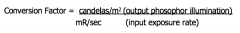
Slide 18. |
|
|
28a. With Cesium Iodide image intensifiers tubes, contrast and resolution are [increased, decreased] compared to Zinc Cadmium Sulfide tubes. 28b. Resolution with Cesium Iodide tubes is _____ lp/mm. |
28a. INCREASED. (Slide 36) 28b. 4 lp/mm. (Page. 408) |
|
|
29. For routine fluoroscopy without ABC/AEC/AERC, the useful dose rate measured at the tabletop may not exceed ________ cGya/min.
|
5 cGya/min. |
|
|
30a. True/False: When comparing a 9" II mode to a 6" II mode, the 9" mode is used to view a larger anatomic area and is considered the regular non-magnification mode. |
30a. TRUE. Slide 19. |
|
|
30b. True/False: The 6" II mode is considered the magnification mode (compared to 9") and this uses more exposure to compensate for the overall decreased brightness of the image and lower overall quality? |
30b. TRUE. Slide 19. |
|
|
31. Which of the following statements concerning a dual mode unit in the 6-inch mode with "ABC/AEC/AERC" are true? a. patient dose is reduced. b. has a reduced flux gain. c. kVp is automatically increased. d. the area used for the output phosphor is decreased. |
a. false. b. true. c. false. d. false. |
|
|
32. A warped image occurs due to the curvature of the input phosphor with non-digital image intensifying units. This warped image is called ________________. |
Pincushion distortion. Slide 59 |
|
|
33. Veiling glare [increases, decreases] contrast because of the light scatter in the image intensifier and an increased "background signal". |
DECREASES. Slide 22. |
|
|
33. Veiling glare is [more, much less] prominent with digital fluoroscopy units. |
MUCH LESS. Not a part of Digital. Slide 22. |
|
|
34. The circuit that interfaces with the x-ray generator to adjust the kVp and mA are known as what? |
Automatic Brightness Control (ABC). Automatic Exposure Control (AEC). or Automatic Exposure Rate Control (AERC). Slide 28. |
|
|
34. True/False: The circuit that interfaces with the x-ray generator to adjust the kVp and mA keep the light input constant and stable to the II input phosphor. |
FALSE. ...it keeps the light OUTPUT constant and stable to the II OUTPUT phosphor. |
|
|
35. Radiation not serving any useful purpose that combines both leakage and scatter radiation is called? |
SECONDARY RADIATION. Slide 46. |
|
|
36. A raster pattern is a TV scanning pattern that scans in a series of [horizontal, vertical] scan lines |
HORIZONTAL. Slide 26. |
|
|
37. How is Flicker (3) in the television picture avoided? |
1. Using interlaced scanning. 2. synchronizing the video signal between the camera and the monitor. 3. scanning the odd lines, then the even lines. |
|
|
38. Grid ratios are generally [higher, lower] with fluoroscopy units as compared to general diagnostic table grid ratios. |
LOWER. |
|
|
39. Patient exposure in cinefluorography is [greater, lesser] than exposure received with other recording systems such as video tape recording. |
GREATER. Increased patient dose. Slide 21. |
|
|
40. When automatic exposure rate control is used, the useful beam exposure rate shall be measured with a phantom equivalent to _____a_____ inches of Lucite or ____b____ inches of water to simulate a patient. |
a. 7 7/8 inches b. 9 inches |
|
|
41. Which of following are characteristics of Charged-Coupled Devices (CCD)? a. have replaced traditional video camera tubes in modern fluoroscopy units. b. useful in high-speed imaging which eliminates lag. c. solid state technology which lowers power consumption. d. useful in cardiac imaging |
All of the above. Slide 35. |
|
|
42. Pulsed fluoroscopy uses short bursts of radiation (<__a___ msec or less) to significantly [increase, decrease] exposure to the patient and personnel in the x-ray room. |
a. <5 msecs or less. DECREASES/REDUCES exposure to the patient and personnel in the x-ray room. |
|
|
43a. With HLC, the maximum tabletop dose rate for high-level fluoroscopy is ______ rads per minute when not using recording devices. |
20 rads per minute. |
|
|
43b. With HLC, this type of fluoroscopy is used for what type of radiography. |
Cardiography and Interventional. |
|
|
43c. True/False, with HLC, an audible signal is optional to be heard when the high level boost is "on". |
FALSE. It is mandatory, a "must." |
|
|
44. How does a dead pixel appear on an image intensified fluoroscopic image on the video monitor? |
Black spot (pixel) on image. Slide 59. |
|
|
45. Under what the two conditions should an unexposed border be visible on the image receptor? |
(1) For automatic collimating devices it should be visible at all heights above the tabletop. (2) When the screen carriage is positioned 14" above the tabletop and collimators are fully open. |
|
|
46. The intensity of the x-ray beam at the tabletop of a fluoroscope should not exceed _______ of operating tube current at a setting of 80 kVp. |
2.2 rads/min for each mA. |
|
|
49. For fluoroscopes with ABC/ABS, the operator must monitor the x-ray tube current (mA) and potential (kVp) at least once each.... (a) [day, week, month] and a written log (b) [must be kept, is not required]. |
a. week. b. must be kept. Slide 29. |
|
|
49. For fluoroscopes with ABC/ABS, it must be evaluated and the dose rate shall be made by a qualified physicist at least once every [year, 3 years]. |
YEAR (annually). Slide 29. |
|
|
49. How long are the logs (monitoring of tube current (mA) and potential (kVp) kept? |
3 YEARS. Slide 29. |
|
|
50. To lower unnecessary dose to the patient, the operator, and other room personnel, the radiographer would: a. [minimize, maximize] the patient-to-image intensifier distance. |
MINIMIZE. |
|
|
50. To lower unnecessary dose to the patient, the operator, and other room personnel, the radiographer would: b. true/false: limit the use of last image hold to view a static image for a longer period of time to reduce "continuous" exposure dose. |
FALSE. Do not limit the use of it, use when necessary. |
|
|
50. To lower unnecessary dose to the patient, the operator, and other room personnel, the radiographer would: c. true/false: take off the spacer on a mobile fluoroscopic unit to maintain proper source-to-skin distance. |
FALSE. Keep the spacer "on". |
|
|
50. To lower unnecessary dose to the patient, the operator, and other room personnel, the radiographer would: d. true/false: use short looks while fluoroscopy unit is on whenever practical to minimize dose. |
TRUE. |
|
|
50. To lower unnecessary dose to the patient, the operator, and other room personnel, the radiographer would: e. true/false: limit the utilization of frame grab. |
FALSE. use frame grab whenever possible. |
|
|
50. To lower unnecessary dose to the patient, the operator, and other room personnel, the radiographer would: f. true/false: use the regular foot switch, not the High-Level Fluoroscopy switch. |
TRUE. |
|
|
51. The source-to-skin distance for mobile fluoroscopy (C-arm) units requires a minimum distance of ______ cm or _____ inches. |
30 cm or 12 inches. Slide 44/55. |
|
|
52. During mobile fluoroscopy, the maximum permissible dose rate of ______ rads/minute may not exceed at 30 cm from the input surface of the fluoroscopic image assembly. |
5 rads/minute. Slide 44. |
|
|
53. When mobile equipment is routinely used in operating rooms in a fixed location, appropriate structural shielding is optional for these rooms. |
FALSE. THEY MUST BE PROVIDED. Slide 44. |
|
|
54. Which of the following statement(s) concerning the spacer on a mobile fluoroscopic equipment is/are correct? a. functions as a lock. b. it should never be removed. c. it is used to maintain the correct 12" source-to-skin distance. |
b and c. |
|
|
55. True or false: Licentiates (physicians) who possess a valid Radiology Supervisor & Operator certificate (radiologist) may be authorized to operate fluoroscopy equipment on human beings in California. |
TRUE. |
|
|
55. True or false: Licentiates (physicians) who possess a valid Fluoroscopy Supervisor & Operator certificate (orthopedist, cardiologist etc) may be authorized to operate fluoroscopy equipment on human beings in California. |
TRUE. |
|
|
55. Who holds the Fluoroscopy Supervisor & Operator certificate? |
Orthopedist, cardiologist etc. |
|
|
55. Who holds the Radiology Supervisor & Operator certificate? |
Radiologist. |
|
|
55. Who holds a valid Fluroscopy permit? |
CRTs. |
|
|
56. When must individuals who hold fluoroscopy permits require to provide their certification document to their employer? |
at every facility where they use fluoroscopy equipment. |
|
|
57. True/False: Radiographers may use fluoroscopic x-ray equipment only under the supervision of a Fluoroscopy Supervisor and Operator. |
TRUE. |
|
|
58. The California Control Regulations states that any person who violates any part of the provisions of these regulations is guilty of a [misdemeanor, felony]. |
MISDEMEANOR. |
|
|
59. True/False: X-Ray Supervisors and Operators may: 1. expose human beings to radiation 2. supervise the activities of techs who hold the technologist fluoroscopy permit. 3. expose human beings to x-radiation only within the scope of their professional licenses. |
ALL OF THE ABOVE. |
|
|
60. True or false: CRT's can use fluoroscopic equipment for fluoroscopic positioning and exposure if they have a fluoroscopy permit. |
TRUE. |
|
|
60. True or false: CRT's are not authorized to report any diagnosis to a patient although ordered by a licentiate of the healing arts. |
FALSE. They may report any diagnosis to patient if ordered to do so by a radiologist, |
|
|
60. True or false: CRT's may not use any title implying the right to practice any of the healing arts |
TRUE. |
|
|
61. Which records must be kept for x-ray machines? a. when a new machine is acquired b. when a machine is transferred or sold. c. when a machine is destroyed or disposed of. d. each calibration, survey, or test. |
a, b, c, d. |
|
|
62. How long should an individual's dosimetry records for occupational workers MUST be kept? |
INDEFINITELY. |
|
|
63. A fluoroscopy permit renewal must be filed [10 days, 15 days, 30 days] prior to the noted expiration date. |
30 days. |
|
|
64. What is a radiation safety officer (RSO)? |
They are responsible for safe use of radiation and regulatory compliance. |
|
|
65. True or False: X-Ray tube housing must be a "diagnostic" type and provide leakage radiation at < 100 mrads/hour at 1 meter. or, < 100 mR per hour @ 1 meter. < 100 mGya per hour @ 1 meter. |
TRUE. |
|
|
66. The fluoroscopy supervisor shall document, in writing, clinical training completion documents to all certified radiologic technologists in CA who have successfully completed the competency based clinical education for the facility where they utilize fluoroscopy. |
TRUE. |
|
|
67. The time required by the eye for recognition of an image is called _____________ time.
This time equals _______ seconds. |
INTEGRATION. Time0.2 seconds.
Slide 5. |
|
|
68. Conventional (older) fluoroscopy uses _________ or night vision. |
SCOPTIC. |
|
|
68. Intensified fluoroscopy uses _________ or daylight vision. |
PHOTOTOPIC. |
|
|
68. Daylight vision uses _______ of the eye. |
CONES. |
|
|
68. Night/dim vision uses _______ of the eye. |
RODS. |
|
|
69. The ability of the eye to resolve separate and distinct objects is called _________ __________. |
Visual Acuity. |
|
|
70. Fluroscopic table tilt for fixed units is ____a____ degrees to the upright position,
and ____b____ in the trendelenberg position. |
a. 90-degrees b. 30-degrees |
|
|
71. Analog conventional fluoroscopy is [more, less] noisy than digital fluorscopy unit because they have [more, less] SNR? |
MORE NOISY. LESS SNR. |
|
|
71. A low SNR equals [gain, loss] of contrast resolution. |
LOSS. |
|
|
72. What is the full name for DAP? |
Dose Area Product. |
|
|
72. What is the full name for KAP? |
Kerma Area Product. |
|
|
72. True/False: Do DAP and KAP assess the absorbed dose risk of an exam compared with the area irradiated. |
FALSE. DAP - absorb. KAP - transfer. |