![]()
![]()
![]()
Use LEFT and RIGHT arrow keys to navigate between flashcards;
Use UP and DOWN arrow keys to flip the card;
H to show hint;
A reads text to speech;
31 Cards in this Set
- Front
- Back
|
Cervical swelling |
Widened region of the spinal cord that corresponds to the attachments of large nerves which supply the upper limbs Extends approximately C3-T2 |
|
|
Lumbar swelling |
Widened region of the spinal cord that corresponds to the attachments of large nerves which supply the lower limbs Extends approximately L1-S2 |
|
|
Conus medullaris |
The tapered lower end of the spinal cord Occurs near L1/L2 |
|
|
Cauda equina |
Think: horse's tail A bundle of spinal nerves that originate in the conus medullaris of the spinal cord |
|
|
Filum terminale |
A thread-like strand of fibrous tissue (pia mater) from the conus medullaris to the back of the coccyx About 20cm in length Gives longitudinal support to the spinal cord |
|
|
Meninges |
The membranes that envelop the brain and spinal cord Dura mater (outer) Arachnoid (middle) Pia mater (inner) |
|
|
Cerebral hemispheres |
The two symmetrical halves of the cerebrum (telencephalon) Connected by the corpus callosum |
|
|
Brain |
Encephalon |
|
|
Diencephalon |
Appears at the upper end of the brainstem Made up of four distinct components: Thalamus, hypothalamus, epithalamus, subthalamus |
|
|
Brainstem |
Midbrain (Mesencephalon) Pons (Metencephalon) Medulla oblongata (Myelencephalon) |
|
|
Cerebellum |
Metencephalon |
|
|
Cerebral aqueduct |
Contains cerebrospinal fluid Connects the 3rd and 4th ventricles of the brain for communication |
|
|
Cerebral peduncles |
Rope-like structures Near the midbrain Bundles of axons Separated by the interpeduncular fossa |
|
|
Medullary pyramids |
Paired white matter structures on the medulla oblongata Contain motor fibres of the corticospinal tracts Fibres of the lateral corticospinal tract decussate (cross) here |
|
|
Olives |
Paired oval structures on the medulla oblongata either side of the pyramids Contains the olivary nuclei |
|
|
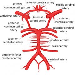
Circle of Willis |
|
|
|
Cranial nerves (names) |
(I) Olfactory n. = smell (II) Optic n. = vision (III) Oculomotor n. = motor control of some eye m. and eyelid (IV) Trochlear n. = motor control of some eye m. (V) Trigeminal n. = chewing m. and some facial sensation (VI) Abducens n. = motor control of some eye m. (VII) Facial n. = taste, salivation and motor control of facial m. (VIII) Vestibulocochlear n. = hearing (X) Vagus n. = think parasympathetic NS effects (XI) Accessory n. = motor impulses to pharynx and shoulder (XIII) Hypoglossal n. = motor control of tongue and some skeletal m. |
|
|
Cranial nerves (types of fibres) |
(I) S (II) S (III) M (IV) M (V) B (VI) M (VII) B (VIII) S (IX) B (X) B (XI) M (XII) M |
|
|
Tympanic membrane |
A thin, semitransparent sheet that separates the external ear from the middle ear At the end of the external acoustic meatus (auditory canal) |
|
|
Ceruminous glands |
Glands along the external acoustic meatus that secrete a waxy material (cerumen) for protection |
|
|
Tympanic cavity |
A synonym for "the middle ear" |
|
|
The auditory ossicles |
Three tiny bones contained in the middle ear |
|
|
Muscles of the middle ear |
Tensor tympani muscle Stapedius muscle Work to reduce the movement of the tympanic membrane, thus protecting it from violent movements under very noisy conditions |
|
|
The fluids that flow through the internal ear |
Perilymph: flows between the bony and membranous labyrinths Endolymph: flows through the membranous labyrinth |
|
|
Cochlear |
The spiral-shaped, bony chamber in the internal ear |
|
|
Muscles of facial expression that operate the eyelid |
Orbicularis oculi: contracts to close the eyelid, innervated by the facial nerve Corrugator supercilii: contracts to draw the eyebrows together, innervated by the facial nerve |
|
|
Muscle fibres in the dorsal and ventral horns, roots, rami, and spinal nerve |
Dorsal horns: sensory Dorsal roots: sensory Dorsal rami: mixed Ventral horns: motor Ventral roots: motor Ventral rami: mixed Spinal nerve: mixed |
|
|
Nerve plexus |
A branching network of intersecting nerves Cervical, brachial, lumbar, and sacral |
|
|
C3, 4, 5 |
Keeps the diaphragm alive! Left and right phrenic nerves innervate the diaphragm |
|
|
C4, T4, L4 |
C4: common corotid artery bifurcates into the internal and external carotid arteries T4: trachea bifurcates into the left and right primary bronchi L4: abdominal aorta bifurcates into the left and right common iliac artery |
|
|
Retroperitoneal organs |
SADPUCKER Spleen, Aorta (and IVC), Duodenum (2 and 3 part), Pancreas (head and neck), Ureters, Colon (ascending and descending), Kidneys, Esophagus, Rectum |