![]()
![]()
![]()
Use LEFT and RIGHT arrow keys to navigate between flashcards;
Use UP and DOWN arrow keys to flip the card;
H to show hint;
A reads text to speech;
52 Cards in this Set
- Front
- Back
|
what is the highest centre of control for motor system |
cerebral cortex |
|
|
what is the cerebral cortex |
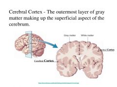
|
|
|
what is the cerebral cortex responsible for |
spontaneous,novel and adaptive behaviour |
|
|
what structures play a rolein the control of motor pathways |
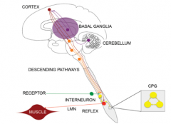
cerebral cortex basal ganglia cerebellum simple reflex arc LMN CPG |
|
|
Inembryological development the brain and spinal cord are derived from what? |
hollow fluid filled neural tube |
|
|
the developing spinal cord isdivided into two regions. what are these two, and how are they divided |

divided into ananterior (ventral) region a posterior (dorsal) region the dividingline being the sulcus limitans |
|
|
which part of the spinal cord has motor neurones |

Theanterior part (to sulcus limitans) is called the basal plate and contains themotor neurones |
|
|
which part of the spinal cord has sensory functions |
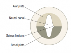
theposterior part (to sulcus limitans) is the alar plate and has sensory function. |
|
|
where are the cell bodies of sensory neurones |
in the dorsal root ganglia |
|
|
where are the cell bodies of motor neurones |
in the basal plate |
|
|
how do mixed spinal roots form |
Motor axons grow out of the anterior partof the spinal cord to form the ventral (motor) spinal nerve roots. Sensoryneurones grow into the posterior aspect of the cord forming the dorsal(sensory) spinal nerve roots. |
|
|
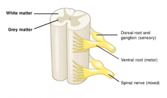
how is the adult spinal cord structured (dorsal horns/ventral horns) |
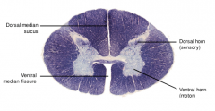
Spinal cordgrey matter in cross section forms an “H” shape which conists of nerve cells(bodies) which are surrounded by a thick layer of white matter which iscomposed of myelinated axons. Spinal cordgrey matter divided into left and right dorsal horns which belong to the alarplate and therefore sensory in function. The ventralhorns derive from the basal plate and contain motor neurones (anterior horncells) |
|
|
what are the laminae of rexed |
comprise a system of ten layers of grey matter, to label portions of the grey columns of the spinal cord |
|
|
what do lamina 1-6 of the lamina of rexed consist of |
make up thedorsal horn, laminae VII and X are mid-region of the cord including the areaaround the fluid-filled central canal. |
|
|
what do the lamina 8-9 of the laminae of rexed consist of |
correspond tothe anterior horn. |
|
|
how are LMN in the spinal cord arranged (what is the medial-lateral gradient) |
thoseinnervating proximal (axial and limb girdle) muscles are found close to themidline motor neurones supplying the distal limb muscles are placedlaterally |
|
|
what is the ventral-dorsalpattern arrangement in the spinal cord |
Neuronessupplying flexor groups are nearer the back of the cord extensors aremore towards the front |
|
|
Somegroups of motor neurones in the spinal cord grey matter have discretefunctional roles and are given specific names. what is the role of the phrenic nucleus from which vertebrae are its spinal roots |
(C3, C4, C5) which supplies the diaphragm via the phrenic nerve and involved in breathing 'C345 keep the diaphragm alive' |
|
|
Some groups of motor neurones in the spinal cord grey matter have discrete functional roles and are given specific names. what is the role of the Spinal accessory nucleus from which vertebrae are its spinal roots |
(C5, C6) Supplies trapezius and sternocleidomastoid |
|
|
Some groups of motor neurones in the spinal cord grey matter have discrete functional roles and are given specific names. What is the role of the Onuf’snucleus from which vertebrae are its spinal roots |
S2, S3, S4 Importantfor continence as it supplies the external urethral and anal sphincters |
|
|
white matter of the spinal cord are divided bythe lines of attachment of the dorsal and ventral nerve roots into threelongitudinal columns what are these columns called |
posterior lateral (clear line) anterior columns (arbitrary) |
|
|
white matter of the spinal cord are divided by the lines of attachment of the dorsal and ventral nerve roots into three longitudinal columns what is the purpose of these columns |
containdescending pathways from the brain (e.g. those carrying instructions related tomovements) contain numerous ascending pathways (e.g. tracts mediating sensationssuch as touch, vibration, pain and temperature). |
|
|
what information do the dorsal columns bring to the brain |
vibration,proprioception and discriminative touch |
|
|
what infomation does the spinothalamic tract bring to the brain |
bringing painand temperature info to the brain. |
|
|
what information does the spinocerebellar tract bring to the brain |
conveys information to the cerebellum about length and tension of muscle fibers (i.e., unconscious proprioceptive sensatio |
|
|
what is the role of the cerebellum |
gathers a vast amount of sensory andother information and uses this to “advise” the motor areas of the frontallobe, helping to ensure that actions are performed in a smooth, precise andcoordinated manner For this reason, cerebellar damage or disease often leadsto clumsiness and poor coordination (termed cerebellar ataxia). |
|
|
what is cerebellar ataxia |
is a disorder that occurs when the cerebellum becomes inflamed or damaged. The cerebellum is the area of the brain responsible for controlling gait and muscle coordination |
|
|
what is the corticospinal tract (where do its neurones originate and synapse) |
a descending tract of the spinal cord which contains bundles of axons which originate in the cerebral cortex and descend to synapse within the brainstem or spinal cord. |
|
|
where is the lateral and anterior column if the cerebrospinal tract |
The lateralcorticospinal tract occupies the lateral column of the cord the anteriorcorticospinal tract and occupies the anterior column of the cord. |
|
|
what is the lateral corticospinal tract |
Lateralcorticospinal tract deals with distal flexors mainly. Lateralcorticospinal tract contains about 90% of the fibres and these are the onesthat crossed over at the level of the foramen magnum. |
|
|
what is the anterior corticospinal tract |
Ant spinaltract deals with proximal extensors Anterior spinaltract is the direct continuation of the pyramids of the medulla. This is why itis situated either side of the midline at the front and doesn’t cross |
|
|
how does the amount of white matter change as you descend the verebral column |

Cross sectionsat different cord levels show that the cervical region contains the most whitematter because of all the descending and ascending pathways present at thislevel. Whereas in thelowermost part of the spinal cord most of the descending pathways are no longerpresent and most of the ascending pathways have yet to join the cord. |
|
|
how does the amount of grey matter change as you descend the vertebral column |
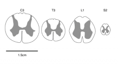
grey matterwhich varies in proportion to the amount of muscle tissue at each level; forthis reason the cervical and lumbosacral regions have the largest amount ofgrey matter (to supply the limbs) whilst the thoracic cord has the least. |
|
|
how can movement be categorised (4 main types) |
reflexive postural rhythmic voluntary |
|
|
how do the different types of movement vary |
vary incomplexity and the degree to which they can be considered voluntary vsautomatic |
|
|
what is voluntary movement (where do these movements originate etc) |
leastautomatic action goal orientatedvoluntary acts that require participation of the cerebral cortex. originatefrom the frontal lobe Involve directconnections from cortex to spinal cord, the lateral corticospinal tract. |
|
|
what is rhythmic movement (where do these movements originate/ how are they coordinated) |
semi-automaticmovements that are mainly rhythmic patterns of activity initiatedand maintained by subcortical structures such as brain stem and spinal cord coordinated bynetworks of neurones called central pattern generators. littleor no contribution from the cerebral hemispheres or cortex |
|
|
what is reflexive movement (how are they triggered, what types of nerve do they involve) |
Reflexesare rapid and automatic responses to particular sensory stimuli and as such arestereotyped varying only in speed, magnitude or duration mostbasic form consists of only two nerve cells (one sensory, one motor) plus anintervening synapse eg the stretch reflex They aretriggered by proprioceptors. These are reflex arcs. can be a lot more complex involvingnumerous nerve cells and synapses extending over several spinal cord segmentsor brain stem levels and in some cases both sides of the body (e.g. limbwithdrawal reflex).eas(0��z� |
|
|
what is the stretch (myotatic) reflex |
muscle spindles are stretch detectors, and found scattered throughout skeletal muscles Each spindlecontains a few striated fibres described as “intrafusal”. Musclespindles are in parallel with the extrafusal fibres so whenever the msucle belly is put under tension causing the muscle spindles to respond. spindles are excited triggering reflex contraction of the homonymous (same) muscle group, resisting change inmuscle length. achieved by asimple spinal cord reflex with a sensory limb, a motor limb and a single synapse,this is the stretch reflex. Antagonistmuscles are inhibited at the same time (reciprocal inhibition) by inhibitoryinterneurones, activated by collaterals of the afferent. |
|
|
how does an UPN lesion affect the stretch reflex |
The stretchreflex is normally dampened down by descending influences from the brain stemto ensure that muscle tone is not excessive. Thismoderating influence tends to be lost in patients with upper motor neuronelesions With the normaldampening down influence lost, the stretch reflex becomes abnormally strong(hyperreflexia) so excessive muscle tone(hypertonia) which manifests as firm resistance to manipulation of joints. Boththe agonist and antagonist muscles start having a tug-of-war trying to contractmaking the muscle stiff, this is spasticity. |
|
|
what are the effects of a LMN lesion |
theperipheral nerve supply to the muscle is interrupted Loss of motornerve supply to the muscle results in flaccid (floppy) paralysis of theaffected muscles accompanied by a complete absence of normal tone (atonia) andloss of deep tendon reflexes (areflexia). so we get gradual wasting(atrophy) of the muscle due to lack of use (disuse atrophy) |
|
|
why may we get twitching in a LMN lesion (fasiculations) |
caused by increased receptor concentration on muscles to compensate for lack of innervation |
|
|
what are the main protective reflexes |
flexor/withdrawalreflex crossed extensor reflex. |
|
|
how do the protective reflexes coordinate eg when you step on a pin |
If you’rewalking along and step on a pin you immediately pull your foot away and you maythink you did this voluntarily but actually it was an automatic reflex.At the sametime you straighten your other leg so you support your body and don’t fallover. The pulling theleg away is the withdrawal reflex, the straightening of the leg is the crossedextensor reflex. These two reflexes coordinate automatic limb withdrawal from anoxious stimulus |
|
|
where does the withdrawal reflex start and what is it triggered by |
The withdrawalreflex is polysynaptic and triggered by nociceptors and is a cutaneous reflex(starts in skin). |
|
|
what is the central pattern generator CPG |
networks of neurones within the spinal grey matter or brain stem involved in coordinating rhythmic motor sequences such as walking, chewing, breathing . |
|
|
how are CPG coordinated |
canbe selected and recruited by descending projections from the brain (‘commandneurons’ operaterelatively independently and autonomously |
|
|
where is the CPG located |
in the spinal cord. command neurones send signals up into the brain |
|
|
what are the basal ganglia loops |
The connections between the motorcortex and the basal ganglia are arranged as a set of “loops” which each arise ina particular region of the frontal lobe and pass through the basal ganglia beforeprojecting back to the cortical region of origin (via a relay in the thalamus) |
|
|
what do the basal ganglia loops do |
involved in (i) the initiation ofvoluntary actions (ii) selection of a particular action among a range ofpossible or potential actions (iii) the learning and performance of variousrepetitive, semi-automatic behaviours (or ‘habits’) |
|
|
what Neurotransmitter is involved in the basal ganglia loops |
dopamine |
|
|
disease and damage to the basal ganglia is associated with what? |
associatedwith a movement disorder, the best example of which is Parkinson’s disease Thebasal ganglia are also involved in cognition, behaviour and emotion due to thepresence of non-motor loops that arise and terminate in other parts of thecerebral cortex. |