![]()
![]()
![]()
Use LEFT and RIGHT arrow keys to navigate between flashcards;
Use UP and DOWN arrow keys to flip the card;
H to show hint;
A reads text to speech;
41 Cards in this Set
- Front
- Back
|
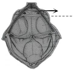
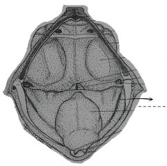
A fold of skin extending over the upper jaw. |
Upper lip fold |
|
|
A groove which runs throughout the extent of the upper jaw, it receives the lower jaw when the mouth is closed. |
Sulcus marginalis |
|
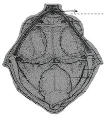
A depression at the mid-anterior portion of the sulcus marginalis. |
Median subrostral fossa |
|
Slight elevations at the side of the median subrostral fossa. |
Pulvinar rostrale |
|
The two depressions in the sulcus marginalis which are adjacent and lateral to the pulvinar rostrale. |
Lateral subrostral fossae |
|
The two small openings which are inward extension of the external nares. |
Internal nares (choanae) |
|
Teeth-like prominences borne by two bones located between the two choanae, one on each side of the mid-dorsal surface of the buccal cavity. |
Vomerine teeth |
|
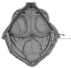
The large opening on each side of the posterior part of the roof of the buccal cavity anterior and dorsal to the angles formed by the upper and lower jaws. |
Eustachian tube |
|
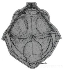
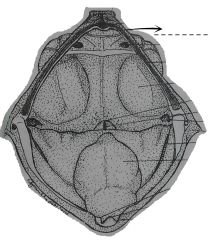
Forms the rigid rim at the tip of the floor of the mouth. |
Lower jaw |
|
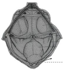
The single elevation at the tip of the lower jaw. This fits into the median subrostral fossa when the mouth is closed. |
Tuberculum prelinguale |
|
Shallow depression immediately at the sides of the tuberculum prelinguale. These receive the pulvinar rostrale of the upper jaw when the mouth is closed. |
Prelingual fossae |
|
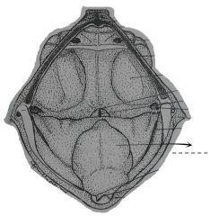
The muscular organ which occupies the greater part of the floor of the buccal cavity. |
Tongue |
|
|
A pair of slit-like openings, one on each side of the posterior end of the floor of the buccal cavity. These are present in male individuals and absent in females. |
Vocal sac openings |
|
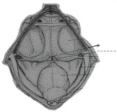
A rounded somewhat elevated area posterior to the free end of the tongue. |
Laryngeal prominence |
|
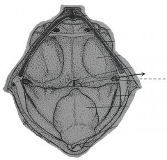
A slit-like opening at the middle of the laryngeal prominence. This is an opening of the larynx or voice box. |
Glottis |
|
|
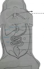
The shiny, thin membrane forming the outermost covering of the visceral organ. |
Visceral peritoneum |
|
|
A thin membrane adhering closely to the inner surface of the body wall, and lines the pleuroparitoneal cavity. |
Parietal peritoneum |
|
|
A thin membrane tissue covering the outermost wall of the heart. |
Visceral pericardium |
|
|
A thin membranosus tissue forming the wall of the pericardinal cavity. |
Parietal pericardium |
|
A narrow short tube just behind the buccal cavity. It connects the pharynx with the stomach. |
Esophagus |
|
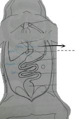
The j-shaped, muscular, slightly curved sac posterior to the esophagus. |
Stomach |
|
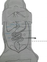
The long, narrow and coiled tube extending from the pylorus to the much dilated large intestine. |
Small intestine |
|

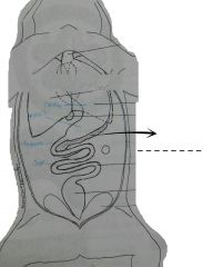
A straight club shaped, usually dark colored tube continuous with the small intestine, and ending at the anus or vent. |
Large intestine |
|
|
A digestive gland, considered as the largest gland in the body, occupying the anterior half of the pleuroperitoneal cavity. This contains three lobes; the left, median, and right. |
Liver |
|
A long, thin, irregular, yellowish organ suspended in the omentum connecting the stomach, duodenum, and liver. The omentum is termed the gastrohepatoduodenal or lesser onentum. |
Pancreas |
|
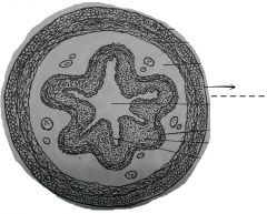
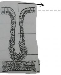
The very thin outermost layer of the stomach. This is only one cell thick and composed of simple squaomus epilethium. |
Tunica serosa (visceral peritoneum) |
|
|
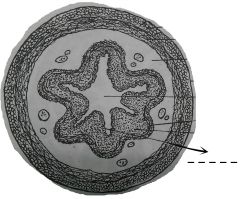
A layer that forms the bulk of the stomach. It is composed of smooth muscles arranged in two layers; longitudinal and circular. |
Tunica muscularis |
|
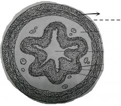
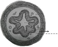
A very thin layer next to the tunica serosa. The fibers are cut crosswise, hence they appear as small circles or discs in the cross-section. |
Longitudinal muscle layer |
|
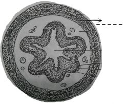
Next and inner to the longitudinal muscle layer and thicker. The fibers are as well as the nuclei are cut longitudinally, so they appear elongated. |
Circular muscle layer |
|
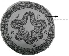
Mainly composed of loose or dense fibrous connective tissues. This coat follows the contour of the inner surface, occupying the outer layer of the ruga. |
Tela submucosa |
|
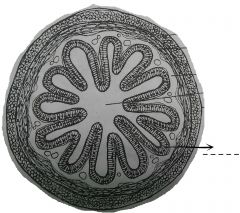
A thick and inner layer of the stomach which is composed of the muscularis mucosa and lamina propria and the simple columnar epilethium. The inner surface of this layer appears to be noticed by numerous depressions, the gastric glands. |
Tunica mucosa |
|
A thin layer of smooth muscle next to the tela submucosa. This is composed of an outer longitudinal and inner circular smooth muscle layer. This thin layer becomes thickened at the head of the ruga. |
Muscularis mucosa |
|
A layer of loose connective tissue wherein the gastric glands are embedded. |
Lamina propria |
|
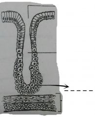
An expanded part of the gastric gland near the base of the tunica mucosa. |
Fundus or body |
|
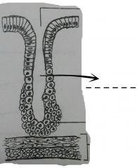
The narrow portion which extends form the base of the inward toward the gastric pits. The neck and body are lined by cuboidal cells. |
Neck |
|
The opening of the gastric gland located at the tip of the neck at the bottom of the gastric pit. |
Mouth |
|
The innermost layer of the tunica mucosa which lines immediately the lumen or cavity of the stomach. |
Simple columnar epilethium |
|
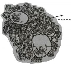
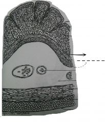
These are numerous cells in the specimen. They have indistinct cell membrane and are arranged in cords or plates. The cells secrete bile or other substances. |
LIver or hepatic cells |
|
Big spaces in the liver which are lined by thin simple squamous epilethium or endolethium. These are usually filled up with blood cells. |
Blood vessels |
|
Small spaces lined by thicker simple cuboidal epilethium. These are usually empty and lie at the sides of the blood vessels. They receive secretory substance from the liver cells, and bring them to bigger ducts. |
Bile ducts |
|
Masses of dark brown structures scattered among the liver cells. |
Pigments |