![]()
![]()
![]()
Use LEFT and RIGHT arrow keys to navigate between flashcards;
Use UP and DOWN arrow keys to flip the card;
H to show hint;
A reads text to speech;
7 Cards in this Set
- Front
- Back
|
THE PELVIS The pelvic girdle is made up of two hip bones (pubic crest & public tubercle) anteriorly & posteriorly that make up the pubic symphisis Sacrum + ilium = sacroiliac joints. The three parts of the pelvic girdle are illium, ischium & pubis Hip bones + pubic symphisis + sacrum + coccynx = bony pelvis False pelvis or greater pelvis- portion of the pelvis superior to the pelvic brim. True pelvis or lesser pelvis- portion of the pelvis inferior to the pelvic brim. |
COMPONENTS OF THE PELVIS The obturator artery branches out to form foveal artery Foveal artery + medial & lateral circumflex arteries supplies the head of femur The fracture of the femoral neck becomes complicated when the blood supply is cut to the femoral head causing avascular necrosis. |
|
|
HIP JOINT Between the head of the femur and the acetabulum. The ligament involved is ligamentum teres. The ligament helps to restrict movement of the femoral head and contributes to the stability of the joint. It also carries a branch of the obturator artery. Type of hip joint- Flexible Ball and Socket Joint Planes of movement- 3o Flexion & Extension, Abduction & Adduction, & Circumflex |
INGINUAL CANAL: INGINUAL LIGAMENT Inguinal ligament runs between the ASIS & the pubic tubercle formed by external oblique aponeurosis. There are two points on the inguinal ligament: 1) The midpoint of the inguinal ligament: halfway between the ASIS and the pubic tubercle. 2) The mid inguinal point: Halfway between the ASIS and pubic symphisis. |
|
|
LACUNAR LIGAMENT The lacunar ligament connectsthe inguinal ligament above to the pectineal line on the pubis below, it too isderived from the external oblique aponeurosis. |
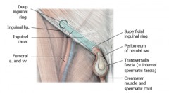
INGUINAL CANAL The main contents of the inguinal canal are the spermatic cord in males and the round ligament of the uterus in females, blood and lympatics, and the illioinguinal nerve. The inguinal canal has two openings: Deep inguinal Ring- Located superior to the middle of the inguinal ligament. formed by an evagination ofthe transversalis fascia →which further along becomes the internal spermatic fascia. Superior Inguinal Ring- formed by a diagonal split in the external oblique aponeurosis. As the aponeurosis splits, there areparts which remain on either side (medial & lateral)of the canal and superficial ring → medial and lateralcrura.These are stabilised by perpendicularfibres running across both crura→ intercruralfibres |
|
|
WALLS OF THE INGUINAL CANAL MALT- MUSCLES, APONEUROSIS, LIGAMENTS, TRANSVERSALIS & TENDON Roof- Internal Oblique & Rectus Abdominis Anterior Wall- External oblique + Internal Oblique laterally Floor- Inguinal & lacunar ligament Posterior Wall- Transversalis Fascia & Cojoint Tendon (inguinal falx) |
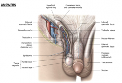
MALE INGUINAL CANAL Males:spermatic cord •Externalspermatic fascia derived from the external oblique aponeurosis •Cremastericfascia & muscle derived from the internal oblique muscle & fascia •Internalspermatic fascia derived from the transversalis fascia •Contentsinclude vas deferens & testicular vessels •Thetunica vaginalis derived from peritoneum •Plus,blood vessels, lymphatics and the ilioinguinalnerve |
|
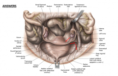
FEMALE INGUINAL CANAL Females:round ligament of the uterus (from the caudal part of the gubernaculum) *cranial part becomes ovarian ligament → So gubernaculum goes from ovary –uterus – inguinal canal – labia majora Plus, blood vessels, lymphatics and the ilioinguinal nerve |
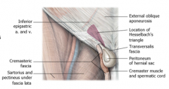
HESSELBERG'S TRIANGLE It is bordered: •Medially by the rectus abdominis muscle. •Inferiorly by the inguinal ligament. •Superolaterally by the inferior epigastric vessels. Hesselbach’striangle is covered by weak fascia and can allow direct inguinal hernias topierce the abdominal wall. |
|
DIRECT INGUINAL HERNIAS
•Directinguinal hernias push directly through the peritoneum→ transversalis fascia → inguinal canal →superficial ringadjacent to the normal inguinal canal contents •Can bepredisposed if the conjoint tendon is not properly formed •Can alsopass around the inguinal canal rather than through it •Accountsfor a third to a quarter of inguinal hernias |
INDIRECT INGUINAL HERNIAS
•Infemales the processusvaginalis also forms the inguinal canal and then normally closes over a period of timeeither before or after birth. •However,in both males & females the processus vaginalis can remain leaving an open connection between the abdominal cavity & the scrotum in males (so abdominal contents can leak into the scrotum via spermatic cord), and the abdominal cavity & labiamajora in females, causing hernias. •Indirectinguinal hernias account for two thirds to three quarters of all inguinalhernias – most common •Cantravel the entire length of the inguinal canal Patent processus vaginalis can cause either Hydrocele (peritoneal fluid in the scrotum or spermatic cord) or Indirectinguinal hernia. The difference between direct and indirect hernias in clinical practice is to apply pressure @ midpoint btw deep inguinal ring and ask them to cough. |
|
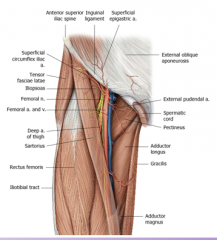
FEMORAL TRIANGLE The subinguinal space liesbelow the inguinal ligament and it is a passageway that connects the maintrunk to the lower limb. The contents of the subinguinal space are divided into 2 compartments; muscular lacuna: iliopsoas muscle & femoral nerve & vascularlacuna: femoral artery, vein & lymphaticsseparated by the iliopectineal arch (a thickening of the iliopsoas fascia). The compartments go from inguinal space → femoral triangle bounded by: • Superiorly by theinguinalligament (ASIS to pubic tubercle) • Medially by the lateral border of theadductorlongus(pubis to linea aspera) • Laterally by thesartorius (ASIS to medial tibia) The roof of the femoral triangle is formed by fascia lata (aponeurosis), fat and skin; the floor of the femoral triangle is formed Mediallyby the pectineusmuscle & Laterallyby the iliopsoasmuscle. Structures of the femoral triangle: NAVEL (f. nerve, f. artery, f. vein, empty space, deep inguinal lymph nodes) The contents of the femora triangle are the femoral nerve (located @ middle of inguinal ligament) & femoral sheath. The contents of the femoral sheath are further divided intocompartments: femoral artery (lateral), femoral vein (intermediate) and femral canal (medial) that contains connective tissue, fat and lymphatics. |
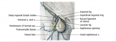
FEMORAL HERNIAS The proximal opening (base) of the femoral canal is the femoral ring. The femoral ring is a weak area on the anterior abdominal wall where femoral hernias begin. The femoral hernias go from femoral ring →femoral canal →saphenous ring →fascia lata →great saphenous vein →femoral vein. Femoral hernias happen more frequently to woman because they have a wider pelvis (bigger space between the inguinal ligament). |