Reading...
![]()
Play button
![]()
Play button
![]()
Use LEFT and RIGHT arrow keys to navigate between flashcards;
Use UP and DOWN arrow keys to flip the card;
H to show hint;
A reads text to speech;
17 Cards in this Set
- Front
- Back

|
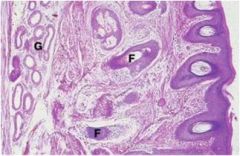
external auditory meatus
Canal from auricle to the tympanic membrane Outer third is cartilage, inner two thirds is temporal bone Lined by skin Coiled tubular ceruminous glands (secrete cerumen) G = ceruminous gland, F = hair follicles |
|

|
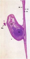
firmly attached to the surrounding bone by a fibrocartilaginous ring.
handle of the malleus is attached to the center of the membrane |
|
|

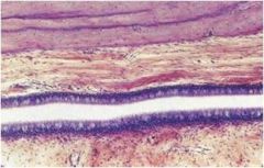
Auditory (eustachian) canal
The auditory canal connects the cavity of the middle ear with the nasopharynx and allows for equalization of air pressure between the middle ear and the external environment. From the middle ear, the tube first passes through bone, but towards the pharynx, the wall is supported on two sides by cartilage and on the remaining two sides by fibrous tissue. Lined by pseudostratified respiratory epithelium with numerous goblet cells particularly towards the pharyngeal end. |
|
|
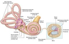
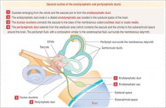
Internal ear. The internal region of the ear is composed of a cavity in the temporal bone, the bony labyrinth, which houses a fluid—filled membranous labyrinth. The membranous labyrinth includes the vestibular organs for the sense of equilibrium and balance (the saccule, utricle, and semicircular ducts) and the cochlea for the sense of hearing.
|
|
|
ducts
|
|
|
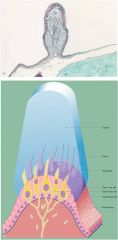
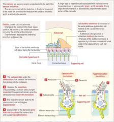
Cristae ampullaris
|
|
|
Stereocilia toward the kinocilium
|

Increased activity of the vestibular nerve
|
|
|
Stereociliar away from the kinocilium
|
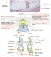
Decreased activity in the vestibular nerve
|
|
|

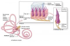
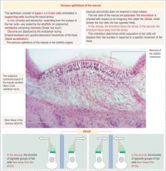
Vestibular maculae and their cells.
(a): Two sensory areas, the maculae, are located in the epithelial walls of the utricle and saccule in the vestibular complex. Both maculae are similar histologically and contain mechanoreceptor cells called hair cells which use gravity and endolymph movement to detect the orientation of the stationary head and linear acceleration of the moving head. (b): A detailed view of a macular wall shows that it is composed of hair cells, supporting cells, and endings of the vestibular branch of the eighth cranial nerve. The apical surface of the air cells is covered by a gelatinous otolithic layer or membrane and the basal ends of the cells have synaptic connections with the nerve fibers. (c): A diagram of a single generalized hair cell shows the numerous straight stereocilia, which contain bundled actin, and a longer single kinocilium, a modified cilium whose tip may be slightly enlarged. |
|

|
Maculae otoliths and kinocilium
|
|
|
Otoliths. Otoliths are crystalline structures in the outer part of the otolithic membrane. Each otolith is a slightly elongated structure, up to 5 by 10 µm in size, and is composed of calcium carbonate on a matrix of proteoglycans. Their presence makes the otolithic membrane considerably heavier than endolymph alone, which facilitates bending of the kilocilia and stereocilia embedded in this membrane by gravity or movement of the head
Present in the muclae in the utricular(horizontal) and saccular (vertical) |
|
|
Striola?
|
|
|

Cochlea
SV = scala vestibuli SM = scala media ST = scala tympani BM = basilar membrane VM = vestibular membrane (Reissner’s membrane) N = afferent nerve fibers Svasc = stria vascularis G = spiral ganglia O = osseous spiral lamina |
|
|
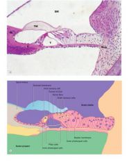
organ of corti; scala media; tectorial membrane
|
|
|
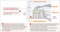
Sound transduction in organ of corti
|
|
|
Isk gene
|
codes a protein of the K channel in the marginal cells of the stria vascularis; if mutated disrupts endolymph production and causes degeneration of the organ of Corti
|
|
|
Connexin 26
|
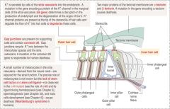
Recycles K ions; mutatoin = deafness
|