Reading...
![]()
Play button
![]()
Play button
![]()
Use LEFT and RIGHT arrow keys to navigate between flashcards;
Use UP and DOWN arrow keys to flip the card;
H to show hint;
A reads text to speech;
84 Cards in this Set
- Front
- Back
|
fxns of skin
|
Protection
– UV light, mechanical, thermal, chemical insults, and microorganisms – Keeps fluid in – prevents dehydration Sensation – Touch, pressure, pain, temperature Temperature regulation – Via hairs, subcutaneous fat, blood flow Secretion/Excretion – Sebum, ‘sweat’, cerumen, milk Metabolic functions – Vitamin D synthesis from UV light – Energy stored in subcutaneous fat |
|
|
epidermis
|
Most superficial layer
Keratinizing stratified squamous epithelium – Keratinocytes (cells w/in epidermis) Sits on top of the dermis |
|
|
components of skin
|
Epidermis
Dermis – Adnexa • Hair follicles • Glands – Sweat glands – Sebaceous glands – Anal glands – Mammary glands Hypodermis (subcutis) |
|
|
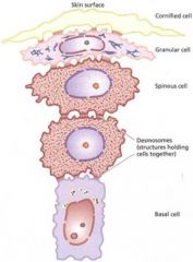
layers of epidermis
|
Stratum corneum (keratin layer)
– Cornified cells • Dead, flattened, eosinophilic cells (squames) • Ortho- or Para-keratosis • Continually shed & replaced – Thickness varies by location Thickest in non-haired regions (footpads, nasal planum) • Thin in inguinal region (Stratum lucidum) – Several layers of translucent, homogenous, dead, squamous cells – Lack nuclei Except in areas with parakeratosis – Primarily foot pads Stratum granulosum – Intracellular purple (keratohyaline) granules – 1-2 cells thick in haired skin – 4-8 cells in non-haired skin or in hair follicles – This is where keratinization begins... – Still composed of dead cells – Only seen in foot pad Stratum spinosum (prickle cell layer) – Can see the desmosomes (intercellular bridges) attaching the cells to each other (spiny/prickly) – Thicker in non-haired skin Stratum basale – Deepest layer – Single layer of cuboidal cells attached to underlying basement membrane – Continually dividing – Provides replacement for layers above it |
|
|
non-epithelial cells in epidermis
|
Melanocytes
– Present in the stratum basale • 1 per 10-20 keratinocytes – Hard to identify with light microscopy – Pass melanin on to adjacent keratinocytes • Not all pigmented cells in the epidermis are melanocytes! Langerhans cells – Present in stratum basale & stratum spinosum – Similar to macrophages • Present antigens to other immune cells to incite an inflammatory response etc – Can’t see with light microscopy Merkel cells – Origin unknown – Clear cells in stratum basale – ‘neuroendocrine’ effects • Hair cycle control – Associated with nerves • Mechanoreceptors |
|
|
the epidermis lacks
|
blood vessels
nerves lymphatics |
|
|
inflammatory cells in the epidermis comes from
|
dermis
|
|
|
Where are dermal scales found?
|
reptiles
non-feathered skin of birds |
|
|
What are dermal scales?
|
extensions of stratum corneum
|
|
|
fxns of the dermis
|
provides tensile strength and elasticity
supports epidermis nourish epidermis – The epidermis has NO blood vessels, so oxygen and inflammatory cells etc come from dermal blood vessels. |
|
|
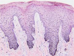
Describe the epidermal/ dermal junction in high friction areas
|
Downward projections of epidermis (rete pegs) interdigitate with upward projections of dermis (dermal papillae)
|
|
|
the dermis is composed of
|
collagen, reticular, and elastic fibers
Ground or interstitial substance - Proteoglycans & electrolytes Blood & lymphatic vessels Nerves Mixed other cells - melanocytes, macrophages, lymphocytes & mast cells |
|
|
What are dermal scales?
|
bony scales arising from dermis
|
|
|
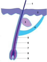
Name the parts of hair follicles
|
1.Hair shaft
2.Epidermis 3.Sebaceous gland 4.Arrector pili muscle 5.Adventitial sheath 6.Outer root sheath 7.Inner root sheath 8.Matrix 9.Dermal papilla 10.Apocrine glands |
|
|
How are wool hairs unique
|
they lack a medulla
|
|
|
1.Hair shaft
2.Epidermis 3.Sebaceous gland 4.Arrector pili muscle 5.Adventitial sheath 6.Outer root sheath 7.Inner root sheath 8.Matrix 9.Dermal papilla 10.Apocrine glands (not shown) |
|
|
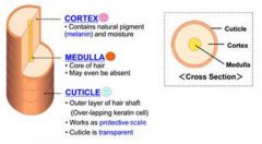
Know the parts of a hair shaft
|
|
|
|
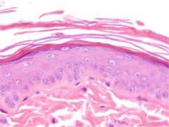
Know the layers of the epidermis
|
|
|
|
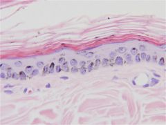
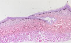
epidermis
- notice stratum corneum (top) |
|
|
foot pad
- large stratum corneum (top) - light stratum lucidum - stratum granulosum beneath that |
|
|
stratum corneum
(orthokeratosis) |
|
|
stratum lucidum
(foot pad) |
|

|
foot pad
Stratum lucidum Stratum granulosum Stratum spinosum Stratum basale |
|
|
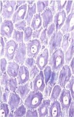
stratum granulosum
|
|
|
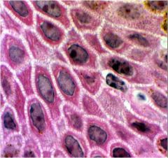
stratum spinosum
|
|
|
stratum spinosum
|
|
|
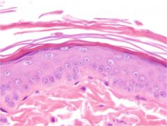
epidermis
- stratum corneum (top) - stratum granulosum (next) - stratum spinosum (next) - stratum basale (notice melanin) - bottom right is mast cell |
|
|
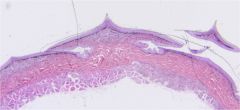
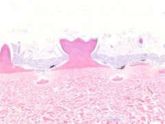
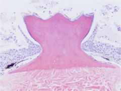
epidermal scales
|
|
|
Epidermal Scales
|
|
|
epidermal dermal junction
|
|
|
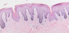
epidermal dermal junction in high friction area (rete pegs & dermal papilla)
|
|
|
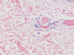
dermis
notice blood vessels on right and few mast cells between the vessels. |
|
|
dermal scales (bony)
|
|
|
dermal scale
notice that it is bone |
|
|
arrector pili muscles
|
|
|
What part of the hair follicle is involved in making hair?
|
Adventitial sheath
Outer root sheath Inner root sheath Matrix Dermal papilla |
|
|
segments of the hair follicle
|
Infundibulum
- Granular layer - Flaky keratin - hair shaft, epidermis Isthmus - Glassy cells - Homogenous keratin - sebaceous gland, arrector pili muscle Suprabulbar & bulbar - Small dark cells (matrical) - adventitial sheath, outer root sheath, matrix, dermal papilla |
|
|
classification of hair follicles
|
Primary
- Large diameter; Deeply rooted in the dermis - Have sebaceous & apocrine glands, & arrector pili muscle Secondary - Smaller diameter; Rooted more superficially - May have a sebaceous gland LACK apocrine glands & arrector pili muscles Simple - single hair comes out Compound - multiple hairs come out |
|
|
Name 3 ways to classify hair follicles
|
Layers
- Inner & Outer root sheaths etc Segments - Infundibular, Isthmus, Suprabulbar/Bulbar Types - Primary vs. Secondary - Simple vs. Compound |
|
|
species differences in hair follicles
|
Horses, Pigs & Cows
- Simple hair follicles Sheep - Wool-growing areas: Compound follicles (both primary & secondary) - Hair-growing areas: Simple follicles Goats - Primary & Secondary follicles Dogs & Cats - Primary (simple) follicles and Secondary (compound) follicles |
|
|
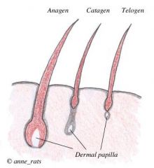
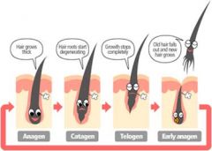
hair cycle
|
Anagen: Growth phase
Catagen: Transitional or involuting phase Telogen: Resting phase The inferior segment (suprabulbar & bulbar) portions are temporary and lost during catagen and telogen The infundibular & isthmus segments of the follicle are permanent |
|
|
know the hair cycle
|
|
|
|
know the hair cycle :)
|
|
|
|
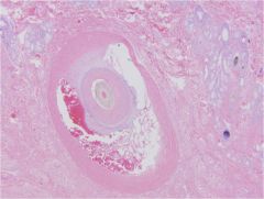
sinus hair
(whisker) |
|
|
describe sinus hair
|
(whisker)
The adventitial sheath (#5) is thickened by connective tissue trabeculae which are filled with blood |
|
|
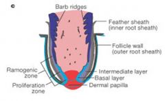
feather follicle
|
|
|
describe a feather follicle
|
|
|
|
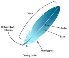
feather anatomy
|
|
|
|
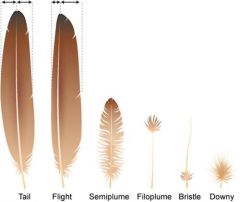
types of feathers
|
|
|
|
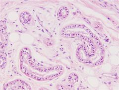
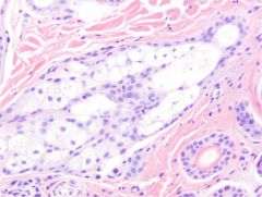
Apocrine sweat glands
|
(epitrichial)
ONLY found on primary follicles NOT secondary follicles Secretory portion: single layer of cuboidal cells surrounded by a layer of myoepithelial cells Secretions mix with sebum for ‘scent’ Thermoregulation in horses and cows only |
|
|
all mammals have _______ sweat glands
|
apocrine & eccrine
|
|
|
Eccrine sweat glands
|
(atrichial)
NOT associated with hair follicles Footpads of dogs & cats ‘frog’ of ungulates (ruminants, camelids) Snout of pigs Medial surface of carpus of pigs Nasal planum of cows Some role in thermoregulation… |
|
|
both types of sweat glands have what shape
|
simple coiled tubular
lined by cuboidal cells and myoepithelium Apocrine (epitrichial) in the skin Eccrine (atrichial) in the foot pad |
|
|
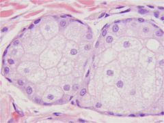
sebaceous glands use what type of secretion
|
Holocrine secretion
The entire cell becomes the secretory product |
|
|
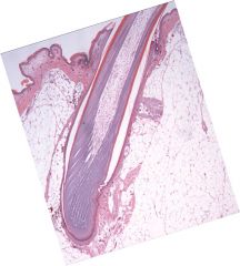
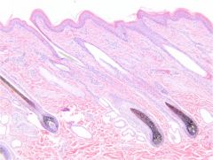
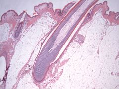
hair follicles
notice associated apocrine glands |
|
|
eccrine (atrichial) glands
|
|
|
sebaceous glands
holocrine secretion |
|
|
sebaceous glands
holocrine secretion notice reserve cells along periphery |
|
|
Where are sebaceous glands found
|
Associated with hair follicles (1o or 2o)
Higher density in some areas - Cats: Chin - Dogs & Cats: Dorsal tail (tail gland) - Goats: Base of horn - Sheep: Infraorbital, inguinal, interdigital NOT on footpads or nasal planum Associated with eyelids (Meibomian glands) |
|
|
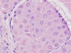
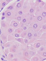
perianal (hepatoid) glands
|
Modified sebaceous glands
Hair follicle associated Resemble hepatocytes (liver cells) = hepatoid Territorial marking... |
|
|
top- epidermis
next- dermis notice follicle in the dermis bottom- perianal (hepatoid) gland |
|
|
perianal (hepatoid) gland
|
|
|
perianal (hepatoid) gland
|
|
|
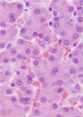
liver (no reserve cells)
|
|
|
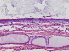
anal glands
|
NOT PERIANAL (HEPATOID)
Specialized apocrine glands Open directly onto anal skin OR Dump into the anal sacs (storage) - Lined by stratified squamous keratinizing epithelium Territorial marking... Found in... - Domestic & wild felids - Ferrets, mink (mustelids) - Raccoons - canids - pigs - rodents |
|
|
anal glands (bottom)
anal sac (top) |
|
|
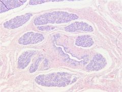
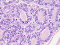
mammary glands
|
Modified sweat glands
Apocrine method of secretion - A piece of the apical (top) cytoplasm pinches off Compound tubuloacinar - Sort of like a bunch of grapes... - Lined by cuboidal to columnar epithelium - Myoepithelium |
|
|
mammary gland secretion pathway
|
Alveolus -> Secretory tubule -> intralobular duct -> lobular duct -> lobar duct -> gland sinus -> teat sinus -> teat canal
|
|
|
Mammary gland
inactive vs. active lactating |
inactive
- cuboidal active - columnar |
|
|
Mammary gland
acini surrounding duct |
|
|
mammary gland lactating
(pink stuff is milk) |
|
|
which species don't have teats?
do they have mammary tissue? |
Rats & horses the males don’t have teats – but they DO have mammary tissue
|
|
|
subcutis (hypodermis)
|
Deepest layer of skin
Thickest layer of skin (in some animals) Site of injections… Absent in some locations: - Lip - Cheek - Eyelid - Ear canal - Anus |
|
|
functions of subcutis
|
Energy storage (fat)
Thermoregulation/Insulation Protective padding Maintains surface contours |
|
|
subcutis is composed of
|
Primarily white adipose tissue
NO lymphatics Blood vessels Nerves Panniculus muscle Attached to underlying muscle & bone (periosteum) |
|
|
chicken feather and subcutis
|
|
|
nose
|
Carnivores: Planum Nasale
- Keratinizing stratified squamous epithelium - Thick keratin layer - No hair follicles or adnexa Cows/Small ruminants: Planum nasolabiale/ Planum nasale - Keratinizing stratified squamous epithelium - Thick keratin layer - No hair follicles - Eccrine glands to moisten the surface |
|
|
planum nasale (dog)
parakeratosis top |
|
|
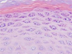
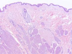
foot pads
|
Epidermis
- Thick keratin layer - Smooth in cats - Papillated in dogs - Thickest epidermis on the body - Highly pigmented - Prominent dermal papillae interdigitate with epidermal rete pegs - Common feature of high-friction sites Dermis - No hair follicles - Eccrine glands Hypodermis with lots of adipose tissue (digital cushion) |
|
|
carnivore toenail
|
Very well developed keratin layer
Supported by bone - Ungual process |
|
|
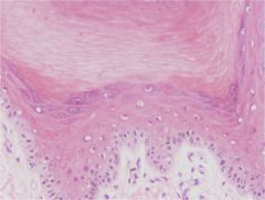
equine foot
|
Hoof:
Insensitive keratinized layer Hoof wall: Epidermis that makes the hoof The epidermis at the junction of skin and hoof is called the periploic epidermis |
|
|
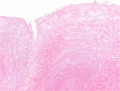
periploic epidermis
|
The epidermis at the junction of skin and hoof
|
|
|
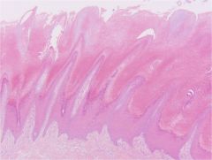
Equine Foot Laminae
|
Epidermal rete pegs and dermal papillae form elongate ridges oriented perpendicular to the ground
There are epidermal and dermal laminae - The epidermal ones are insensitive - The dermal ones are sensitive There are primary and secondary laminae - Primary laminae are the main rete pegs/dermal papillae - Secondary laminae branch off the primary laminae at an acute angle These can be epidermal or dermal SDL PDL SEL PEL – keratinize centrally to make the hoof |
|
|
ruminant & porcine claws
|
Similar to the horse...
NO secondary laminae More primary laminae |