Reading...
![]()
Play button
![]()
Play button
![]()
Use LEFT and RIGHT arrow keys to navigate between flashcards;
Use UP and DOWN arrow keys to flip the card;
H to show hint;
A reads text to speech;
34 Cards in this Set
- Front
- Back
|
Define fertilization
|
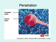
Human fertilization is the union of a human egg and sperm, usually occurring in the ampulla of the uterine tube
|
|
|
Define zygote
|

A zygote (from Greek ζυγωτός zygōtos "joined" or "yoked", from ζυγοῦν zygoun "to join" or "to yoke"),[1] or zygocyte, is the initial cell formed when two gamete cells are joined by means of sexual reproduction.
|
|
|
Define cleavage - when does this first occur?
|
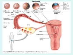
In embryology, cleavage is the division of cells in the early embryo. The zygotes of many species undergo rapid cell cycles with no significant growth, producing a cluster of cells the same size as the original zygote. The different cells derived from cleavage are called blastomeres and form a compact mass called the morula. Cleavage ends with the formation of the blastula.
For testing purposes, this usually happens within the first two days. |
|
|
Define gastrulation
|

Gastrulation is a phase early in the embryonic development of most animals, during which the single-layered blastula is reorganized into a trilaminar ("three-layered") structure known as the gastrula. These three germ layers are known as the ectoderm, mesoderm, and endoderm.
|
|
|
What is a morula? When does it develop?
|

A morula (Latin "morus", mulberry) is an embryo at an early stage of embryonic development, consisting of cells (called blastomeres) in a solid ball contained within the zona pellucida.[1]
For testing purposes, it forms after 3 days. |
|
|
What is a blastula?
|

The blastula (from Greek βλαστός (blastos), meaning "sprout") is a solid sphere of cells formed during an early stage of embryonic development in animals. The blastula is created when the zygote undergoes the cell division process known as cleavage.[1] The blastula is preceded by the morula and is followed by the gastrula in the developmental sequence.
For testing purposes, it occurs after four days. |
|
|
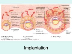
What is implantation?
|
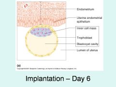
In humans, implantation is the beginning of pregnancy, in which the embryo adheres to the wall of the uterus. At this stage of prenatal development, the embryo is a blastocyst. It is by this adhesion that the fetus receives the oxygen and the nutrients from the mother to be able to grow.
For testing purposes, this occurs between days six and seven |
|
|
What is the difference between an embryo and a fetus?
|
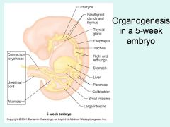
An embryo is still differentiating, whereas a fetus has all major organs, though they may not be fully developed - functionally, an embryo becomes a fetus after organogenesis, approximately 9 weeks after fertilization.
|
|
|
Name the three primary germ layers, and identify the major organs that develop from each.
|
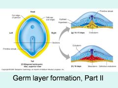
Ectoderm (Protection and feeling)
Skin Nerves Endoderm (Tubing) GI tract Lining of respiratory tract Mesoderm (Everything else) Muscles Bone |
|
|
What is the difference between a blastula and a gastrula?
|

A blastula has a single layer of cells. A gastrula has begun differentiating into the three layers of ectoderm, mesoderm, and endoderm
|
|
|
What is a blastocyst?
|

The blastocyst is a structure formed in the early embryogenesis of mammals, after the formation of the morula. It is a specifically mammalian example of a blastula.[citation needed] It possesses an inner cell mass (ICM), or embryoblast, which subsequently forms the embryo, and an outer layer of cells, or trophoblast, which later forms the placenta. The trophoblast surrounds the inner cell mass and a fluid-filled blastocyst cavity known as the blastocoele or the blastocystic cavity. The human blastocyst comprises 70-100 cells.
|
|
|
What is an inner cell mass, and where would it be located?
|

In early embryogenesis of most eutherian mammals, the inner cell mass (abbreviated ICM and also known as the embryoblast or pluriblast, the latter term being applicable to all mammals) is the mass of cells inside the primordial embryo that will eventually give rise to the definitive structures of the fetus. This structure forms in the earliest steps of development, before implantation into the endometrium of the uterus has occurred. The ICM lies within the blastocoele (more correctly termed "blastocyst cavity," as it is not strictly homologous to the blastocoele of anamniote vertebrates) and is entirely surrounded by the single layer of cells called trophoblast.
|
|
|
What is a trophoblast, and where would it be located?
|

Trophoblasts (from Greek trephein: to feed, and blastos: germinator) are cells forming the outer layer of a blastocyst, which provide nutrients to the embryo and develop into a large part of the placenta. They are formed during the first stage of pregnancy and are the first cells to differentiate from the fertilized egg. This layer of trophoblasts is also collectively referred to as "the trophoblast", or, after gastrulation, the trophectoderm, as it is then contiguous with the ectoderm of the embryo.
|
|
|
What is an amnion, and where would it be located?
|
The amnion is a membrane building the amniotic sac that surrounds and protects an embryo. It is developed in reptiles, birds, and mammals, which are hence called “Amniota”; but not in amphibians and fish (Ichthyopsida), which are consequently termed “Anamniota”. The primary role of this is the protection of the embryo for its development. It stems from parts of the mesoderm on the outer side and the ectoderm on the inner side.
|
|
|
What is an allantois, and where is it located?
|

Allantois (plural allantoides or allantoises) is a part of a developing animal conceptus (which consists of all embryonic and extra-embryonic tissues). It helps the embryo exchange gases and handle liquid waste.
The allantois, along with the amnion and chorion (other embryonic membranes), identify humans as amniotes, along with reptiles, dinosaurs, birds, and other mammals. Of the vertebrates, only Ichthyopsidas (fish and amphibians) lack this structure. |
|
|
What is a yolk sac, and where would you find it?
|

The yolk sac is a membranous sac attached to an embryo, providing early nourishment in the form of yolk in bony fishes, sharks, reptiles, birds, and primitive mammals. It functions as the developmental circulatory system of the human embryo, before internal circulation begins.
|
|
|
What are chorionic villi, and where would you find them?
|

Chorionic villi are villi that sprout from the chorion in order to give a maximum area of contact with the maternal blood.
Embryonic blood is carried to the villi by the branches of the umbilical arteries, and after circulating through the capillaries of the villi, is returned to the embryo by the umbilical veins. Thus, the villi are part of the border between maternal and fetal blood during pregnancy. |
|
|
Describe the process and timing of implantation in humans.
|

In humans, implantation is the beginning of pregnancy, in which the embryo adheres to the wall of the uterus. At this stage of prenatal development, the embryo is a blastocyst. It is by this adhesion that the fetus receives the oxygen and the nutrients from the mother to be able to grow.
For testing purposes, the slides indicate initial implantation at day 6, penetration by the syncytiotrophoblast on day 7, and completion of implantation at day 16 |
|
|
Define decidua basalis - where is it located?
|

That part of the decidua that interacts with the trophoblast is the decidua basalis. The remainder of the decidua is termed the decidua parietalis or decidua vera. Also, there is the decidua capsularis, which grows over the embryo on the luminal side, enclosing it into the endometrium and surrounding the embryo together with decidua basalis.
|
|
|
Define decidua capsularis - where is it located?
|

That part of the decidua that interacts with the trophoblast is the decidua basalis. The remainder of the decidua is termed the decidua parietalis or decidua vera. Also, there is the decidua capsularis, which grows over the embryo on the luminal side, enclosing it into the endometrium and surrounding the embryo together with decidua basalis.
|
|
|
Describe the gross anatomy and general function of the human placenta.
|
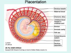
The placenta is an organ that connects the developing fetus to the uterine wall to allow nutrient uptake, waste elimination, and gas exchange via the mother's blood supply. Placentas are a defining characteristic of eutherian or "placental" mammals, but are also found in some snakes and lizards with varying levels of development up to mammalian levels.[1] The word placenta comes from the Latin for cake, from Greek plakóenta/plakoúnta, accusative of plakóeis/plakoús – πλακόεις, πλακούς, "flat, slab-like",[2] in reference to its round, flat appearance in humans. Prototherial (egg-laying) and metatherial (marsupial) mammals produce a choriovitelline placenta that, while connected to the uterine wall, provides nutrients mainly derived from the egg sac. The placenta develops from the same sperm and egg cells that form the fetus, and functions as a fetomaternal organ with two components, the fetal part (Chorion frondosum), and the maternal part (Decidua basalis).
|
|
|
Identify the three basic activities that ensure the formation of a viable offspring.
|

• Increase in cell number and cell growth
• Cellular specialization • Morphogenesis |
|
|
A fetus has umbilical arteries and veins running from the mother to it - how many of each?
|
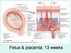
There are usually two umbilical arteries present together with one umbilical vein in the cord.
The umbilical arteries are the only arteries in the human body, aside from the pulmonary arteries, that carry deoxygenated blood. Note that several models in the lab are colored incorrectly for oxygenation - disregard the color on the model and count instead to differentiate. |
|
|
What is an allele?
|
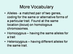
a matched pair of two genes, coding for the same or alternative forms of a particular trait. Found at the same location (locus) on homologous chromosomes.
|
|
|
What is dominance, from a genetic perspective?
|

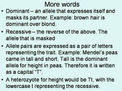
Dominant – an allele that expresses itself and masks its partner. Example: brown hair is dominant over blond.
|
|
|
What is a genotype?
|

the actual alleles an organism has is it’s genotype. In our heterozygote pea plant that would be Tt.
|
|
|
What does it mean to be heterozygous?
|

Heterozygous – having different alleles for the same trait.
|
|
|
What does it mean to be homoozygous?
|

Homozygous - having the same alleles for a trait
|
|
|
What is incomplete dominance?
|
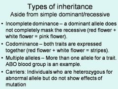
Incomplete dominance – a dominant allele does not completely mask the recessive (red flower + white flower = pink flower).
|
|
|
What is a phenotype?
|
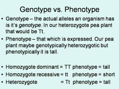
Phenotype – that which is expressed. Our pea plant maybe genotypically heterozygotic but phenotypically it is tall.
|
|
|
What is a recessive trait?
|
A trait that may be masked by a dominant trait
|
|
|
What is a Punnett square?
|
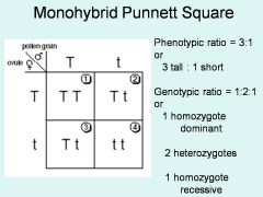
A grid describing all possible combinations from genetic donors
|
|
|
What is meant by sex-linked inheritance?
|
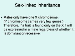
Males only have one X chromosome. (Y chromosome carries very few genes.) Therefore, if a trait is found only on the X it will be expressed in a male regardless of whether it is dominant or recessive.
|
|
|
What is the difference between autosomes and sex chromosomes?
|
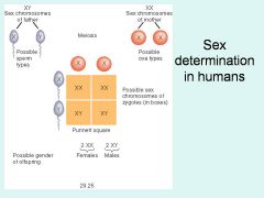
An autosome is a chromosome that is not a sex chromosome, or allosome; that is to say, there is an equal number of copies of the chromosome in males and females.[1] For example, in humans, there are 22 pairs of autosomes. In addition to autosomes, there are sex chromosomes, to be specific: X and Y. So, humans have 23 pairs of chromosomes.
|